. 2024 Jun 20;9(2):108.
doi: 10.3390/jfmk9020108.

Upper-Limb Muscle Fatigability in Para-Athletes Quantified as the Rate of Force Development in Rapid Contractions of Submaximal Amplitude

Affiliations

Upper-Limb Muscle Fatigability in Para-Athletes Quantified as the Rate of Force Development in Rapid Contractions of Submaximal Amplitude

Gennaro Boccia et al. J Funct Morphol Kinesiol. .

Abstract

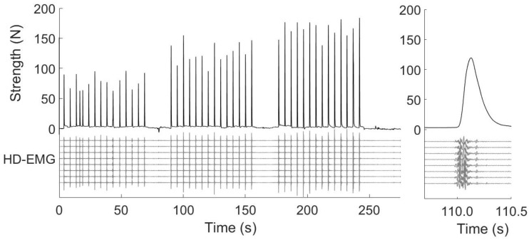
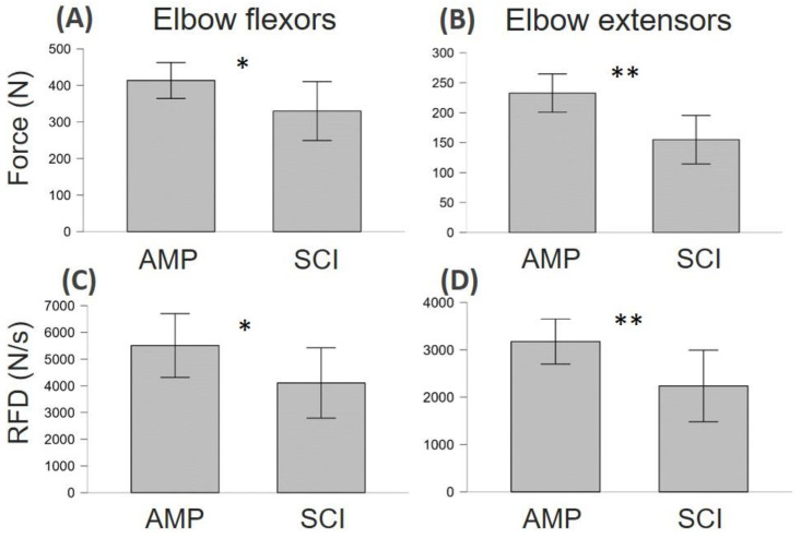
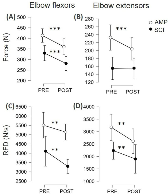
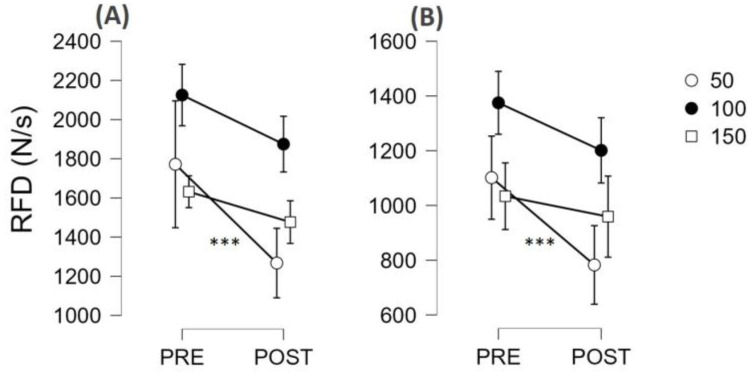
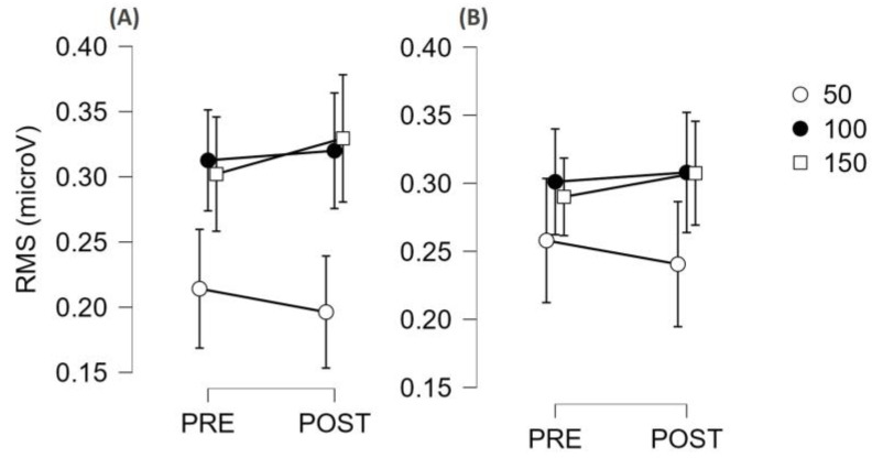
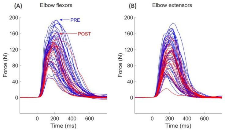
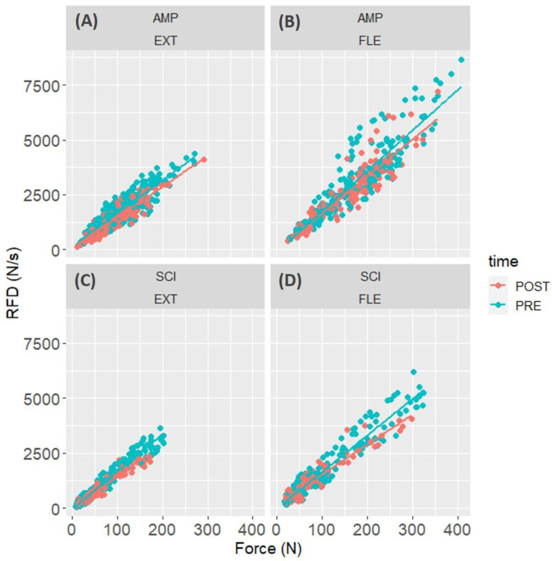
This study aimed to compare neuromuscular fatigability of the elbow flexors and extensors between athletes with amputation (AMP) and athletes with spinal cord injury (SCI) for maximum voluntary force (MVF) and rate of force development (RFD). We recruited 20 para-athletes among those participating at two training camps (2022) for Italian Paralympic veterans. Ten athletes with SCI (two with tetraplegia and eight with paraplegia) were compared to 10 athletes with amputation (above the knee, N = 3; below the knee, N = 6; forearm, N = 1). We quantified MVF, RFD at 50, 100, and 150 ms, and maximal RFD (RFDpeak) of elbow flexors and extensors before and after an incremental arm cranking to voluntary fatigue. We also measured the RFD scaling factor (RFD-SF), which is the linear relationship between peak force and peak RFD quantified in a series of ballistic contractions of submaximal amplitude. SCI showed lower levels of MVF and RFD in both muscle groups (all p values ≤ 0.045). Despite this, the decrease in MVF (Cohen's d = 0.425, p < 0.001) and RFDpeak (d = 0.424, p = 0.003) after the incremental test did not show any difference between pathological conditions. Overall, RFD at 50 ms showed the greatest decrease (d = 0.741, p < 0.001), RFD at 100 ms showed a small decrease (d = 0.382, p = 0.020), and RFD at 150 ms did not decrease (p = 0.272). The RFD-SF decreased more in SCI than AMP (p < 0.0001). Muscle fatigability impacted not only maximal force expressions but also the quickness of ballistic contractions of submaximal amplitude, particularly in SCI. This may affect various sports and daily living activities of wheelchair users. Early RFD (i.e., ≤50 ms) was notably affected by muscle fatigability.

Keywords: disability; explosive strength; fatigability; sport.


Conflict of interest statement

The authors declare no conflicts of interest.

Figures

Figure 1
Figure 1
In the left panel, the upper part of the graph shows a representative example of the force signal recorded during the evaluation of the elbow extensor muscles of an amputee subject. The lower part of the graph shows the electromyographic (HD-EMG) signals from one of the four columns of electrodes that are part of the array placed on the triceps brachii (lateral head). In the right panel, the magnification of one repetition is plotted. As can be seen, the active phase of the muscle contraction lasts about 200 ms.
Figure 2
Figure 2
The level of maximum voluntary force (MVF) and rate of force development (RFD) of the elbow flexor and extensor muscles in athletes with amputation (AMP) and athletes with a spinal cord injury (SCI) for elbow flexors (A,C) and elbow extensors (B,D). * p < 0.05, ** p < 0.01.
Figure 3
Figure 3
Maximum voluntary force (MVF) and rate of force development (RFDpeak) of the elbow flexor and extensor muscles in athletes with amputation (AMP) and athletes with a spinal cord injury (SCI) are reported for PRE and POST for elbow flexors (A,C) and elbow extensors (B,D). ** p < 0.01, *** p < 0.001.
Figure 4
Figure 4
Rate of force development (RFD) values (mean and 95% CI) before (PRE) and after (POST) a maximal arm ergometer test (fatiguing task) are reported. Values are shown for the elbow flexor (A) and elbow extensor (B) muscles and are reported separately for 50, 100, and 150 ms time intervals. As the condition did not emerge as a significant factor, the two groups (AMP and SCI) were merged. *** p < 0.001.
Figure 5
Figure 5
Electromyographic signal amplitude (RMS) values (mean and 95% CI) before (PRE) and after (POST) a maximal arm ergometer test (fatiguing task) are reported. Values are shown for the elbow flexor (A) and elbow extensor (B) muscles and are reported separately for 50, 100, and 150 ms time intervals. As the condition did not emerge as a significant factor, the two groups (AMP and SCI) were merged.
Figure 6
Figure 6
Representative example of the force recorded during all explosive muscle contractions performed before (blue) and after (red) the fatiguing task in an amputee subject in elbow flexors (A) and extensors (B).
Figure 7
Figure 7
Peak rate of force development (RFD) is plotted against the peak force reached in each contraction of the RFD-SF protocol before (PRE) and after (POST) the fatiguing task. Values are shown for the elbow extensor (EXT, (A,C)) and elbow flexor muscles (FLE, (B,D)) separately for subjects with amputation (AMP, (A,B)) and with spinal cord injury (SCI, (C,D)).

Similar articles

References

    1. Qi L., Zhang L., Lin X.-B., Ferguson-Pell M. Wheelchair propulsion fatigue thresholds in electromyographic and ventilatory testing. Spinal Cord. 2020;58:1104–1111. doi: 10.1038/s41393-020-0470-2. - DOI - PubMed
    1. Van der Woude L.H., de Groot S., Janssen T.W. Manual wheelchairs: Research and innovation in rehabilitation, sports, daily life and health. Med. Eng. Phys. 2006;28:905–915. doi: 10.1016/j.medengphy.2005.12.001. - DOI - PubMed
    1. Rodgers M.M., McQuade K.J., Rasch E.K., Keyser R.E., Finley M.A. Upper-limb fatigue-related joint power shifts in experienced wheelchair users and nonwheelchair users. J. Rehabil. Res. Dev. 2003;40:27–38. doi: 10.1682/JRRD.2003.01.0027. - DOI - PubMed
    1. Gandevia S.C. Spinal and supraspinal factors in human muscle fatigue. Physiol. Rev. 2001;81:1725–1789. doi: 10.1152/physrev.2001.81.4.1725. - DOI - PubMed
    1. Maffiuletti N.A., Aagaard P., Blazevich A.J., Folland J., Tillin N., Duchateau J. Rate of force development: Physiological and methodological considerations. Eur. J. Appl. Physiol. 2016;116:1091–1116. doi: 10.1007/s00421-016-3346-6. - DOI - PMC - PubMed

LinkOut - more resources