. 2024 Jun 18;13(6):514.
doi: 10.3390/pathogens13060514.

Adaptive Cellular Responses following SARS-CoV-2 Vaccination in Primary Antibody Deficiency Patients

Affiliations

Adaptive Cellular Responses following SARS-CoV-2 Vaccination in Primary Antibody Deficiency Patients

Sudhir Gupta et al. Pathogens. .

Abstract

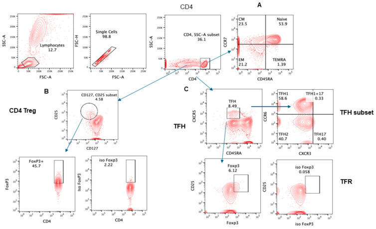
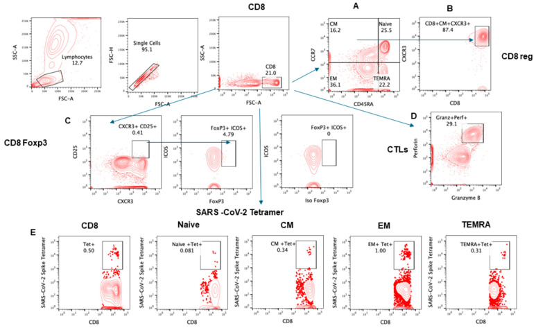
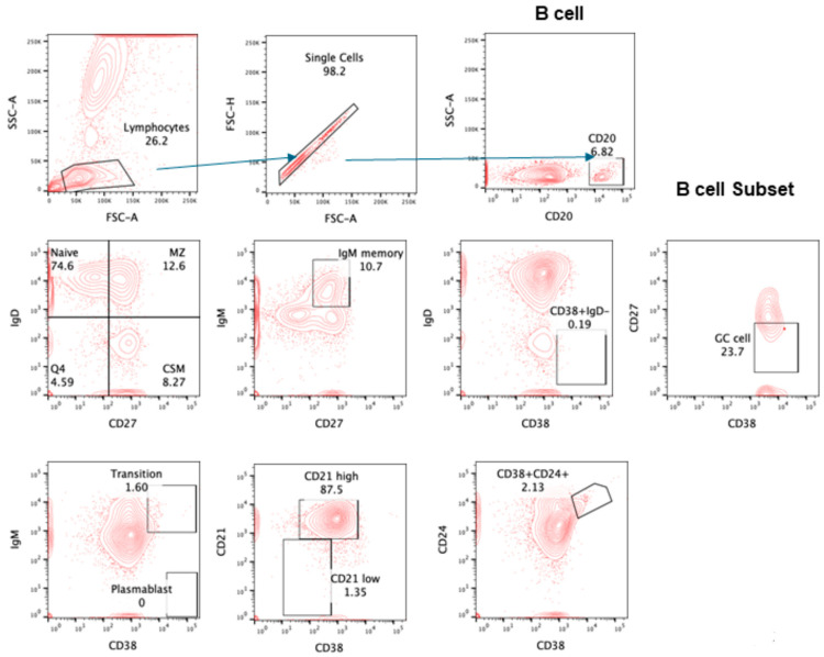
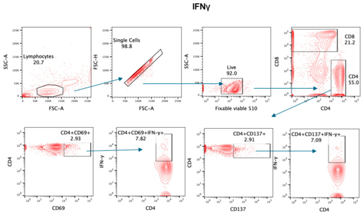
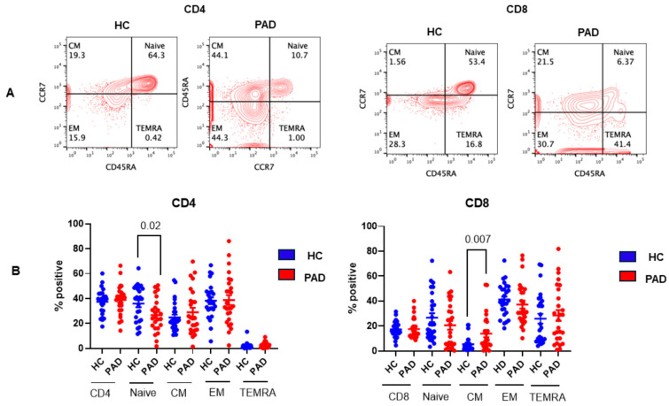
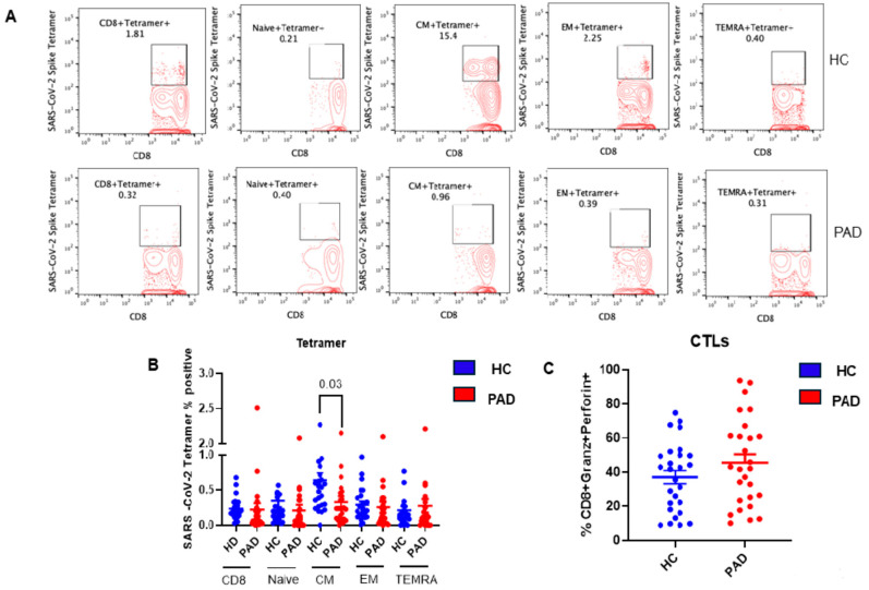
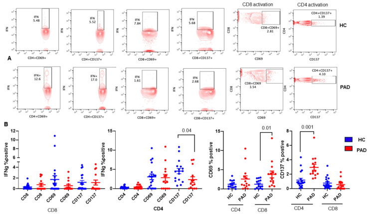
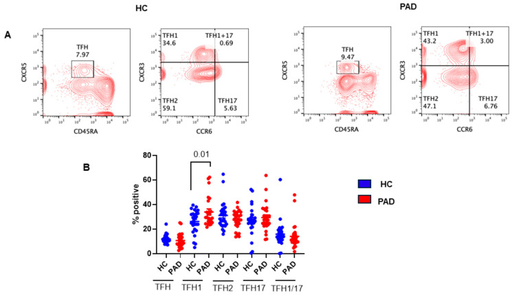
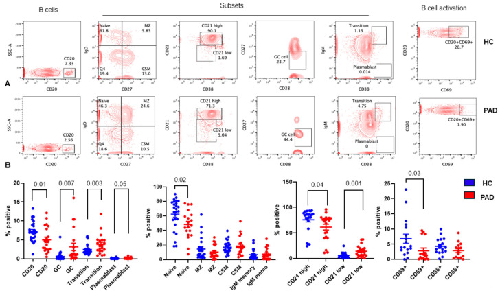
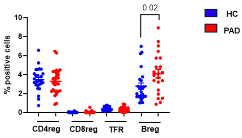
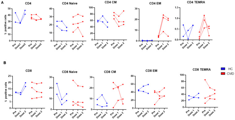
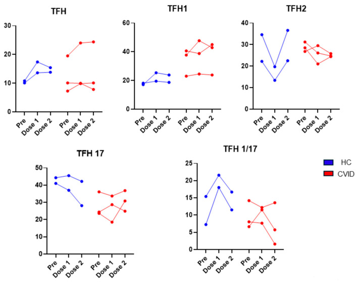
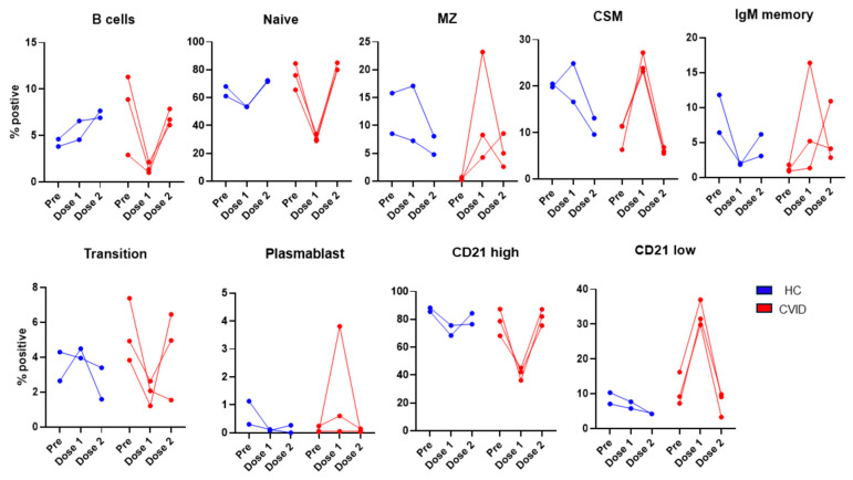
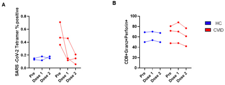
Since the start of the COVID-19 pandemic, in a short span of 3 years, vaccination against SARS-CoV-2 has resulted in the end of the pandemic. Patients with inborn errors of immunity (IEI) are at an increased risk for SARS-CoV-2 infection; however, serious illnesses and mortality, especially in primary antibody deficiencies (PADs), have been lower than expected and lower than other high-risk groups. This suggests that PAD patients may mount a reasonable effective response to the SARS-CoV-2 vaccine. Several studies have been published regarding antibody responses, with contradictory reports. The current study is, perhaps, the most comprehensive study of phenotypically defined various lymphocyte populations in PAD patients following the SARS-CoV-2 vaccine. In this study, we examined, following two vaccinations and, in a few cases, prior to and following the 1st and 2nd vaccinations, subsets of CD4 and CD8 T cells (Naïve, TCM, TEM, TEMRA), T follicular helper cells (TFH1, TFH2, TFH17, TFH1/17), B cells (naïve, transitional, marginal zone, germinal center, IgM memory, switched memory, plasmablasts, CD21low), regulatory lymphocytes (CD4Treg, CD8Treg, TFR, Breg), and SARS-CoV-2-specific activation of CD4 T cells and CD8 T cells (CD69, CD137), SARS-CoV-2 tetramer-positive CD8 T cells, and CD8 CTL. Our data show significant alterations in various B cell subsets including Breg, whereas only a few subsets of various T cells revealed alterations. These data suggest that large proportions of PAD patients may mount significant responses to the vaccine.

Keywords: Breg; CD4 Treg; CD8 Treg; COVID-19 vaccine; CVID; Cytotoxic T cells; IFNγ; TFR.


Conflict of interest statement

All authors declare no conflicts of interest. The funder had no role in the design of the study; in the collection, analyses, or interpretation of data; in the writing of the manuscript; or in the decision to publish the results in Pathogens.

Figures

Figure 1
Figure 1
Gating strategy for subsets of CD4+ T cells. Contour plot was used for gating strategy; gated lymphocytes were analyzed for singlet and CD4-expressing cells. These CD4+ cells were further analyzed for (A) CD4 subsets naïve: CCR7+CD45RA+, central memory (CM): CCR7+CD45RA−, effector memory (EM): CCR7−CD45RA−, and T effector memory: CD45RA+ (TEMRA) CCR7− CD45RA+. (B) Treg CD25+CD127 low cells expressing FoxP3. (C) Follicular helper cells (TFH) CXCR5+CD45RA−; subsets of TFH cells were further analyzed—TFH1: CXCR3+CCR6−, TFH2: CXCR3−CCR6−, TFH17: CXCR3−CCR6+.
Figure 2
Figure 2
Gating strategy for subsets of CD8+ T cells. Contour plot was used for gating strategy; gated lymphocytes were analyzed for singlet and CD8-expressing cells. These CD8+ cells were further analyzed for (A) CD4 subsets naïve: CCR7+CD45RA+, central memory (CM): CCR7+CD45RA−, effector memory (EM): CCR7−CD45RA−, and T effector memory RA+ (TEMRA): CCR7−CD45RA+. (B) CD8Treg CXCR3 expressing CM: CCR7+CD45RA−. (C) FoxP3 and ICOS+ expression in CD25+CXCR3+ CD8 cells. (D) CD8+ cells were analyzed for granzyme B and perforin expression. (E) SARS-CoV-2 spike protein-specific, tetramer-positive cells in total CD8 and subsets.
Figure 3
Figure 3
Gating strategy for subsets of CD20+ B cells. Contour plot was used for gating strategy; gated lymphocytes were analyzed for singlet and CD20-expressing B cells. These B cells were further analyzed for naïve: IgD+CD27−, MZ: IgD+CD27+, CSM: IgD−CD27+, IgM memory: IgM+CD27+, transition B cells: IgM+CD38+, plasma blast: IgM−CD38+, CD21High and CD21Low, Breg: CD24+CD38+, and GC cells: IgD−CD38+CD27−.
Figure 4
Figure 4
Gating for intracellular IFNγ. Representative contour plot was used for gating strategy; gated lymphocytes were analyzed for singlet, live, and CD4/CD8-expressing cells. These CD4+ cells were further analyzed for IFNγ in CD69+ and CD137+ cells.
Figure 5
Figure 5
Subsets of CD4+ and CD8+ T cells in the HC and PAD patients following two doses of SARS-CoV-2 vaccine. A representative contour flow cytograph is shown (A). Cumulative data show significantly decreased naïve CD4+ T cells and significantly decreased CD8TCM cells in PAD patients as compared to the HC (B).
Figure 6
Figure 6
SARS-CoV-2-specific, tetramer-positive CD8+ T cell subsets and CD8 CLT cells following two doses of SARS-CoV-2 vaccine. A representative contour flow cytograph for IFNγ+ in various subsets of CD8+ T cells in the HC and PAD patients (A). Cumulative data for the HC and PAD patients for IFNγ+ CD8 T cell subsets (B) and CD8 CTL cells (C) show significantly decreased tetramer-positive CD8 TCM cells in the PAD patients (B).
Figure 7
Figure 7
Effect of in vitro activation with SARS-CoV-2 protein on activation of CD4 and CD8 T cells and IFNγ+ CD4 and CD8 T cells. PBMC were activated with SARS-CoV-2 spike protein, activation of CD4 and CD8 was examined by the expression of CD69 and CD137, and intracellular IFNγ was examined in CD69+ and CD137+ CD4 and CD8 T cells. A representative contour flow cytograph for HC and PAD is shown (A). Cumulative data show significantly increased CD8+CD69+ T cells and CD4+CD137+ T cells and significantly decreased C4+CD137+IFNγ+ T cells in the PAD group (B).
Figure 8
Figure 8
TFH cells and subsets of TFH cells in HC and PAD following two doses of SARS-CoV-2 vaccine. A representative contour flow cytograph in HC and PAD (A). Cumulative data show a significant increase in TFH1 cells (B).
Figure 9
Figure 9
B cells and B cell subsets in HC and PAD following two doses of SARS-CoV-2 vaccine. Various subsets of B cells and activation by the expression of CD69 and CD86 on CD20+ B cells were examined. A representative contour flow cytograph is shown in (A). Cumulative data (B) show significantly reduced naïve B cells and CD20+ B cells and significantly increased transitional B cells, GC B cells, and plasmablasts in the PAD group as compared to the HC.
Figure 10
Figure 10
Regulatory lymphocytes following two doses of SARS-CoV-2 vaccine. CD4Treg, CD8Treg, TFR, and Breg were examined in the HC and CVID patients. Data show significantly increased Breg cells in CVID patients as compared to the HC.
Figure 11
Figure 11
Subsets of CD4 (A) and CD8 (B) prior to and following 1st dose and 2nd doses of SARS-CoV-2 vaccine. Though variable changes were observed in both HC and CVID, CVID patients exhibited increased CD4TEM and CD4TEMRA as compared to HC (A). CD8 responses were highly variable in both HC and CVID.
Figure 12
Figure 12
Subsets of TFH cells prior to and following 1st dose and 2nd doses of SARS-CoV-2 vaccine. Basal levels of TFH1 were higher and remained high after vaccination in CVID. TFH2 were markedly reduced after the 1st dose in HC, whereas they were modestly reduced in CVID. TFH1/17 increased after the 1st dose in HC, whereas in CVID, two of three subjects showed no increase.
Figure 13
Figure 13
Subsets of B cells prior to and following 1st dose and 2nd dose of SARS-CoV-2 vaccine. CD20+ B cells increased after 1st and 2nd doses of vaccine, whereas CVID patients showed a marked decrease following 1st dose and recovered, to a large extent, following 2nd dose. Following 1st dose of vaccine, MZ, CSM, IgM memory, plasmablasts, and CD21low increased markedly, and transitional cells decreased markedly in CVID as compared to HC.
Figure 14
Figure 14
Regulatory lymphocytes prior to and following 1st dose and 2nd dose of SARS-CoV-2 vaccine. CD4Treg increased after 2nd dose in HC; however, no such increase was observed in CVID. TFR cells increased after 1st and 2nd doses in both HC and CVID. A reverse effect was observed for CD8Treg. In HC, CD8Treg markedly increased whereas in CVID CD8Treg markedly decreased after 1st dose of vaccine.
Figure 15
Figure 15
SARS-CoV-2 tetramer-positive (A) and CTL CD8 T cells (B) prior to and following 1st dose and 2nd dose of SARS-CoV-2 vaccine. SARS-CoV-2+ CD8+ T cells markedly decreased following 1st and 2nd vaccinations, and only minor changes were observed in HC (A). CTL CD8 T cells were similar in both groups.

Similar articles

Cited by

References

    1. Baden L.R., El Sahly H.M., Essink B., Kotloff K., Frey S., Novak R., Diemert D., Spector S.A., Rouphael N., Creech C.B., et al. Efficacy and Safety of the mRNA-1273 SARS-CoV-2 Vaccine. N. Engl. J. Med. 2021;384:403–416. doi: 10.1056/NEJMoa2035389. - DOI - PMC - PubMed
    1. Anderson E.J., Rouphael N.G., Widge A.T., Jackson L.A., Roberts P.C., Makhene M., Chappell J.D., Denison M.R., Stevens L.J., Pruijssers A.J., et al. Safety and Immunogenicity of SARS-CoV-2 mRNA-1273 Vaccine in Older Adults. N. Engl. J. Med. 2020;383:2427–2438. doi: 10.1056/NEJMoa2028436. - DOI - PMC - PubMed
    1. McMenamin M.E., Nealon J., Lin Y., Wong J.Y., Cheung J.K., Lau E.H.Y., Wu P., Leung G.M., Cowling B.J. Vaccine effectiveness of one, two, and three doses of BNT162b2 and CoronaVac against COVID-19 in Hong Kong: A population-based observational study. Lancet Infect. Dis. 2022;22:1435–1443. doi: 10.1016/S1473-3099(22)00345-0. - DOI - PMC - PubMed
    1. Sette A., Crotty S. Adaptive immunity to SARS-CoV-2 and COVID-19. Cell. 2021;184:861–880. doi: 10.1016/j.cell.2021.01.007. - DOI - PMC - PubMed
    1. Tarke A., Sidney J., Kidd C.K., Dan J.M., Ramirez S.I., Yu E.D., Mateus J., da Silva Antunes R., Moore E., Rubiro P., et al. Comprehensive analysis of T cell immunodominance and immunoprevalence of SARS-CoV-2 epitopes in COVID-19 cases. Cell Rep. Med. 2021;2:100204. doi: 10.1016/j.xcrm.2021.100204. - DOI - PMC - PubMed

Grants and funding