

Antimicrobial Peptide Reduces Cytotoxicity and Inflammation in Canine Epidermal Keratinocyte Progenitor Cells Induced by Pseudomonas aeruginosa Infection
- PMID: 38921982
- PMCID: PMC11209461
- DOI: 10.3390/vetsci11060235
Antimicrobial Peptide Reduces Cytotoxicity and Inflammation in Canine Epidermal Keratinocyte Progenitor Cells Induced by Pseudomonas aeruginosa Infection
Abstract
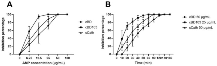
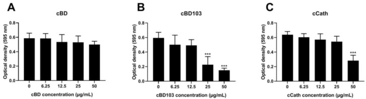
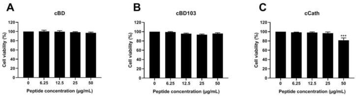
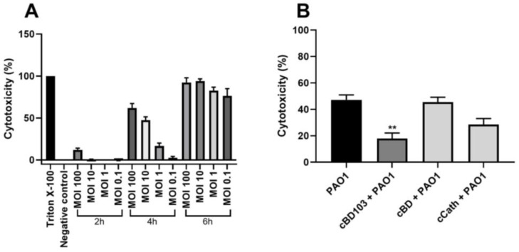
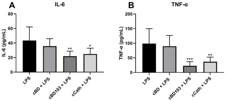
The direct effects and antimicrobial activity of synthetic antimicrobial peptides (AMPs) obtained from dogs, including cBD, cBD103, and cCath, against P. aeruginosa wild-type strain PAO1 and canine keratinocytes were analyzed. Antibacterial effects on planktonic bacteria were assessed by determining the minimum bactericidal concentrations (MBCs) of AMPs and by a time-kill assay. Antibiofilm effects were assessed using the microtiter plate assay. We also evaluated the effects of AMPs on cell cytotoxicity and host immune response induced by stimulating canine epidermal keratinocyte progenitor (CPEK) cells with PAO1 and its LPS. cBD, cBD103, and cCath all exhibited dose-dependent antimicrobial and antibiofilm effects. In particular, 25 μg/mL cBD103 showed rapid bactericidal activity within 60 min and inhibited biofilm formation. In addition, pretreatment with cBD103 (25 µg/mL) and cCath (50 µg/mL) 1 h before stimulation significantly reduced the cytotoxicity of the CPEK cells by PAO1 and LPS-induced IL-6 and TNF-a expressions. cBD had little effect on the response to PAO1 and LPS in the cells. These results indicate the therapeutic potential of AMPs in P. aeruginosa skin infections. However, further studies on the mechanism of action of AMPs in keratinocytes and clinical trials are needed.
Keywords: antibacterial activity; antibiofilm effect; antimicrobial peptides; dogs; keratinocytes; lipopolysaccharides; pseudomonas aeruginosa.
Conflict of interest statement
The authors declare no conflicts of interest.
Figures
Similar articles
-
Evaluation of the in vitro effect of Boldo and Meadowsweet plant extracts on the expression of antimicrobial peptides and inflammatory markers in canine keratinocytes.Res Vet Sci. 2017 Dec;115:255-262. doi: 10.1016/j.rvsc.2017.05.021. Epub 2017 May 17. Res Vet Sci. 2017. PMID: 28549300
-
Evaluation of canine antimicrobial peptides in infected and noninfected chronic atopic skin.Vet Dermatol. 2013 Feb;24(1):39-47.e10. doi: 10.1111/j.1365-3164.2012.01091.x. Vet Dermatol. 2013. PMID: 23331678
-
Inhibitory effect of a novel chicken-derived anti-biofilm peptide on P. aeruginosa biofilms and virulence factors.Microb Pathog. 2020 Dec;149:104514. doi: 10.1016/j.micpath.2020.104514. Epub 2020 Sep 22. Microb Pathog. 2020. PMID: 32976967
-
Microbiome-derived antimicrobial peptides offer therapeutic solutions for the treatment of Pseudomonas aeruginosa infections.NPJ Biofilms Microbiomes. 2022 Aug 29;8(1):70. doi: 10.1038/s41522-022-00332-w. NPJ Biofilms Microbiomes. 2022. PMID: 36038584 Free PMC article.
-
Inhibitory Effects of Antimicrobial Peptides on Lipopolysaccharide-Induced Inflammation.Mediators Inflamm. 2015;2015:167572. doi: 10.1155/2015/167572. Epub 2015 Nov 3. Mediators Inflamm. 2015. PMID: 26612970 Free PMC article. Review.
References
LinkOut - more resources
Full Text Sources
