

Vaulting further: cranial vault expansion for craniocerebral disproportion without primary craniosynostosis
- PMID: 38922367
- PMCID: PMC11579096
- DOI: 10.1007/s00381-024-06517-2
Vaulting further: cranial vault expansion for craniocerebral disproportion without primary craniosynostosis
Abstract
Purpose: Treatment of subjects with refractory idiopathic intracranial hypertension (IIH) or shunted hydrocephalus with chronic shunt complications is challenging. What is the role for cranial vault expansion, particularly utilizing posterior vault distraction osteogenesis (PVDO), in these cases? This study assesses medium-term efficacy of cranial vault expansion in this unique patient population.
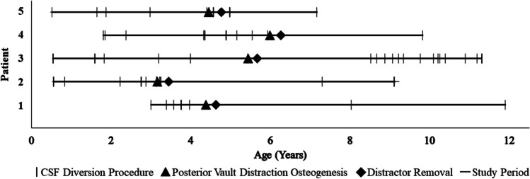
Methods: A retrospective review was conducted of patients who underwent cranial vault expansion from 2008 to 2023 at the Children's Hospital of Philadelphia. Subjects who did not have a diagnosis of primary craniosynostosis were included in the study. Demographic information, medical history, and perioperative details were collected from medical records. Primary outcomes were the rate of CSF diversion procedures and resolution of presenting signs and symptoms. Secondary outcomes were perioperative and 90-day complications and reoperation requirement.
Results: Among 13 included subjects, nine (69.2%) patients had a primary diagnosis of shunted hydrocephalus and 4 (30.8%) patients had IIH. Twelve (92.3%) subjects underwent posterior vault distraction osteogenesis (PVDO) and one (7.7%) underwent posterior vault remodeling (PVR). All 4 patients with IIH demonstrated symptomatic improvement following PVDO, including resolution of headaches, vomiting, and/or papilledema. Among 9 patients with shunted hydrocephalus, CSF diversion requirement decreased from 2.7 ± 1.6 procedures per year preoperatively to 1.2 ± 1.8 per year following cranial vault expansion (p = 0.030). The mean postoperative follow-up was 4.1 ± 2.1 years and four (30.8%) patients experienced complications within 90 days of surgery, including infection (n = 2), CSF leak (n = 1), and elevated ICP requiring lumbar puncture (n = 1). Four (30.8%) patients underwent repeat cranial vault expansion for recurrence of ICP-related symptoms. At most recent follow-up, 7 of 9 patients with shunted hydrocephalus demonstrated symptomatic improvement.
Conclusion: Cranial vault expansion reduced intracranial hypertension-related symptomology as well as the rate of CSF diversion-related procedures in patients with refractory IIH and shunted hydrocephalus without craniosynostosis, and should be considered in those who have significant shunt morbidity.
Keywords: Cranial vault expansion; Craniocerebral disproportion; Idiopathic intracranial hypertension; PVDO; Shunted hydrocephalus.
© 2024. The Author(s).
Conflict of interest statement
Declarations. Competing interests: The authors declare no competing interests. Conflict of interest: The authors have no relevant financial or non-financial interest to disclose. No funding was received to assist with the preparation of this manuscript.
Figures

References
-
- Utria AF, Mundinger GS, Bellamy JL et al (2015) The importance of timing in optimizing cranial vault remodeling in syndromic craniosynostosis. Plast Reconstr Surg 135(4):1077–1084. 10.1097/PRS.0000000000001058 - PubMed
-
- Bartels MC, Vaandrager JM, de Jong TH, Simonsz HJ (2004) Visual loss in syndromic craniosynostosis with papilledema but without other symptoms of intracranial hypertension. J Craniofac Surg. 15(6):1019–22; discussion 1023–4. 10.1097/00001665-200411000-00026 - PubMed
-
- Auricchio AM, Bohnen A, Nichelatti M, Cenzato M, Talamonti G (2022) Management of slit ventricle syndrome: a single-center case series of 32 surgically treated patients. World Neurosurg 158:e352–e361. 10.1016/j.wneu.2021.10.183 - PubMed
-
- Ko MW, Liu GT (2010) Pediatric idiopathic intracranial hypertension (pseudotumor cerebri). Horm Res Paediatr 74(6):381–389. 10.1159/000321180 - PubMed
MeSH terms
LinkOut - more resources
Full Text Sources
Medical
Research Materials
