

Evaluation of Neonatal Cerebral Circulation Under Hypoxic Ischemic Risk Factors Based on Quantitative Analysis of Cerebral Veins with Magnetic Resonance Susceptibility Weighted Imaging
- PMID: 38922421
- PMCID: PMC11564194
- DOI: 10.1007/s00062-024-01432-0
Evaluation of Neonatal Cerebral Circulation Under Hypoxic Ischemic Risk Factors Based on Quantitative Analysis of Cerebral Veins with Magnetic Resonance Susceptibility Weighted Imaging
Abstract
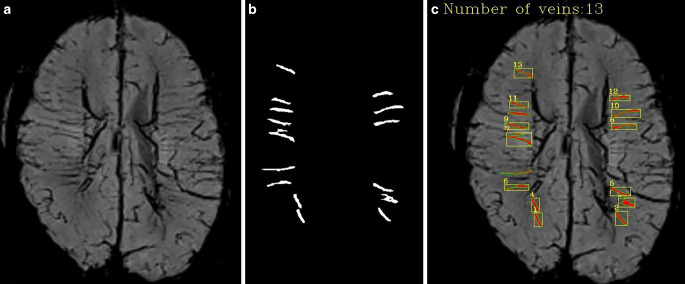
Purpose: To observe the regulation of cerebral circulation in vivo based on image segmentation algorithms for deep learning in medical imaging to automatically detect and quantify the neonatal deep medullary veins (DMVs) on susceptibility weighted imaging (SWI) images. To evaluate early cerebral circulation self-rescue for neonates undergoing risk of cerebral hypoxia-ischaemia in vivo.
Methods: SWI images and clinical data of 317 neonates with or without risk of cerebral hypoxia-ischaemia were analyzed. Quantitative parameters showing the number, width, and curvature of DMVs were obtained using an image segmentation algorithm.
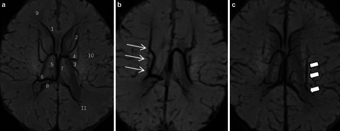
Results: The number of DMVs was greater in males than in females (p < 0.01), and in term than in preterm infants (p = 0.001). The width of DMVs was greater in term than in preterm infants (p < 0.01), in low-risk than in high-risk group (p < 0.01), and in neonates without intracranial extracerebral haemorrhage (ICECH) than with ICECH (p < 0.05). The curvature of DMVs was greater in term than in preterm infants (P < 0.05). The width of both bilateral thalamic veins and anterior caudate nucleus veins were positively correlated with the number of DMVs; the width of bilateral thalamic veins was positively correlated with the width of DMVs.
Conclusion: The DMVs quantification based on image segmentation algorithm may provide more detailed and stable quantitative information in neonate. SWI vein quantification may be an observable indicator for in vivo assessment of cerebral circulation self-regulation in neonatal hypoxic-ischemic brain injury.
Keywords: DMVs; Image Segmentation Algorithm; MRI; Neonatal Hypoxic-Ischemia; SWI.
© 2024. The Author(s).
Conflict of interest statement
Figures
Similar articles
-
Texture analysis of deep medullary veins on susceptibility-weighted imaging in infants: evaluating developmental and ischemic changes.Eur Radiol. 2020 May;30(5):2594-2603. doi: 10.1007/s00330-019-06618-6. Epub 2020 Feb 5. Eur Radiol. 2020. PMID: 32025833
-
The relationship between deep medullary veins score and the severity and distribution of intracranial microbleeds.Neuroimage Clin. 2019;23:101830. doi: 10.1016/j.nicl.2019.101830. Epub 2019 Apr 22. Neuroimage Clin. 2019. PMID: 31039526 Free PMC article.
-
Incidence and clinical analysis of asymptomatic intracranial hemorrhage in neonates with cerebral hypoxic-ischaemic risk based on multisequence MR images.Sci Rep. 2024 Jun 26;14(1):14721. doi: 10.1038/s41598-024-62473-6. Sci Rep. 2024. PMID: 38926428 Free PMC article.
-
Deep medullary vein engorgement and superficial medullary vein engorgement: two patterns of perinatal venous stroke.Pediatr Radiol. 2021 May;51(5):675-685. doi: 10.1007/s00247-020-04846-3. Epub 2020 Oct 22. Pediatr Radiol. 2021. PMID: 33090246 Review.
-
Susceptibility weighted imaging in acute cerebral ischemia: review of emerging technical concepts and clinical applications.Neuroradiol J. 2017 Apr;30(2):109-119. doi: 10.1177/1971400917690166. Epub 2017 Jan 1. Neuroradiol J. 2017. PMID: 28424015 Free PMC article. Review.
References
-
- Kinney HC. The near-term (late preterm) human brain and risk for periventricular leukomalacia: a review. Semin Perinatol. 2006;30(2):81–8. 10.1053/j.semperi. - PubMed
-
- Verschuuren S, Poretti A, Buerki S, Lequin MH, Huisman TA. Susceptibility-weighted imaging of the pediatric brain. Am J Roentgenol. 2012;198(5):W440–9. 10.2214/AJR.11.8049. - PubMed
MeSH terms
Grants and funding
LinkOut - more resources
Full Text Sources
Medical
