Review
. 2024 Jul;18(7):1695-1718.
doi: 10.1002/1878-0261.13691. Epub 2024 Jun 22.

Metabolic dialogues: regulators of chimeric antigen receptor T cell function in the tumor microenvironment

Affiliations
Review

Metabolic dialogues: regulators of chimeric antigen receptor T cell function in the tumor microenvironment

Josquin Moraly et al. Mol Oncol. 2024 Jul.

Abstract

Tumor-infiltrating lymphocytes (TILs) and chimeric antigen receptor (CAR) T cells have demonstrated remarkable success in the treatment of relapsed/refractory melanoma and hematological malignancies, respectively. These treatments have marked a pivotal shift in cancer management. However, as "living drugs," their effectiveness is dependent on their ability to proliferate and persist in patients. Recent studies indicate that the mechanisms regulating these crucial functions, as well as the T cell's differentiation state, are conditioned by metabolic shifts and the distinct utilization of metabolic pathways. These metabolic shifts, conditioned by nutrient availability as well as cell surface expression of metabolite transporters, are coupled to signaling pathways and the epigenetic landscape of the cell, modulating transcriptional, translational, and post-translational profiles. In this review, we discuss the processes underlying the metabolic remodeling of activated T cells, the impact of a tumor metabolic environment on T cell function, and potential metabolic-based strategies to enhance T cell immunotherapy.

Keywords: T cells; anti‐tumor immunotherapy; chimeric antigen receptor; immunometabolism; nutrient transporters; tumor microenvironment.


Conflict of interest statement

The authors declare no conflict of interest.

Figures

Fig. 1
Fig. 1
Crosstalk between T cell receptor signaling and metabolic pathways. TCR/CD28 signaling induces the expression of SLC transporters that regulate the entry of a large array of metabolites—glucose, glutamine, leucine, arginine, methionine, and iron, among others—enabling a massive induction of downstream pathways. The crosstalk between TCR signaling and intracellular metabolites modulates multiple critical cellular processes including, but not limited to, mTOR signaling, calcium/NFAT signaling, protein translation, N‐glycosylation, and epigenome remodeling. Ac, acetyl group; AKT, protein kinase C; ATP, adenosine triphosphate; Ca2+, calcium; CD28, cluster of differentiation 28; CytC, cytochrome C; EIF5A, elongation initiation factor 5A; ETC electron transport chain; Fe, iron; GAPDH, glyceraldehyde‐3‐phosphate dehydrogenase; IFNγ, interferon gamma; JMDJ, jumonji C‐domain lysine demethylases; LDHA, lactate dehydrogenase A; Me, methyl group; MGAT, mannose glycoprotein acetylglucosaminyltransferase; mTOR, mammalian target of rapamycin; NFAT, nuclear factor of activated T cells; PEP, phosphoenol pyruvate; PI3K, phosphoinositide 3‐kinase; ROS, reactive oxygen species; SAM, S‐adenosyl methionine; SERCA, sarcoendoplasmic reticulum calcium ATPase; SLC, solute carrier; TCA, tricarboxylic acid; TCR, T cell receptor; TET, ten‐eleven translocation; Tf, transferrin; VitC, vitamin C; α‐KG, alpha‐ketoglutarate.
Fig. 2
Fig. 2
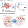
An immune hostile tumor microenvironment: Impact on T cell function. (A) The high uptake of diverse nutrients by tumor cells frequently results in the selective depletion of glutamine, asparagine, aspartate, serine, and arginine within the TME, leading to their decreased availability for anti‐tumor T lymphocytes. Moreover, arginase‐1, produced by tumor‐infiltrating TAM and MDSC, catabolizes arginine to ornithine, further limiting the utilization of arginine by tumor‐infiltrating cytotoxic T cells. The TME is often further compromised by suboptimal gas exchange, resulting in low oxygen concentrations (left). A limited accessibility to nutrients results in decreased mTOR signaling in tumor‐infiltrating T cells which in turn results in lower levels of cell surface metabolite transporters such as GLUT1. Decreases in glycolytic enzymes such as enolase result in the reduced generation of PEP, attenuating Ca2+‐NFAT activity via SERCA activity (right). (B) An abundance of tumor‐related metabolites suppresses T cell function. These include cholesterol, fumarate, lactate, low pH (secondary to high lactate secretion by tumor cells), and kynurenine – converted from tryptophan by IDO. PGE2 inhibits cDC function, thereby impairing T cell immune responses. Furthermore, ATP and ADP, released from dying cells, are converted to AMP and eADO by CD39 and CD73, respectively, on the surface of exhausted T cells. eADO as well as potassium from necrotic tumor tissue suppress T cell activity in the TME. ADO, aldehyde deformylating oxygenase; ADP, adenosine diphosphate; AMP, adenosine monophosphate; ATP, adenosine triphosphage; CD39, cluster of differentiation 39; CD73, cluster of differentiation 73; cDC, conventional dendritic cells; eADO, extracellular adenosine; ER, endoplasmic reticulum; GLUT1, glucose transporter; IDO, indoleamine 2, 3‐dioxygenase; MDSC, myeloid‐derived suppressor cell; mTOR, mammalian target of rapamycin; NFAT, nuclear factor of activated T cells; PEP, phosphoenolpyruvate; PGE2, prostaglandin E2; SERCA, sarcoendoplasmic reticulum calcium ATPase; TAM, tumor‐associated macrophage; TME, tumor microenvironment.
Fig. 3
Fig. 3
Potential metabolic interventions to optimize CAR T cell efficacy. (A) Diverse pharmacological manipulations have been employed during the ex vivo generation of CAR T cells in an attempt to improve their in vivo cytotoxicity and persistence. Engineered IL‐2, a partial agonist of the IL‐2 receptor, PI3K inhibitors (Duvelisib, Idelalisib, LY294002), AKT inhibitors, and rapamycin‐like molecules increase mitochondrial OXPHOS by modulating the PI3K‐AKT–mTOR signaling axis. Metformin stimulates AMPK, modulating mitochondrial OXPHOS; MPC inhibitors result in increased generation of acetyl‐CoA and histone acetylation with glutamine/fatty acid‐driven mitochondrial metabolism; and MCT‐1 inhibitors block lactate export, potentiating mitochondrial activity. IL‐7, IL‐15, IL‐21, WNT, and NOTCH‐mediated signal transductions also facilitate mitochondrial OXPHOS. Upregulation of mitochondrial reprogramming generally induces memory T cell formation and enhanced anti‐tumor activity. (B) Genetic manipulation of CAR T cells can be exploited to enhance anti‐tumor function. The type of costimulatory domain on the CAR construct will modulate its metabolic state. Regnase‐1 KO, PRODH2 OE, and PGC1α OE facilitate mitochondrial OXHOS and memory formation as well as increased transcription of effector function‐related genes. OE of metabolite transporters as well as enhanced expression of enzymes involved in their generation/catabolism, such as ASS, OTC, ARG1, and ARG2, may also stimulate anti‐tumor CAR T cell cytotoxicity. The harmful effects of tumor metabolites, such as fumarate and adenosine, in CAR T cells can be suppressed by overexpression of fumarate hydratase and knockdown of the adenosine A2A receptor, respectively. A2AR, adenosine A2A receptor; ADO, adenosine; AKT, protein kinase B; AMPK, 5′ adenosine monophosphate‐activated protein kinase; ARG1‐2, arginase 1–2; ASS, arginosuccinate synthase; CAR, chimeric antigen receptor; CD19, cluster of differentiation 19; CD28, cluster of differentiation 28; eADO, extracellular adenosine; FH, fumarate hydratase; IL‐15, interleukin 15; IL‐2, interleukin 2; IL‐21, interleukin 21; IL‐2R, interleukin 2 receptor; IL‐7, interleukin 7; MCT‐1, monocarboxylate transporter 1; MCT1i, monocarboxylate transporter 1 inhibitor; MPC, mitochondrial pyruvate carrier; MPCi, mitochondrial pyruvate carrier inhibitor; mTOR, mammalian target of rapamycin; OE, overexpression; OTC, ornithine transcarboxylase; OXPHOS, oxidative phosphorylation; PGC1α, pparg coactivator 1 alpha; PI3K, phosphoinositide 3‐kinase; PRODH2, proline dehydrogenase 2; TCA, tricarboxylic acid; TCR, T cell receptor.

References

    1. Almeida L, Lochner M, Berod L, Sparwasser T. Metabolic pathways in T cell activation and lineage differentiation. Semin Immunol. 2016;28(5):514–524. - PubMed
    1. Geltink RIK, Kyle RL, Pearce EL. Unraveling the complex interplay between T cell metabolism and function. Annu Rev Immunol. 2018;36:461–488. - PMC - PubMed
    1. Makowski L, Chaib M, Rathmell JC. Immunometabolism: from basic mechanisms to translation. Immunol Rev. 2020;295(1):5–14. - PMC - PubMed
    1. Yong CS, Abba Moussa D, Cretenet G, Kinet S, Dardalhon V, Taylor N. Metabolic orchestration of T lineage differentiation and function. FEBS Lett. 2017;591(19):3104–3118. - PubMed
    1. MacIver NJ, Michalek RD, Rathmell JC. Metabolic regulation of T lymphocytes. Annu Rev Immunol. 2013;31:259–283. - PMC - PubMed

Substances

LinkOut - more resources