. 2024 Aug;11(32):e2309307.
doi: 10.1002/advs.202309307. Epub 2024 Jun 25.

Circulating Small Extracellular Vesicles Involved in Systemic Regulation Respond to RGC Degeneration in Glaucoma

Affiliations

Circulating Small Extracellular Vesicles Involved in Systemic Regulation Respond to RGC Degeneration in Glaucoma

Tong Li et al. Adv Sci (Weinh). 2024 Aug.

Erratum in

Abstract

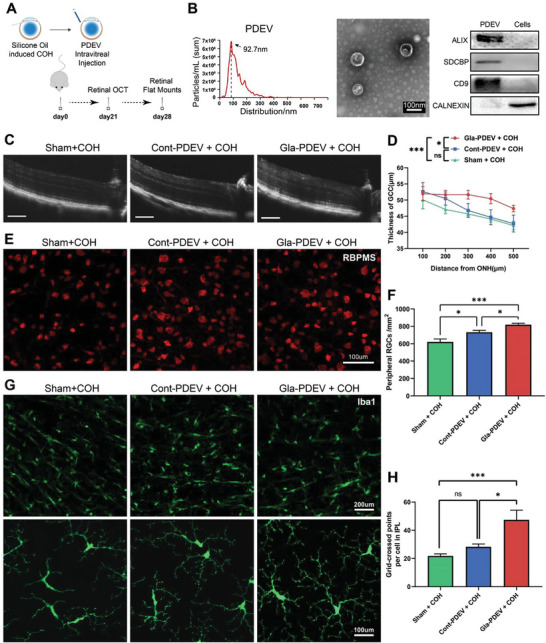
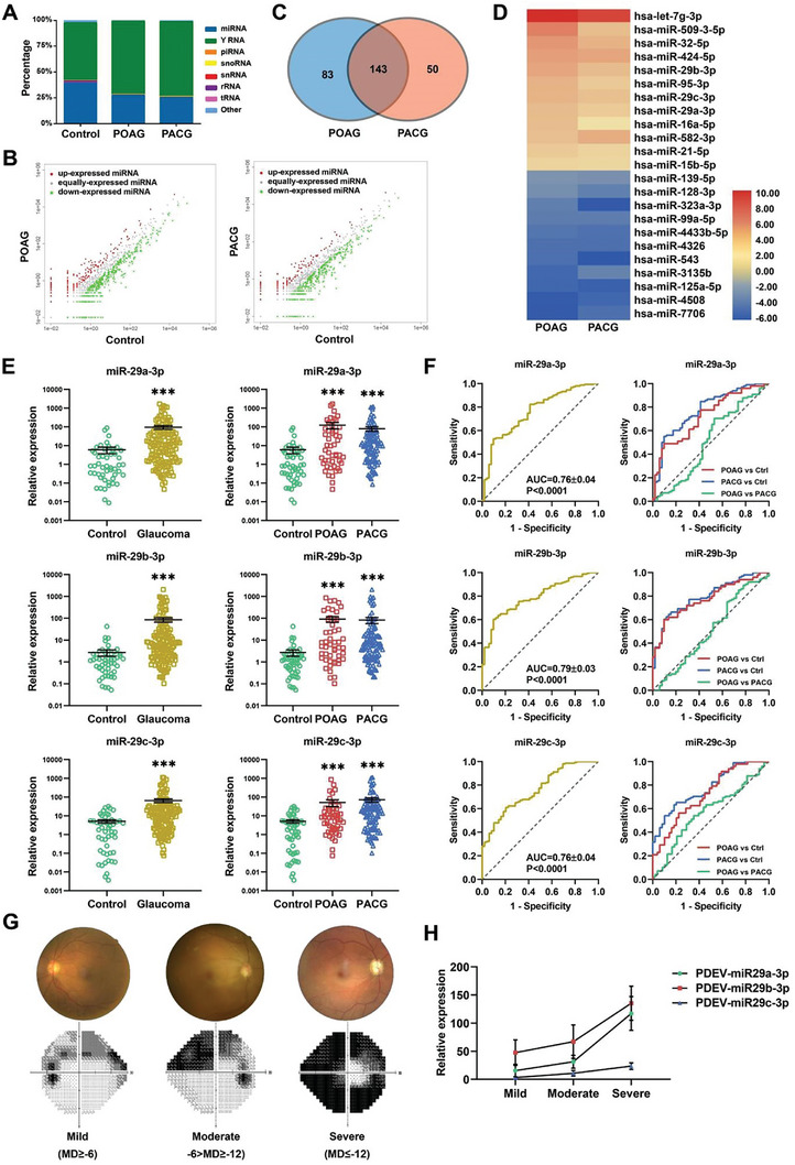
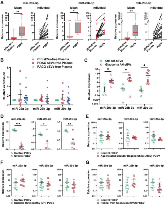
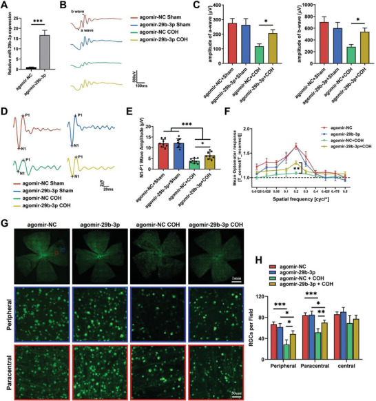
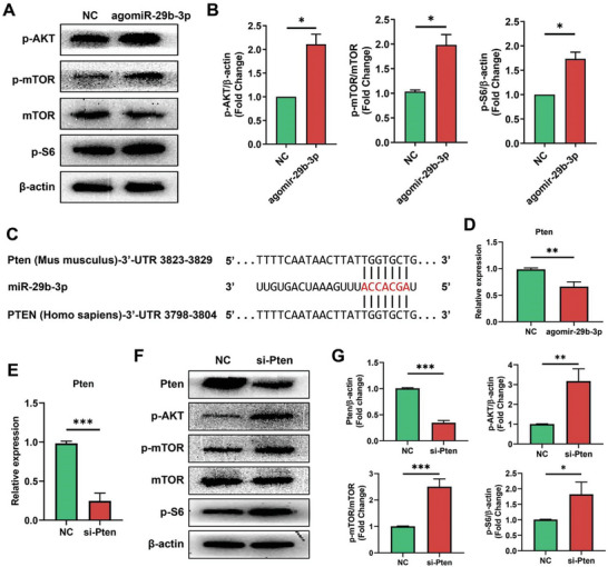
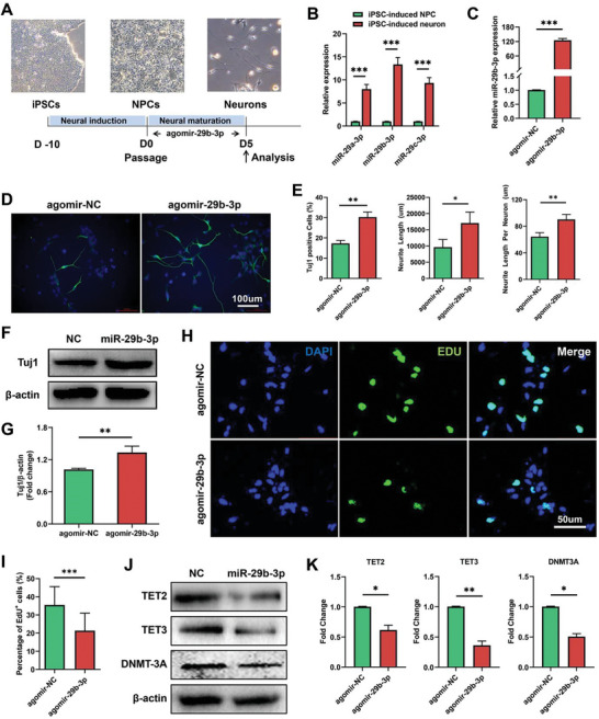
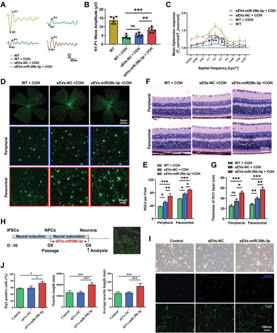
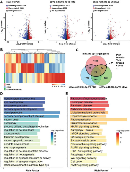
Glaucoma is a leading cause of irreversible blindness worldwide and is characterized by progressive retinal ganglion cell (RGC) degeneration and vision loss. Since irreversible neurodegeneration occurs before diagnosable, early diagnosis and effective neuroprotection are critical for glaucoma management. Small extracellular vesicles (sEVs) are demonstrated to be potential novel biomarkers and therapeutics for a variety of diseases. In this study, it is found that intravitreal injection of circulating plasma-derived sEVs (PDEV) from glaucoma patients ameliorated retinal degeneration in chronic ocular hypertension (COH) mice. Moreover, it is found that PDEV-miR-29s are significantly upregulated in glaucoma patients and are associated with visual field defects in progressed glaucoma. Subsequently, in vivo and in vitro experiments are conducted to investigate the possible function of miR-29s in RGC pathophysiology. It is showed that the overexpression of miR-29b-3p effectively prevents RGC degeneration in COH mice and promotes the neuronal differentiation of human induced pluripotent stem cells (hiPSCs). Interestingly, engineered sEVs with sufficient miR-29b-3p delivery exhibit more effective RGC protection and neuronal differentiation efficiency. Thus, elevated PDEV-miR-29s may imply systemic regulation to prevent RGC degeneration in glaucoma patients. This study provides new insights into PDEV-based glaucoma diagnosis and therapeutic strategies for neurodegenerative diseases.

Keywords: RGCs degeneration; engineered sEVs; glaucoma; miR‐29; plasma‐derived sEVs.


Conflict of interest statement

The authors declare no conflict of interest.

Figures

Figure 1
Figure 1
Intravitreal injection of Gla‐PDEV alleviated glaucomatous optic neuropathy in COH mice. A) Schematic illustration of the in vivo experiment. Mice were injected with silicone oil immediately followed by PDEV injection. Retinal OCT and flat‐mount imaging were conducted 3 and 4 weeks after COH. B) NTA revealed the size distribution of PDEV, TEM showed the typical morphology of PDEV, and WB confirmed the expression of specific markers (ALIX, SDCBP, CD9, and calnexin) in PDEV. C,D) Representative OCT images and quantification of retinal thickness after different treatments in COH mice (Bar = 100 µm, n = 9‐12). E,F) Representative flat‐mount confocal images and quantification of RGC numbers showing surviving RBPMS‐positive (red) RGCs in the peripheral retina (n = 8). G,H) Flat‐mount confocal images showing the distribution of GFP‐positive microglia in the 3 groups and quantification of the grid‐crossed points per microglia in the IPL (n = 6). (ns > 0.05, *< 0.05, ***< 0.001).
Figure 2
Figure 2
Identification and validation of candidate PDEV‐miRNAs with glaucoma specificity. A) Distribution of small RNAs detected by RNA‐seq in PDEV (n = 20 for each group; mt‐tRNA, transfer RNA located in the mitochondrial genome; rRNA, ribosomal RNA; piRNA, PIWI‐interacting RNA; snoRNA, small nucleolar RNA; snRNA, small nuclear RNA). B) Scatter plot showing differentially expressed PDEV‐miRNAs in glaucoma patients compared to controls (fold change = 2 as the threshold). C) A Venn diagram showing that 143 differentially expressed PDEV‐miRNAs overlapped between POAG and PACG patients. D) Twenty‐three candidate miRNAs were selected from the RNA‐seq data, including the top 12 upregulated miRNAs and the top 11 downregulated miRNAs. E) qPCR analysis of individual patient samples confirmed that miR‐29s were significantly upregulated in both POAG and PACG patients (n = 51 for control and POAG, n = 102 for PACG). F) ROC analysis showing the diagnostic performance of PDEV‐miR‐29a‐3p, miR‐29b‐3p, and miR‐29c‐3p in POAG and PACG. G,H) Glaucoma patient classification based on MD values and PDEV‐miR‐29s expression increased with disease severity. (***< 0.001).
Figure 3
Figure 3
Expression of miR‐29s in sEVs‐free plasma and AH‐sEVs and detection of PDEV‐miR‐29s in other eye disease types. A) The expression of miR‐29s in PDEV was significantly greater than that in sEVs‐free plasma at both the average and individual levels (n = 21). B) The expression of miR‐29s in sEVs‐free plasma did not differ between the glaucoma patients and controls (n = 7). C) The expression of miR‐29s in AH‐sEVs from glaucoma patients was significantly increased (n = 7). D–G) The expression of PDEV‐miR‐29s in the uveitis (D), AMD (E), DR (F), and RVO (G) groups was compared with that in the control group (n = 6 for the uveitis group, n = 14 for the AMD, DR, and RVO groups). (*< 0.05, **< 0.01, ***< 0.001).
Figure 4
Figure 4
Overexpression of miR‐29b‐3p in the retina prevents retinal degeneration in COH mice. A) Intravitreal injection of agomir‐29b‐3p significantly increased miR‐29b‐3p levels in the mouse retina. The a‐ and b‐wave amplitudes under scotopic conditions B,C) (n = 12) and the N1‐P1 amplitude of FVEP D,E) (n = 10) significantly increased in miR‐29b‐3p‐overexpressing mice 4 weeks after COH. F) Visual function under a spatial frequency of 0.2 cyc/d was significantly improved in miR‐29b‐3p‐overexpressing mice 4 weeks after COH (n = 6). G,H) RGC survival in the peripheral and paracentral areas was greater in whole flat‐mounted retinas from miR‐29b‐3p‐overexpressing mice than in those from control mice (n = 5). (*< 0.05, **< 0.01, ***< 0.001).
Figure 5
Figure 5
miR‐29b‐3p regulates the Pten‐dependent AKT/mTOR pathway. A) Western blots showing high levels of phosphorylated AKT, mTOR and S6 in retinas overexpressing miR‐29b‐3p. B) Quantification of the data in A, n = 3. C) Alignment between the binding sites of miR‐29b‐3p and Pten. D) qPCR showed a significant decrease in the level of the Pten mRNA in miR‐29b‐3p‐overexpressing cells. E) qPCR showing efficient knockdown of Pten mRNA in retinas after Pten‐specific siRNA transfection. F) Western blots showing decreased Pten protein levels in Pten‐knockdown cells, which resulted in increased levels of phosphorylated AKT, mTOR, and S6. G) Quantification of the data in F, n = 3. (*< 0.05, **< 0.01, ***< 0.001).
Figure 6
Figure 6
Overexpression of miR‐29b‐3p promotes the neuronal differentiation of ESC‐induced NPCs. A) Schematic diagram of neuronal differentiation. B) The expression of miR‐29s was significantly increased during differentiation. C) Agomir‐29b‐3p transfection efficiently increased miR‐29b‐3p levels in NPCs. D,E) miR‐29b‐3p significantly increased the number of Tuj1‐positive cells, total length, and average length of neurites after 5 days of differentiation. F,G) miR‐29b‐3p significantly increased the protein level of Tuj1 in differentiated cells. H,I) miR‐29b‐3p inhibited the proliferation of NPCs, as detected by an EdU assay. J,K) The expression of the DNA methylation‐related genes TET2, TET3, and DNMT3A significantly decreased in miR‐29b‐3p‐overexpressing cells. (*< 0.05, **< 0.01, ***< 0.001).
Figure 7
Figure 7
sEVs‐mediated delivery of miR‐29b‐3p mitigated RGC degeneration in COH mice and enhanced in vitro neuronal differentiation efficiency. A,B) The N1‒P1 amplitude of FVEP significantly increased in sEVs‐miR‐29b‐3p‐injected mice 3 weeks after COH (n = 6). C) The visual function of sEVs‐miR‐29b‐3p‐injected mice was significantly improved (n = 6). D,E) RGC survival in the peripheral and paracentral areas was increased in whole flat‐mounted retinas of sEVS‐miR‐29b‐3p‐injected mice (n = 7). F,G) The thickness of the ganglion cell complex (GCC) layer was restored in sEVs‐miR‐29b‐3p‐injected retinas (n = 6). H) Schematic diagram of sEVsthe effect of sEVs‐miR‐29b‐3p treatment on neuronal differentiation. I,J) Increased numbers of Tuj1‐positive cells and increased total length and average length of neuronal neurites were observed in the sEVS‐miR29b‐3p‐treated group. (ns > 0.05, *< 0.05, **< 0.01, ***< 0.001).
Figure 8
Figure 8
Transcriptomic analysis of sEVs‐miR‐29b‐3p‐treated retinas from COH mice. A) Volcano plots showing the number of downregulated and upregulated genes among the 3 groups. B) Heatmap of DEGs in the control, sEVs‐treated, and sEVs‐miR‐29b‐3p‐treated groups. C) Venn diagram for the analysis of genes that were differentially expressed in the sEVs‐miR‐29b‐3p group and verified miR‐29b‐3p targets. D,E) GO and KEGG pathway enrichment analyses of DEGs between the sEVs‐miR‐29b‐3p‐treated group and the PBS‐treated group.

References

    1. Jonas J. B., Aung T., Bourne R. R., Bron A. M., Ritch R., Panda‐Jonas S., Lancet 2017, 390, 2183. - PubMed
    1. Nickells R. W., Howell G. R., Soto I., John S. W., Annu. Rev. Neurosci. 2012, 35, 153. - PubMed
    1. Nugent R. B., Lee G. A., Surv. Ophthalmol. 2015, 60, 406. - PubMed
    1. Shtein R. M., Shen J. F., Kuo A. N., Hammersmith K. M., Li J. Y., Weikert M. P., Ophthalmology 2020, 127, 128. - PubMed
    1. Okumura Y., Inomata T., Fujimoto K., Fujio K., Zhu J., Yanagawa A., Shokirova H., Saita Y., Kobayashi Y., Nagao M., Nishio H., Sung J., Midorikawa‐Inomata A., Eguchi A., Nagino K., Akasaki Y., Hirosawa K., Huang T., Kuwahara M., Murakami A., Br. J. Ophthalmol. 2022, 108, 37. - PubMed