. 2024 Jul 23;43(7):114405.
doi: 10.1016/j.celrep.2024.114405. Epub 2024 Jun 25.

CK2 phosphorylation of CMTR1 promotes RNA cap formation and influenza virus infection

Affiliations

CK2 phosphorylation of CMTR1 promotes RNA cap formation and influenza virus infection

Radoslaw Lukoszek et al. Cell Rep. .

Abstract

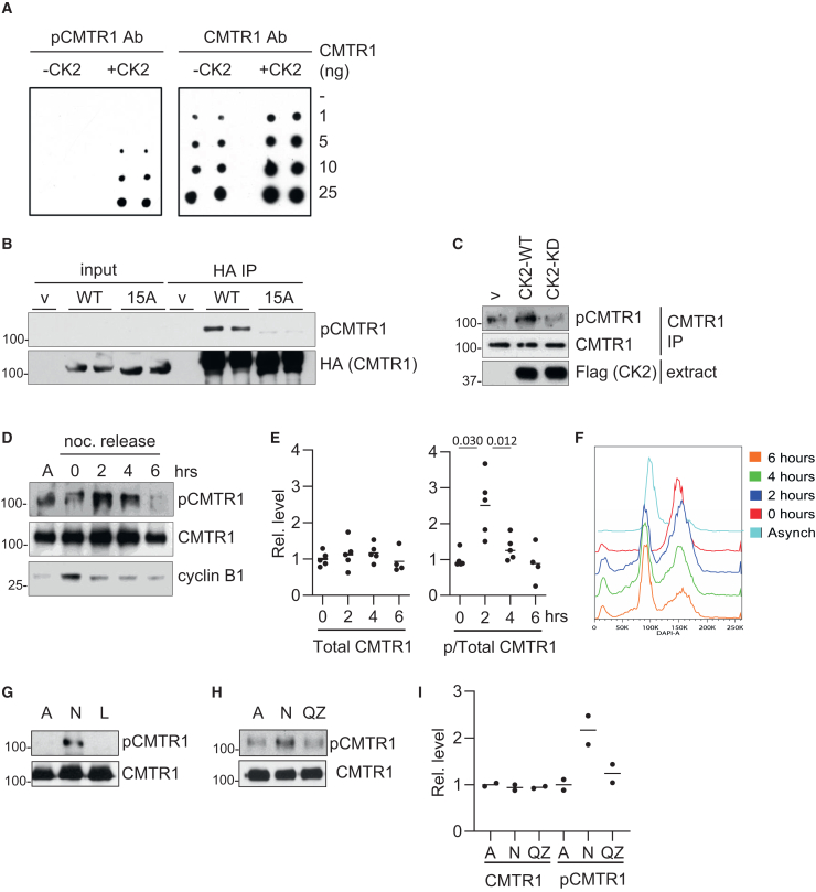
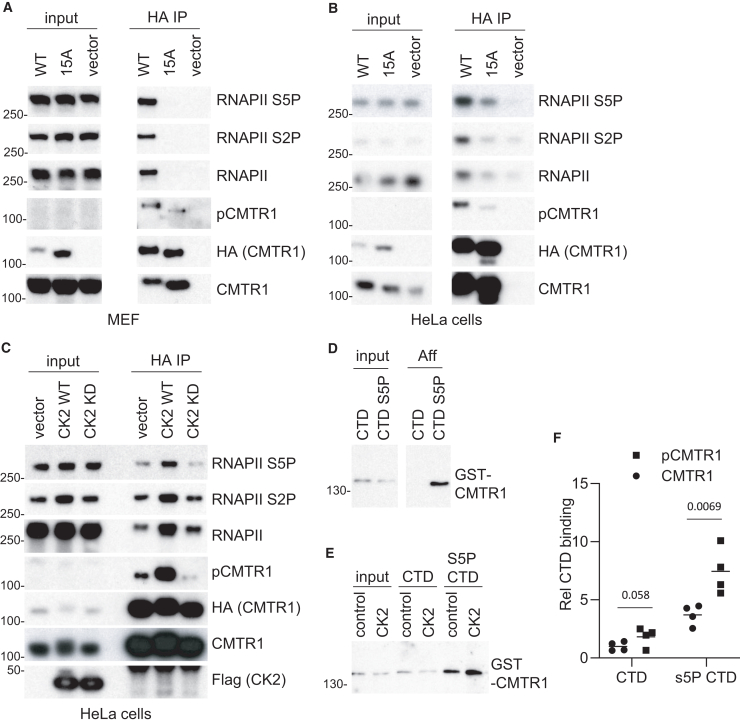
The RNA cap methyltransferase CMTR1 methylates the first transcribed nucleotide of RNA polymerase II transcripts, impacting gene expression mechanisms, including during innate immune responses. Using mass spectrometry, we identify a multiply phosphorylated region of CMTR1 (phospho-patch [P-Patch]), which is a substrate for the kinase CK2 (casein kinase II). CMTR1 phosphorylation alters intramolecular interactions, increases recruitment to RNA polymerase II, and promotes RNA cap methylation. P-Patch phosphorylation occurs during the G1 phase of the cell cycle, recruiting CMTR1 to RNA polymerase II during a period of rapid transcription and RNA cap formation. CMTR1 phosphorylation is required for the expression of specific RNAs, including ribosomal protein gene transcripts, and promotes cell proliferation. CMTR1 phosphorylation is also required for interferon-stimulated gene expression. The cap-snatching virus, influenza A, utilizes host CMTR1 phosphorylation to produce the caps required for virus production and infection. We present an RNA cap methylation control mechanism whereby CK2 controls CMTR1, enhancing co-transcriptional capping.

Keywords: CMTR1; CP: Microbiology; CP: Molecular biology; RNA; RNA cap; cell proliferation; influenza virus; innate immunity; ribosomes; transcription; translation.


Conflict of interest statement

Declaration of interests The authors declare no competing interests.

Figures

None
Graphical abstract
Figure 1
Figure 1
CMTR1 contains a highly phosphorylated P-Patch at the N terminus (A) Diagram of human CMTR1. Domains and positions indicated. P-Patch is amino acids 26–89 (red). High-confidence phosphorylation sites are labeled “P.” (B) HA-CMTR1 was expressed in HeLa cells and immunoprecipitated via the HA tag. Phospho-peptides were identified by mass spectrometry, and the mass was reported. High-confidence phosphorylation sites are stated in “possible sites” with the percentage of probability given. For peptides with several potential sites, the most likely are stated and all are underlined. (C) In vitro phosphorylation of recombinant CMTR1 and OTUB1 by CK2. Reaction constituents indicated. Phospho-analysis of proteins (top). Coomassie blue staining of proteins (bottom). (D) In vitro phosphorylation of recombinant CMTR1, CMTR1Δ143, and OTUB1 over a time course, as above. “c” indicates a reaction without CK2. (E) Quantitation of moles of ATP incorporated into moles of substrates in (D). See also Figures S1 and S2.
Figure 2
Figure 2
CMTR1 is phosphorylated by CK2 during G1 phase (A) Recombinant CMTR1 was in vitro phosphorylated with CK2 and a titration was blotted onto PVDF (ng indicated). Blots were probed with pCMTR1 antibody or (pan) CMTR1 antibody. (B) HA-CMTR1 WT, HA-CMTR1 15A, or empty vector (v) were transiently expressed in HeLa cells. HA-CMTR1 proteins were immunoprecipitated via the HA tag and analyzed by western blot. (C) FLAG-CK2 WT, D156A (kinase dead [KD]), or empty vector (v) were transiently expressed in HeLa cells. Endogenous CMTR1 was immunoprecipitated and analyzed by western blot. (D) HeLa cells were arrested in G2/M phase using nocodazole and released into the cell cycle by replacement with fresh medium. CMTR1 was immunoprecipitated over a time course of nocodazole release or from asynchronous cells (A) and analyzed by western blot for phospho-CMTR1 and total CMTR1 (representative shown). Cyclin B expression was analyzed. (E) pCMTR1/CMTR1 and total CMTR1 were quantitated. Dots indicate data for 4 or 5 independent experiments. Line indicates the average. Student’s t test was performed, and p values are stated. (F) Cell cycle progression in (D) was analyzed by flow cytometry using DAPI DNA stain. The proportion of cells in each stage of the cell cycle is indicated. (G) Cells released from nocodazole block for 2 h were untreated (N), treated with lambda phosphatase (L), or asynchronous (A). (H) As in (G) except cells were treated with quinalizarin (QZ). (I) Detection of pCMTR1/CMTR1 and total CMTR1 was quantitated for 2 independent experiments. Dots indicate data, and line indicates the average. See also Figures S3 and S4.
Figure 3
Figure 3
CK2 phosphorylation of CMTR1 increased interaction with RNA Pol II (A and B) HA-CMTR1 WT, 15A, or vector control. Dots indicate data were transiently expressed in (A) MEFs or (B) HeLa cells. HA-CMTR1 WT or 15A was immunoprecipitated from cell extracts via the HA tag. Western blots were performed on input material and immunoprecipitates (IPs). (C) HA-CMTR1 was transiently co-expressed in HeLa cells with CK2 WT, CK2 KD, or vector control. Western blots were performed on input material and HA-CMTR1 IPs for the antigens indicated. (D) Recombinant GST-CMTR1 was incubated with biotinylated CTD peptide, unphosphorylated (CTD), or phosphorylated on serine-5 (CTD S5P). CMTR1 was detected by western blot in inputs and streptavidin pull-downs (Aff). (E) As in (D) except GST-CMTR1 was in vitro phosphorylated by incubation with CK2 prior to CTD and CTD-S5P pull-downs. (F) GST-CMTR1 binding to CTD was quantitated for 4 independent experiments Dots indicate data, and line indicates the average. Student’s t test was performed, and p values are stated.
Figure 4
Figure 4
Intramolecular interactions of CMTR1 (A) Diagram of CMTR1 mutants used. (B) HA-CMTR1-WW was transiently co-expressed with the GFP-CMTR1 WT and mutants indicated above blots in HeLa cells. GFP-CMTR1 proteins were immunoprecipitated using GFP nanobodies and western blots performed to detect HA-WW (anti-HA antibody) and GFP-tagged protein (anti-GFP antibodies). (C) As in (B) except HA-CMTR1-GT-WW was co-expressed with GFP-tagged CMTR1 mutants. (D) The predicted structure of CMTR1 by AlphaFold2. The domains of interest are indicated. (E and F) HA-CMTR1-GT-WW was expressed with GFP-CMTR1 WT, 15A, or GFP alone. (E) GFP-CMTR1 proteins immunoprecipitated with nanobodies. (F) HA-GT-WW was immunoprecipitated using anti-HA antibodies. RNA Pol II S5P and other antigens were detected by western blot.
Figure 5
Figure 5
CMTR1 phosphorylation is required for RNA cap methylation and gene expression MEF lines were created to express HA-CMTR1 WT, 15A, and vector control. Cre recombinase was expressed to delete the Cmtr1 gene. (A) Relative abundance of cap structures in Cmtr1 knockout (KO) MEFs expressing HA-CMTR1 WT, 15A, or vector control. Data presented are from three independent experiments, and bar indicates the average. (B) MA plots of transcript levels (log2RPKM [reads per kilobase per million mapped reads]) and log2 fold change of CMTR1 WT vs. KO (left) and CMTR1 15A vs. KO (right). Genes significantly down-/up-regulated (EdgeR exactTest, false discovery rate [FDR]-adjusted p < 0.05) are highlighted. (C) Venn diagram of numbers of genes increased by expression of CMTR1 WT and 15A, defined as those gene transcripts significantly up-regulated relative to Cmtr1 KO. (D) Volcano plots indicating the relationship between log2FC and –log10 FDR-adjusted p value for CMTR1 WT vs. KO (left) and CMTR1 15A vs. KO (right). Ribosomal protein genes (79 genes) are highlighted. (E) MEF lines with and without Cre-directed Cmtr1 deletion. MEFs were plated and counted each day. Data shown are for 3 experiments, and bar indicates the average. Student’s t test was performed, and p values are stated. See also Figures S5 and S6.
Figure 6
Figure 6
Induction of interferon-stimulated genes is dependent on CMTR1 P-Patch phosphorylation (A) Cmtr1 KO MEF lines expressing HA-CMTR1 WT, 15A, and vector control were incubated in 400 U/mL interferon for 1, 2, and 4 h. RNA was harvested and RNAs detected by PCR. Data are from 3 independent experiments, and bar indicates the average. Student’s t test was performed, and p values are indicated. (B and C) MEFs were (B) incubated with 400 U/mL interferons for 0, 4, 8, and 24 h and (C) transfected with 5 μg/mL polyI:C for 17 and 24 h. IFIT3 and ISG15 proteins were analyzed by western blot. Actin was used as a loading control. (D) WT MEFs were incubated with 10 μM QZ for 0.5–3 h and 400 U/mL interferon for a subsequent 4 h. RT-PCR was performed as above. Data are from 4 independent experiments. Student’s t test was performed, and p values are indicated. (E) Cmtr1 WT and KO MEF lines expressing HA-CMTR1 WT, 15A, and vector control (con) were incubated with PR8 ColorFlu expressing a mCherry reporter gene. Cells producing virus were detected by fluorescence-activated cell sorting at 17 and 24 h post-infection. Data are from 3 independent wells. Student’s t test was performed, and p values are indicated. (F) For mCherry-positive cells, the geometric mean of red fluorescence was reported. A sample histogram is presented for Cmtr1−/− MEFs expressing HA-CMTR1 WT, 15A, and vector control, normalized to the mode.

References

    1. Dohnalkova M., Krasnykov K., Mendel M., Li L., Panasenko O., Fleury-Olela F., Vågbø C.B., Homolka D., Pillai R.S. Essential roles of RNA cap-proximal ribose methylation in mammalian embryonic development and fertility. Cell Rep. 2023;42 doi: 10.1016/j.celrep.2023.112786. - DOI - PubMed
    1. Inesta-Vaquera F., Cowling V.H. Regulation and function of CMTR1-dependent mRNA cap methylation. Wiley Interdiscip. Rev. RNA. 2017;8 doi: 10.1002/wrna.1450. - DOI - PMC - PubMed
    1. Belanger F., Stepinski J., Darzynkiewicz E., Pelletier J. Characterization of hMTr1, a human Cap1 2'-O-ribose methyltransferase. J. Biol. Chem. 2010;285:33037–33044. doi: 10.1074/jbc.M110.155283. - DOI - PMC - PubMed
    1. Smietanski M., Werner M., Purta E., Kaminska K.H., Stepinski J., Darzynkiewicz E., Nowotny M., Bujnicki J.M. Structural analysis of human 2'-O-ribose methyltransferases involved in mRNA cap structure formation. Nat. Commun. 2014;5:3004. doi: 10.1038/ncomms4004. - DOI - PMC - PubMed
    1. Pelletier J., Schmeing T.M., Sonenberg N. The multifaceted eukaryotic cap structure. Wiley Interdiscip. Rev. RNA. 2021;12 doi: 10.1002/wrna.1636. - DOI - PubMed

Publication types