. 2024 Sep;115(9):3013-3025.
doi: 10.1111/cas.16258. Epub 2024 Jun 26.

RECK/GPR124-driven WNT signaling in pancreatic and gastric cancer cells

Affiliations

RECK/GPR124-driven WNT signaling in pancreatic and gastric cancer cells

Hai Yu et al. Cancer Sci. 2024 Sep.

Abstract

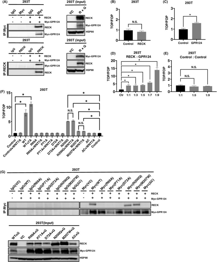
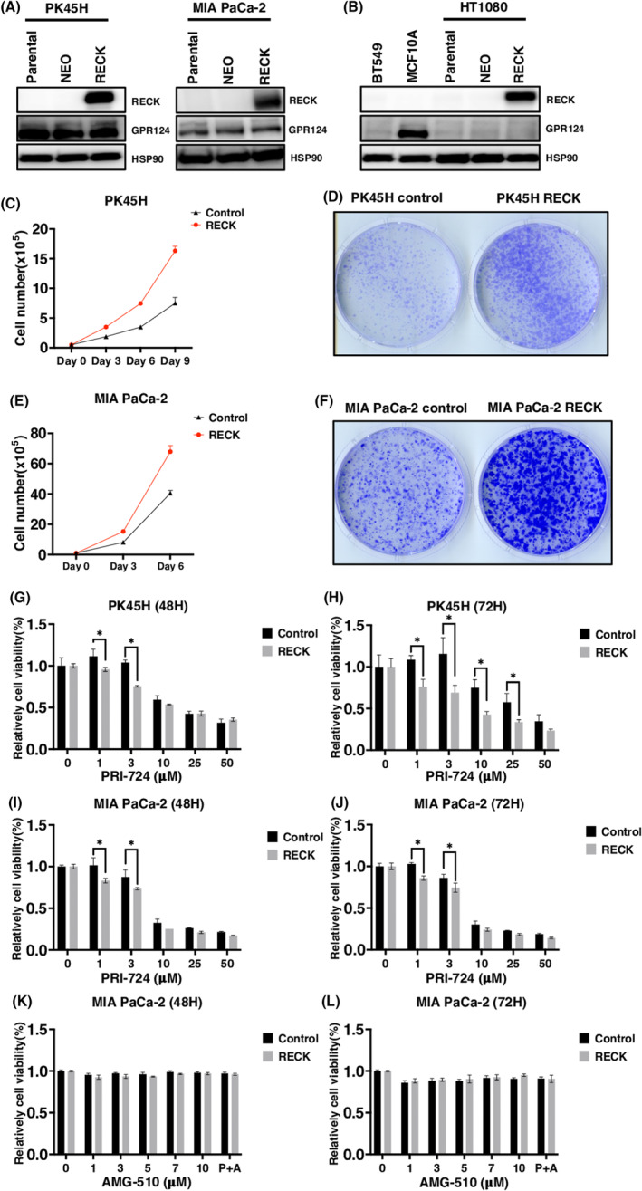
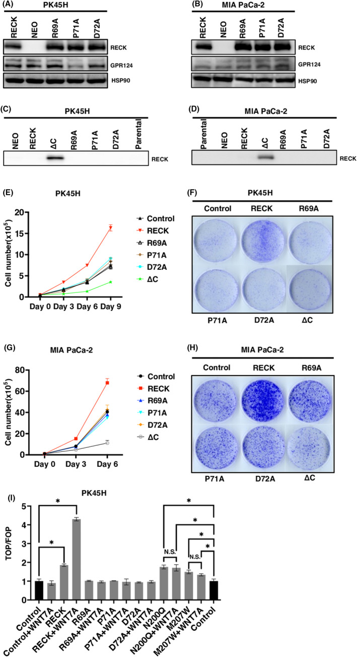
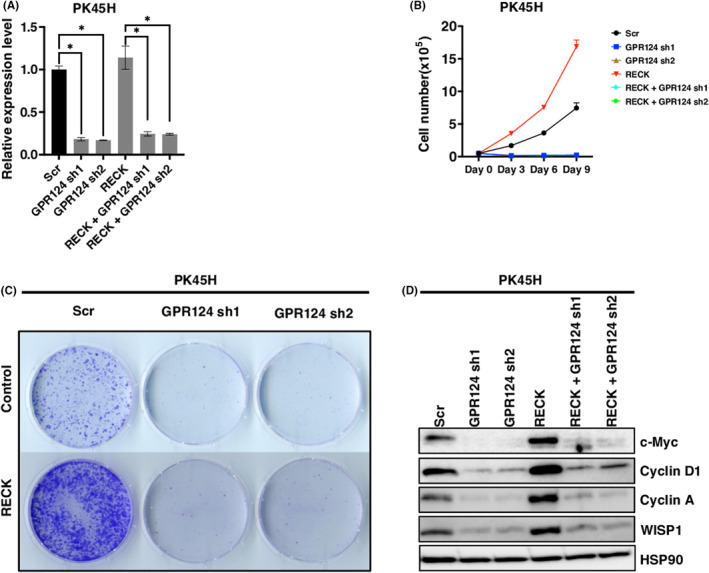
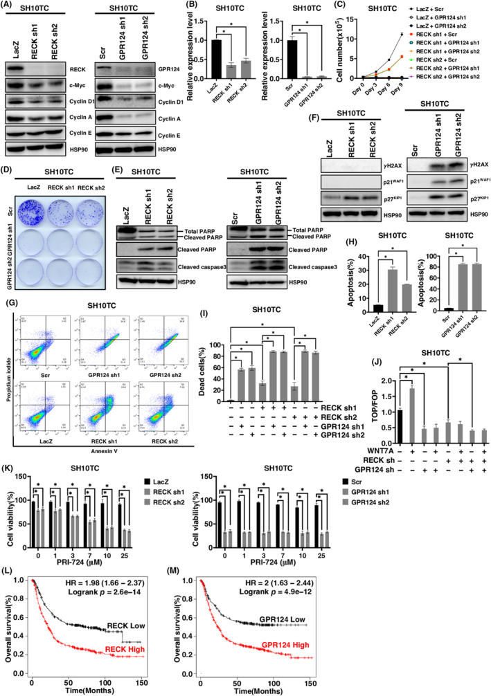
RECK has been described to modulate extracellular matrix components through negative regulation of MMP activities. Recently, RECK was demonstrated to bind to an orphan G protein-coupled receptor GPR124 to mediate WNT7 signaling in nontumor contexts. Here, we attempted to clarify the role of RECK in driving WNT signaling in cancer cells. RECK and GPR124 formed a complex in 293T cells, and when both were expressed, WNT signaling was significantly enhanced in a WNT7-dependent manner. This cooperation was abolished when RECK mutants unable to bind to GPR124 were transduced. RECK stimulated the growth of KRAS-mutated pancreatic ductal adenocarcinoma (PDAC) cells with increased sensitivity to WNT inhibitor in a GPR124-dependent manner. A gastric cancer cell line SH10TC endogenously expresses both RECK and GPR124 under regular culture conditions. In this cell line, inhibited cell growth and WNT signaling as well as increased apoptosis in the GPR124 depletion was dominantly found over those in the RECK deletion. These findings suggest that RECK promotes tumor cell growth by positively modulating WNT signaling through GPR124. This study proposes that the RECK/GPR124 complex might be a good therapeutic target in PDAC and gastric cancer.

Keywords: GPR124; RECK; WNT7; gastric cancer; pancreatic cancer.


Conflict of interest statement

The authors have no financial interest to disclose. Chiaki Takahashi is an editorial board member of Cancer Science.

Figures

FIGURE 1
FIGURE 1
The RECK/GPR124 complex mediates WNT signaling. (A) RECK and Myc‐tagged GPR124 simultaneously expressed in 293T cells and immunoprecipitated (IP) by antibodies indicated. The precipitants were then immunoblotted (IB) by antibodies indicted (left). The expression of proteins in the input whole‐cell lysates (right). (B) TOP/FOP canonical WNT/β‐catenin reporter assay of 293T cells transduced with pCXN2‐RECK (N = 3). (C) TOP/FOP assay of 293T cells transduced with Myc‐tagged GPR124 in 293T cells (N = 3). (D) TOP/FOP assay of 293T cells transduced with pCXN2‐RECK and pcDNA3.1 Myc‐GPR124 mixed at the indicated ratio (N = 3). (E) TOP/FOP assay of 293T cells transduced with pCXN2‐NEO and pcDNA3.1 mixed at the indicated ratio (N = 3). (F) TOP/FOP assay of 293T cells transduced with pCXN2‐RECK, RECK mutants and pcDNA3.1 Myc‐GPR124 mixed at the ratio of 1:9 and treated with or without 10 ng/mL of WNT7A (N = 3). Control columns appear twice for the convenience of assessing significance between control and N200Q and M207W columns. (G) RECK (WT and mutants) and Myc‐tagged GPR124 were simultaneously expressed in 293T cells and immunoprecipitated (IP) by the antibodies indicated. The precipitants were then immunoblotted (IB) by the antibodies indicated (upper). The expression of proteins in the input whole‐cell lysates were detected by the corresponding antibodies (lower). * p < 0.05 against vector control; N.S., not significant.
FIGURE 2
FIGURE 2
RECK stimulates cell proliferation in a WNT signal‐dependent manner when induced in GPR124‐positive pancreatic ductal adenocarcinoma (PDAC) cells. (A, B) Immunoblotting (IB) of the indicated proteins in the indicated cells parental or transduced with PCXN2‐NEO or PCXN2‐RECK. (C) Cell proliferation of PK45H transduced with PCXN2‐NEO or PCXN2‐RECK (N = 3). (D) Representative image of colony formation by PK45H cells transduced with PCXN2‐NEO or PCXN2‐RECK (N = 3). (E) Cell proliferation of MIA PaCa‐2 cells transduced with PCXN2‐NEO or PCXN2‐RECK (N = 3). (F) Representative image of colony formation by MIA PaCa‐2 transduced with PCXN2‐NEO or PCXN2‐RECK (N = 3). (G–L) Relative viability of the indicated cells transduced with PCXN2‐NEO or PCXN2‐RECK and treated with the indicated concentration of the indicated chemicals for the indicated period. The viability of cells without treatment was set to 100% (N = 3). *p < 0.05 against vector control; N.S., not significant.
FIGURE 3
FIGURE 3
RECK mutants lacking binding to GPR124 failed to stimulate growth in pancreatic ductal adenocarcinoma (PDAC) cells. (A, B) Immunoblotting (IB) of the indicated proteins in the indicated cells. (C, D) Immunoblotting of soluble RECK in the conditioned media prepared from the indicated cells transduced with the indicated protein. Conditioned media were prepared by cultivating cells with serum‐free culture medium for 48 h and filtered through a 0.45‐μM filter. (E, G) Cell growth in the indicated cells transduced with the indicated protein. (F, H) Representative image of colony formation by the indicated cells transduced with the indicated protein. (I) TOP/FOP assay of PK45H cells transduced with pCXN2‐RECK, RECK mutants and pcDNA3.1 Myc‐GPR124 mixed at the ratio of 1:9 and treated with or without 10 ng/mL of WNT7A (N = 3). Control columns appear twice for the convenience of assessing significance between control and N200Q and M207W columns. *p < 0.05 against vector control; N.S., not significant.
FIGURE 4
FIGURE 4
Depletion of GPR124 induces growth arrest which cannot be rescued by RECK. (A) Relative mRNA expression levels of GPR124 in PK45H cells transduced with pCXN2‐NEO or pCXN2‐RECK and then infected with lentivirus carrying shRNA targeting GPR124 or control (N = 3) (B) Cell proliferation curve of PK45H cells established in (A). (C) Representative image of colony formation by PK45H cells established in (A) (N = 3). (D) Immunoblotting (IB) of the indicated proteins in PK45H cells established in (A). *p < 0.05 against vector control; N.S., not significant.
FIGURE 5
FIGURE 5
The effect of RECK and GPR124 depletion in SH10TC cells. (A) Immunoblotting (IB) of the indicated proteins in RECK‐depleted (left) or GPR124‐depleted (right) SH10TC cells. (B) Relative mRNA expression levels of the indicated gene in RECK‐depleted (left) or GPR124‐depleted (right) SH10TC cells (N = 3). (C) Cell proliferation curve of SH10TC cells infected with lentivirus carrying shRNA targeting the indicated gene in the indicated combination. (D) Representative images of colony formation by SH10TC cells analyzed in (C). (E, F) Immunoblotting of the indicated proteins in RECK‐depleted (left) or GPR124‐depleted (right) SH10TC cells. (G) Representative results of flow cytometry analysis of SH10TC cells infected with the indicated lentivirus. (H) Quantitation of flow cytometry analyses of RECK‐depleted (left) and GPR124‐depleted cells (right) (N = 3). (I) The percentage of dead SH10TC cells infected with the indicated lentivirus assessed by trypan blue staining (N = 3). (J) TOP/FOP canonical WNT/β‐catenin reporter assay of SH10TC cells infected with the indicated lentivirus and treated with or without WNT7A. (K) Viability of SH10TC cells infected with the indicated lentivirus and treated with the indicated concentration of PRI‐724 for 72 h (N = 3). (L) Kaplan–Meier survival curves for patients with gastric cancer with high and low RECK expression. p = 2.6 × 10−14 by long‐rank test. (M) Kaplan–Meier survival curves for patients with gastric cancer with high and low GPR124 expression. p = 4.9 × 10−12 by long‐rank test. *p < 0.05 against vector control; N.S., not significant.

References

    1. Noda M, Selinger Z, Scolnick E, Bassin R. Flat revertants isolated from Kirsten sarcoma virus‐transformed cells are resistant to the action of specific oncogenes. Proc Natl Acad Sci USA. 1983;80:5602‐5606. - PMC - PubMed
    1. Kitayama H, Sugimoto Y, Matsuzaki T, Ikawa Y, Noda M. A ras‐related gene with transformation suppressor activity. Cell. 1989;56:77‐84. - PubMed
    1. Takahashi C, Sheng Z, Horan TP, et al. Regulation of matrix metalloproteinase‐9 and inhibition of tumor invasion by the membrane‐anchored glycoprotein RECK. Proc Natl Acad Sci USA. 1998;95:13221‐13226. - PMC - PubMed
    1. Chang HC, Cho CY, Hung WC. Silencing of the metastasis suppressor RECK by RAS oncogene is mediated by DNA Methyltransferase 3b–induced promoter methylation. Cancer Res. 2006;66:8413‐8420. - PubMed
    1. Sasahara RM, Takahashi C, Noda M. Involvement of the Sp1 site in ras‐mediated downregulation of the RECK metastasis suppressor gene. Biochem Biophys Res Commun. 1999;264:668‐675. - PubMed

MeSH terms