. 2024 Jun 28;10(26):eadl6543.
doi: 10.1126/sciadv.adl6543. Epub 2024 Jun 26.

Terahertz control and timing correlations in a transmission electron microscope

Affiliations

Terahertz control and timing correlations in a transmission electron microscope

Joel Kuttruff et al. Sci Adv. .

Abstract

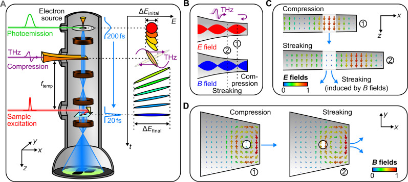
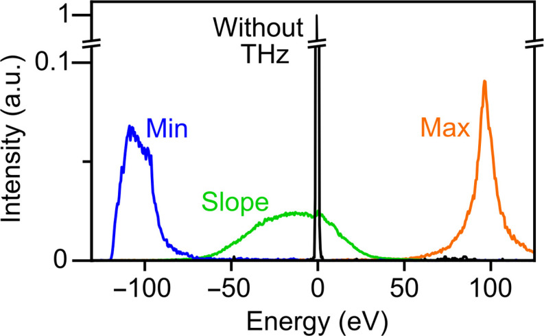
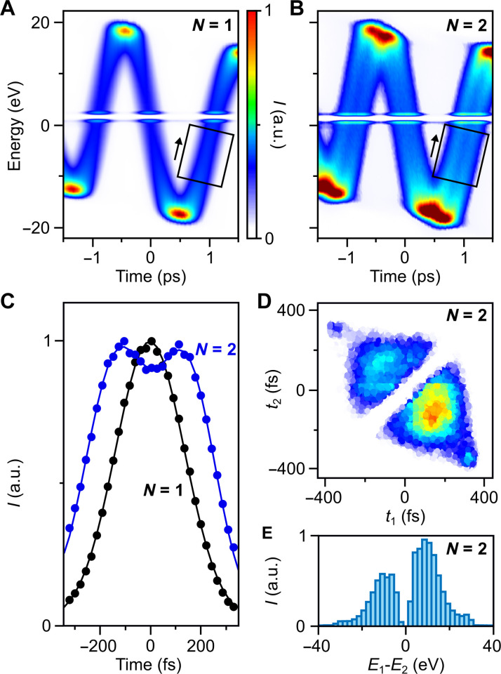
Ultrafast electron microscopy provides a movie-like access to structural dynamics of materials in space and time, but fundamental atomic motions or electron dynamics are, so far, too quick to be resolved. Here, we report the all-optical control, compression, and characterization of electron pulses in a transmission electron microscope by the single optical cycles of laser-generated terahertz light. This concept provides isolated electron pulses and merges the spatial resolution of a transmission electron microscope with the temporal resolution that is offered by a single cycle of laser light. We also report the all-optical control of multi-electron states and find a substantial two-electron and three-electron anticorrelation in the time domain. These results open up the possibility to visualize atomic and electronic motions together with their quantum correlations on fundamental dimensions in space and time.


Figures

Fig. 1.
Fig. 1.. All-optical electron beam control in a transmission electron microscope.
(A) Experimental setup and pulse compression concept. A photoemission laser (green) creates short electron pulses (blue) that intersect with the electric or magnetic anti-nodes of a metal waveguide (orange) under terahertz illumination (violet). A pump laser or secondary terahertz beam (red) excites a material and pump-probe, or streaking images are measured on a screen (green). Inset, phase space diagram of terahertz compression in the time-energy domain. If there are no space charge effects (23) and all forces are sufficiently linear in time (25), the initial electron pules (red) become as much shorter in time as they broaden in the energy domain (blue). (B) Generation of electric and magnetic anti-nodes by reflection of terahertz pulses at a displaced waveguide end. The purely electric (red) and purely magnetic (blue) anti-nodes are ideal for pulse compression or streaking of electron beams at positions ① or ②, respectively. (C) Simulated longitudinal electric fields inside the waveguide at ideal time delays for compression (top) and streaking (bottom). (D) Simulated in-plane magnetic fields at ideal time delays for compression (left) and streaking (right). The circle depicts the subwavelength entrance and exit holes for the electron beam.
Fig. 2.
Fig. 2.. Energy modulation and standing-wave terahertz interaction.
(A) Energy spectrum of electron pulses after interaction with the compression waveguide as a function of terahertz delay. (B) Measured energy modulation (black) by electric fields in comparison to the measured transversal deflection by magnetic fields (green). Shaded area indicates standard deviation of the deflection data. Time I is ideal for pulse compression, time II produces tilted pulses, and time III is ideal for stretching electron pulses in time. (C) Measured (left) and simulated (right) central energy modulation of compressed electrons (I) as a function of position x and y across the beam. (D) Measured (left) and simulated (right) central energy modulation of the electron beam at an improper interaction time (II). a.u., arbitrary units.
Fig. 3.
Fig. 3.. Characterization of electron pulses by streaking and generation of 19-fs pulses.
(A) Terahertz streaking curve of uncompressed electron pulses. (B) Streaking data at the steepest slope. (C) Streaking data for electron pulses after terahertz compression. (D) Measured time-dependent electron current for uncompressed electron pulses and Gaussian fit (red). (E) Measured time-dependent electron current for compressed electron pulses and Lorentzian fit (red).
Fig. 4.
Fig. 4.. Transmission electron microscopy and diffraction with terahertz-compressed electron pulses.
(A) Microscope images at a magnification of 200,000 of gold nanoparticles without (left) and with (right) terahertz compression. (B) Line cuts at the dotted lines for uncompressed (orange) and compressed 19-fs electron pulses (green). Feature sizes of 3 nm can be resolved in both cases. (C) Diffraction pattern of crystalline silicon recorded without (left) and with (right) terahertz compression. (D) Measured beam sizes of the uncompressed (orange) and compressed (green) electron pulses as a function of the microscope objective lens defocus. The focus shifts by 20 μm.
Fig. 5.
Fig. 5.. Electron acceleration and deceleration.
Measured electron energy spectra for initial electron pulses (black), maximum acceleration (orange), maximum deceleration (blue), and pulse compression (green).
Fig. 6.
Fig. 6.. Multi-electron phase-space characterization and control.
(A) Measured spectrogram for one-electron states (N = 1). (B) Measured spectrogram for two-electron states (N = 2). Black rectangles, regions of interest for our further analysis; white lines, artefacts from our detector system. (C) Measured electron arrival times for exactly one electron (black) and exactly two electrons per pulse (blue). (D) Measured pair density as a function of arrival times t1 and t2 for electrons 1 and 2 within a pair. Data are recorded at terahertz time delay of 1.1 ps. (E) Measured histogram of energy differences E1-E2 within a pair state after terahertz-driven energy magnification. The initial separation of ~2 eV (49, 50) becomes ~15 eV.

References

    1. Zewail A. H., Four-dimensional electron microscopy. Science 328, 187–193 (2010). - PubMed
    1. Feist A., Bach N., da Silva N. R., Danz T., Möller M., Priebe K. E., Domröse T., Gatzmann J. G., Rost S., Schauss J., Strauch S., Bormann R., Sivis M., Schäfer S., Ropers C., Ultrafast transmission electron microscopy using a laser-driven field emitter: Femtosecond resolution with a high coherence electron beam. Ultramicroscopy 176, 63–73 (2017). - PubMed
    1. Caruso G. M., Houdellier F., Weber S., Kociak M., Arbouet A., High brightness ultrafast transmission electron microscope based on a laser-driven cold-field emission source: Principle and applications. Adv. Phys. X 4, 1660214 (2019). - PubMed
    1. Nabben D., Kuttruff J., Stolz L., Ryabov A., Baum P., Attosecond electron microscopy of sub-cycle optical dynamics. Nature 619, 63–67 (2023). - PubMed
    1. Grinolds M. S., Lobastov V. A., Weissenrieder J., Zewail A. H., Four-dimensional ultrafast electron microscopy of phase transitions. Proc. Natl. Acad. Sci. U S A 103, 18427–18431 (2006). - PMC - PubMed