. 2024 Jun 26;16(753):eadl3758.
doi: 10.1126/scitranslmed.adl3758. Epub 2024 Jun 26.

Transcobalamin receptor antibodies in autoimmune vitamin B12 central deficiency

Affiliations

Transcobalamin receptor antibodies in autoimmune vitamin B12 central deficiency

John V Pluvinage et al. Sci Transl Med. .

Abstract

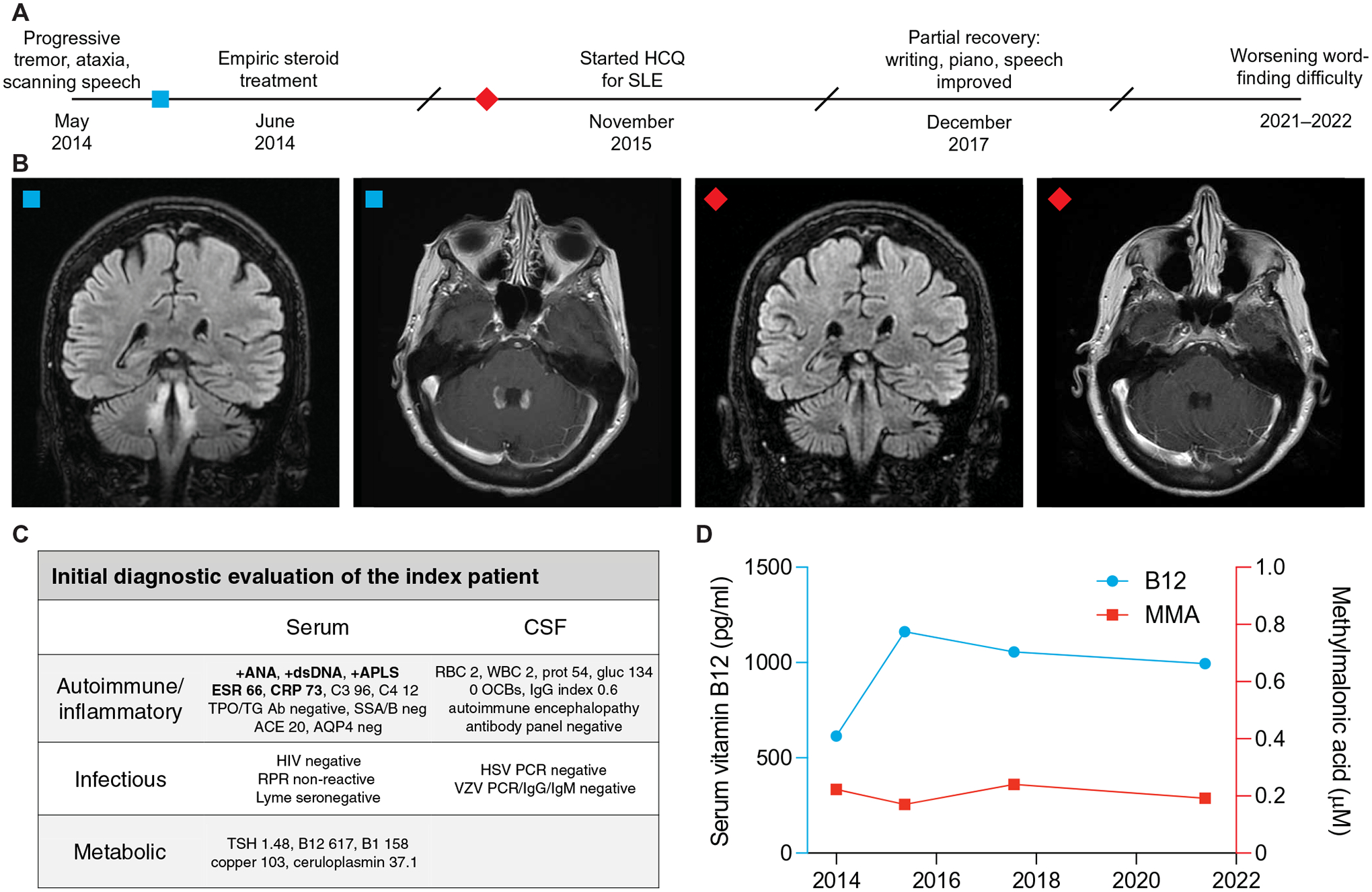
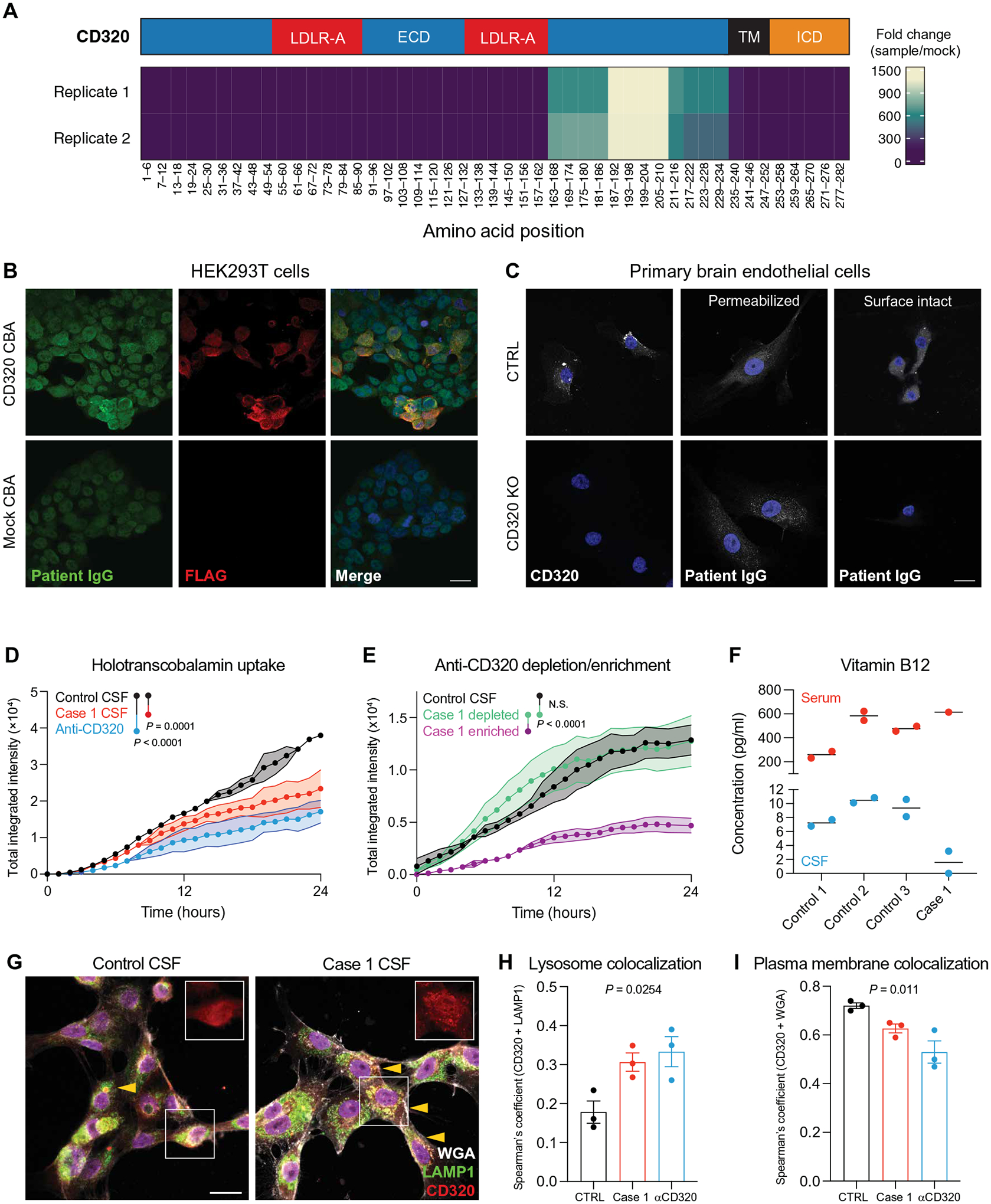
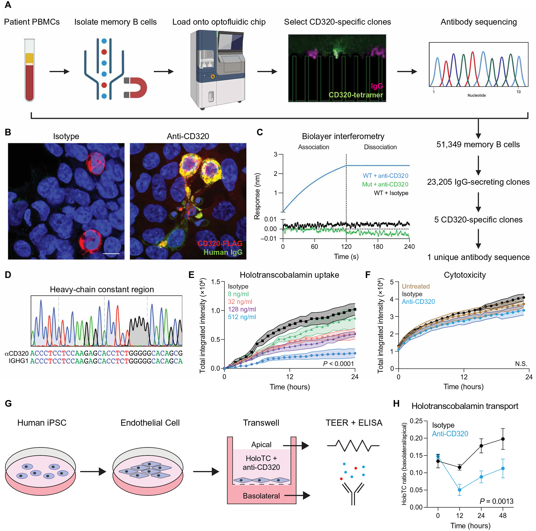
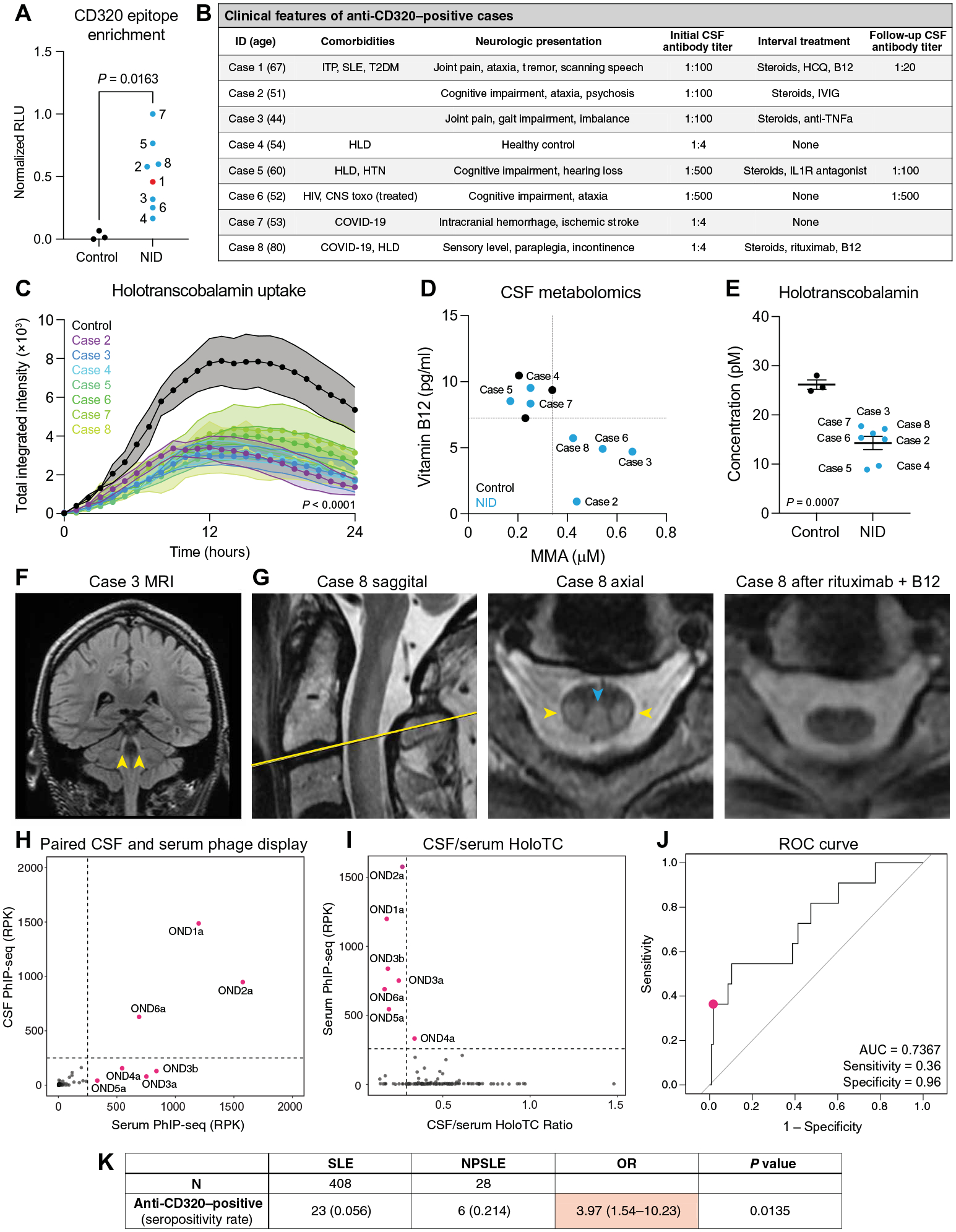
Vitamin B12 is critical for hematopoiesis and myelination. Deficiency can cause neurologic deficits including loss of coordination and cognitive decline. However, diagnosis relies on measurement of vitamin B12 in the blood, which may not accurately reflect the concentration in the brain. Using programmable phage display, we identified an autoantibody targeting the transcobalamin receptor (CD320) in a patient with progressive tremor, ataxia, and scanning speech. Anti-CD320 impaired cellular uptake of cobalamin (B12) in vitro by depleting its target from the cell surface. Despite a normal serum concentration, B12 was nearly undetectable in her cerebrospinal fluid (CSF). Immunosuppressive treatment and high-dose systemic B12 supplementation were associated with increased B12 in the CSF and clinical improvement. Optofluidic screening enabled isolation of a patient-derived monoclonal antibody that impaired B12 transport across an in vitro model of the blood-brain barrier (BBB). Autoantibodies targeting the same epitope of CD320 were identified in seven other patients with neurologic deficits of unknown etiology, 6% of healthy controls, and 21.4% of a cohort of patients with neuropsychiatric lupus. In 132 paired serum and CSF samples, detection of anti-CD320 in the blood predicted B12 deficiency in the brain. However, these individuals did not display any hematologic signs of B12 deficiency despite systemic CD320 impairment. Using a genome-wide CRISPR screen, we found that the low-density lipoprotein receptor serves as an alternative B12 uptake pathway in hematopoietic cells. These findings dissect the tissue specificity of B12 transport and elucidate an autoimmune neurologic condition that may be amenable to immunomodulatory treatment and nutritional supplementation.


Conflict of interest statement

J.V.P., J.L.D., S.J.P., and M.R.W. are coinventors on a patent application related to this work (PCT/US2024/018105, “Compositions and methods related to transcobalamin receptor autoantibodies”). M.R.W. receives unrelated research grant funding from Roche/Genentech and Novartis; received speaking honoraria from Genentech, Takeda, WebMD, and Novartis; and is a founder and paid consultant for Delve Bio Inc. J.L.D. is a founder and paid consultant for Delve Bio Inc. and a paid consultant for the Public Health Company and Allen & Co. M.R.W. and J.L.D. receive licensing fees from CDI Laboratories. C.M.B. is a physician consultant for the Neuroimmune Foundation. J.M.G. receives research support from Hoffman LaRoche and Vigil Neurosciences for clinical trials and is a paid consultant for Arialys and Ventyx Bio. S.L.H. currently serves on the scientific advisory board of Accure, Alector, and Annexon; has previously consulted for BD, Moderna, NGM Bio, and Pheno Therapeutics; previously served on the board of directors of Neurona; and has received travel reimbursement and writing support from F. Hoffmann-La Roche and Novartis AG for anti-CD20 therapy–related meetings and presentations. A.J.G. is an Associate Editor for JAMA Neurology and received advisory board fees from Pipeline Therapeutics outside the submitted work. J.Y. has received grant support from Gilead, Aurinia, and BMS Foundation and performed consulting for Astra Zeneca, Pfizer, and UBC. The other authors declare that they have no competing interests.

Figures

Fig. 1.
Fig. 1.. Clinical course and initial diagnostic investigation of the index patient.
(A) Timeline of evaluation and treatment of the index patient. (B) Coronal slices of T2-weighted fluid-attenuated inversion recovery (FLAIR) MRI and axial slices of T1-weighted post-gadolinium MRI before (left two images) and 9 months after (right two images) steroid treatment. (C) Initial diagnostic evaluation of serum and CSF samples. Bolded items were found to be abnormal. (D) Serum vitamin B12 (blue) and MMA (red) concentrations over the clinical timecourse.
Fig. 2.
Fig. 2.. Discovery and validation of a functional transcobalamin receptor autoantibody.
(A) Epitope map of CD320 peptides enriched by patient CSF antibodies. Coverage is divided into five amino acid bins and aligned to the full-length protein (NP_057663.1). The blue region represents the extracellular domain (ECD), the red regions represent LDLR-A domains within the ECD, the black region represents the transmembrane (TM) domain, and the orange region represents the intracellular domain (ICD). (B) Immunore-activity of patient CSF IgG (green) to HEK293T cells overexpressing a FLAG-tagged CD320 construct (red). Staining dilution = 1:25. Scale bar, 20 μm. (C) Control and CD320 KO primary human brain endothelial cells stained with a commercial CD320 antibody (left column) or patient CSF (middle and right columns) and DAPI (blue) to label nuclei. Scale bar, 20 μm. (D) Holotranscobalamin uptake in cells treated with case 1 CSF (red) or a commercial CD320 antibody (blue) compared with cells treated with healthy control CSF (black) (n = 2, paired one-way ANOVA with Tukey’s multiplicity correction, means ± SE). (E) Holotranscobalamin uptake in cells treated with control CSF (black) or case 1 CSF-enriched (magenta) or CSF-depleted (green) of anti-CD320 by affinity purification (n = 3, one-way ANOVA with Tukey’s multiplicity correction, means ± SE). (F) Vitamin B12 concentration in serum (red) and CSF (blue) of three healthy controls and case 1. (G) Representative images of brain endothelial cells treated with control or patient CSF, followed by staining for CD320 (red), wheat germ agglutinin (WGA, a plasma membrane marker; gray), and lysosomal-associated membrane protein (LAMP1; green). Arrows indicate yellow puncta where CD320 (red) colocalizes with lysosomes (LAMP1, green; scale bar = 20 μm). Quantification of CD320 colocalization with the LAMP1 (H) or WGA (I) in cells treated with control CSF (black), case 1 CSF (red), or a commercial anti-CD320 antibody (blue) (n = 3; one-way ANOVA; means ± SE).
Fig. 3.
Fig. 3.. Isolation of a patient-derived monoclonal autoantibody.
(A) Schematic of the Beacon optofluidic autoantibody discovery workflow. Memory B cells were magnetically isolated from PBMCs, loaded onto an optofluidic chip, and screened for antibody secretion (IgG, magenta) and antigen specificity (CD320-tetramers, green). CD320-reactive clones were exported for cDNA synthesis and immunoglobulin sequencing. (B) HEK293T cells transfected with CD320-FLAG (red) were stained with an isotype control antibody (left) or the patient-derived monoclonal antibody (green, right). Yellow signal represents colocalization of human IgG with the CD320-FLAG construct. Scale bar, 20 μm. (C) Kinetic analysis of anti-CD320 binding to wild-type (WT) CD320 peptide (blue), anti-CD320 binding to a mutant CD320 peptide with the autoepitope replaced with a stretch of 10 alanines (green), or isotype control antibody binding wild-type CD320 peptide (black). (D) Sanger sequencing of the heavy-chain constant region compared with the immunoglobulin heavy constant gamma 1 (IGHG1) consensus sequence. (E) Dose response of anti-CD320 on holotranscobalamin uptake (n = 3, one-way ANOVA with Tukey’s multiplicity correction, means ± SE). (F) Cytotoxicity in untreated HEK293T cells (brown) or HEK293T cells treated with isotype control antibody (black) or anti-CD320 (blue) (n = 3, one-way ANOVA, means ± SE). (G) Schematic of the in vitro model of the human BBB. iPSCs were differentiated into endothelial cells and plated on a semi-permeable transwell membrane, and B12 transport was measured in the presence of anti-CD320 or isotype control by serial sampling of holotranscobalamin in the apical and basolateral media. (H) Ratio of basolateral to apical holotranscobalamin concentration (measured by ELISA) in isotype control (black)– or anti-CD320 (blue)– treated wells over 48 hours. A higher ratio represents greater B12 transport (n = 3, two-way ANOVA, means ± SD). N.S., not significant.
Fig. 4.
Fig. 4.. Identification of additional cases with CD320 autoantibodies.
(A) Enrichment of the CD320 target peptide by CSF immunoglobulin from three healthy controls (black) and eight individuals enrolled in a neuroinflammatory disease study (NID; blue), including the index patient (red), using a split-luciferase binding assay (two-sided t test). (B) Clinical characteristics of individuals harboring CSF anti-CD320 autoantibodies. Autoantibody titer was determined by cell-based assay. HLD, hyperlipidemia; HTN, hypertension. (C) Holotranscobalamin uptake by cells treated with CSF from the seven additional cases compared with treatment with healthy control CSF (n = 3, one-way ANOVA with Tukey’s multiplicity correction, means ± SE). (D) Total vitamin B12 (y axis) and MMA (x axis) concentrations in CSF from the seven cases enrolled in a neuroinflammatory disease study (blue dots) and three healthy controls (black dots). Dotted lines show the lowest CSF vitamin B12 or highest MMA level among healthy controls. (E) Holotranscobalamin concentrations in CSF from healthy controls and cases in the neuroinflammatory disease cohort (two-sided t test; means ± SE). (F) Coronal T2-weighted FLAIR MRI of case 3. Yellow arrows indicate symmetric T2-FLAIR hyperintensities involving the bilateral cerebellar peduncles. (G) Sagittal and axial cuts of the T2-weighted C-spine MRI for case 8 before (left two images) and axial cut after (right image) treatment with rituximab and B12 supplementation. Blue arrow indicates T2 hyperintensity in the dorsal columns. Yellow arrows indicate T2 hyperintensity in the lateral corticospinal tracts. (H) CD320 enrichment of paired serum and CSF samples from 132 patients with MS or other neurologic diseases. Enrichment determined by PhIP-seq, where RPK represents depth-normalized sequencing reads. Labeled samples exhibit CD320 enrichment equal to or greater than 250 RPK (dotted line). (I) CSF/serum holotranscobalamin (HoloTC) ratio versus serum anti-CD320 enrichment. The CSF/serum holotranscobalamin threshold (0.2) was chosen on the basis of previously reported reference ranges in each biofluid (4, 49). (J) Receiver operator characteristic (ROC) curve showing the performance of serum anti-CD320 seropositivity for predicting a low CSF/serum holotranscobalamin level. RPK threshold was chosen to stratify seropositive versus seronegative and is not a proxy for autoantibody titer. (K) Contingency analysis of anti-CD320 seropositivity in non-neurologic SLE and NPSLE. Fisher’s exact test, 95% confidence interval. RLU, relative light unit; AUC, area under the curve.
Fig. 5.
Fig. 5.. LDLR is an alternative B12 uptake receptor.
(A) Schematic of a CRISPRi screen for genetic modifiers of holotranscobalamin uptake. K562 cells expressing dCas9-KRAB were infected with a library of sgRNAs targeting the transcriptional start sites of all protein coding genes. This pool of single knockdown cells was then incubated with holotranscobalamin conjugated to a pH-sensitive fluorescent dye. Cells with high (top 5% of red fluorescent signal) and low (bottom 5%) holoTC uptake were separated by FACS and then sequenced and analyzed using casTLE. Screen was performed in technical duplicate. (B) Volcano plot of the genes which when repressed impair (blue) or promote (red) holoTC uptake. The top 12 hits are labeled, meeting an adjusted P value of <0.00001 (Benjamini-Hochberg). (C) Flow cytometry confirmation of control (black), CD320 KO (blue), LDLR KO (red), and double KO (brown) K562 cells. (D) Holotranscobalamin uptake by control (black), CD320 KO (blue), LDLR KO (red), or double KO (brown) K562 cells (n = 3, one-way ANOVA with Tukey’s multiplicity correction, means ± SE). (E) Flow cytometry confirmation of control (black), CD320 KO (blue), LDLR KO (red), and double KO (brown) primary human brain endothelial cells. (F) Holotranscobalamin uptake by control (black), CD320 KO (blue), LDLR KO (red), or double KO (brown) primary brain endothelial cells (n = 2, one-way ANOVA with Tukey’s multiplicity correction, means ± SE). APC, allophycocyanin; PE, phycoerythrin.

References

    1. Green R, Allen LH, Bjørke-Monsen A-L, Brito A, Guéant J-L, Miller JW, Molloy AM, Nexo E, Stabler S, Toh B-H, Ueland PM, Yajnik C, Vitamin B12 deficiency. Nat. Rev. Dis. Primers 3, 17040 (2017). - PubMed
    1. Stabler SP, Vitamin B12 deficiency. N. Engl. J. Med 368, 149–160 (2013). - PubMed
    1. Hooshmand B, Appold F, Fissler P, Perneczky R, Otto M, Tumani H, Kivipelto M, von Arnim CAF, Markers of vitamin B12 status in relation to cerebrospinal fluid biomarkers of Alzheimer’s disease and cognitive performance. Ann. Neurol 94, 223–231 (2023). - PubMed
    1. Christine CW, Auinger P, Saleh N, Tian M, Bottiglieri T, Arning E, Tran NK, Ueland PM, Green R, Parkinson study group-DATATOP investigators, relationship of cerebrospinal fluid vitamin B12 status markers with Parkinson’s disease progression. Mov. Disord 35, 1466–1471 (2020). - PMC - PubMed
    1. Oki R, Izumi Y, Fujita K, Miyamoto R, Nodera H, Sato Y, Sakaguchi S, Nokihara H, Kanai K, Tsunemi T, Hattori N, Hatanaka Y, Sonoo M, Atsuta N, Sobue G, Shimizu T, Shibuya K, Ikeda K, Kano O, Nishinaka K, Kojima Y, Oda M, Komai K, Kikuchi H, Kohara N, Urushitani M, Nakayama Y, Ito H, Nagai M, Nishiyama K, Kuzume D, Shimohama S, Shimohata T, Abe K, Ishihara T, Onodera O, Isose S, Araki N, Morita M, Noda K, Toda T, Maruyama H, Furuya H, Teramukai S, Kagimura T, Noma K, Yanagawa H, Kuwabara S, Kaji R, Efficacy and safety of ultrahigh-dose methylcobalamin in early-stage amyotrophic lateral sclerosis. JAMA Neurol 79, 575–583 (2022). - PMC - PubMed

Publication types