. 2024 Jul;33(7):e5081.
doi: 10.1002/pro.5081.

Antigen-binding fragments with improved crystal lattice packing and enhanced conformational flexibility at the elbow region as crystallization chaperones

Affiliations

Antigen-binding fragments with improved crystal lattice packing and enhanced conformational flexibility at the elbow region as crystallization chaperones

Heather A Bruce et al. Protein Sci. 2024 Jul.

Abstract

It has been shown previously that a set of three modifications-termed S1, Crystal Kappa, and elbow-act synergistically to improve the crystallizability of an antigen-binding fragment (Fab) framework. Here, we prepared a phage-displayed library and performed crystallization screenings to identify additional substitutions-located near the heavy-chain elbow region-which cooperate with the S1, Crystal Kappa, and elbow modifications to increase expression and improve crystallizability of the Fab framework even further. One substitution (K141Q) supports the signature Crystal Kappa-mediated Fab:Fab crystal lattice packing interaction. Another substitution (E172G) improves the compatibility of the elbow modification with the Fab framework by alleviating some of the strain incurred by the shortened and bulkier elbow linker region. A third substitution (F170W) generates a split-Fab conformation, resulting in a powerful crystal lattice packing interaction comprising the biological interaction interface between the variable heavy and light chain domains. In sum, we have used K141Q, E172G, and F170W substitutions-which complement the S1, Crystal Kappa, and elbow modifications-to generate a set of highly crystallizable Fab frameworks that can be used as chaperones to enable facile elucidation of Fab:antigen complex structures by x-ray crystallography.

Keywords: crystal lattice packing interactions; phage display technology; protein domain swapping; surface entropy reduction; x‐ray crystallography.


Figures

FIGURE 1
FIGURE 1
Fab library design and results. (a) FabS1CE‐F1 (PDB entry 8T7I shown as a representative structure) (Bruce et al., 2023) with the heavy chain and light chain colored light blue or gray, respectively. Constant and variable domains of the light chain (CL/VL) and heavy chain (CH/VH) are indicated. In addition, residue positions in key regions shown as spheres are colored as follows: S1 (dark blue), Crystal Kappa (green), elbow (magenta), KGP site (cyan), FPE site (orange), GLY site (yellow). (b) The principal Fab molecule heavy chain is colored light blue, while the packing Fab molecule heavy chain and light chain are colored dark blue or green, respectively. The principle Fab light chain is omitted. (Left panel) In the crystal lattice, the Crystal Kappa mediated β‐sheet stacking interaction (Lieu et al., 2020) occurs between the CH domain (from the principal Fab in this representation) and CL′ domain of the packing Fab molecule (indicated by prime symbol). (Middle panel) Due to their proximity, the Crystal Kappa substitution (green) in the packing Fab, and the elbow substitution (magenta) in the principle Fab, form a contiguous crystal lattice packing interface. Residues Phe136H, Asn137H, Gln138H, and Ile139H of the elbow region form weak intermolecular interactions with residues Thr155H′, Ser156H′, and Gly157H′ (light blue) in the CH domain of the packing Fab molecule. The KGP site, which directly follows the elbow region, is colored cyan. (Right panel) Rotating the packing Fab molecules in the crystal lattice by 180° reveals a sub‐optimal crystal lattice packing site; residues Lys141H, Gly142H, and Pro143H of the KGP site do not participate in the Fab:Fab packing interaction at the Crystal Kappa:elbow junction. The flexible side chain of Lys141H projects away from the crystal lattice packing site toward the solvent. (c) Sequences of Fab variants selected from the phage‐displayed library, The WT sequence (S1CE) is shown at the top and variant sequences (S1CE1–7) are shown below with dashes indicating identities. The number of times each variant was observed amongst 94 sequenced clones (N) is shown to the right of each sequence, followed by the protein yield from transient transfection of mammalian cell cultures, and the melting temperature (T m ) determined by differential scanning fluorimetry.
FIGURE 2
FIGURE 2
Crystal screening results for Fabs and Fab:antigen complexes. Crystal hits for FabS1CE‐EPR‐1 with substitution E172G or F170W, with or without K141Q, are plotted for (a) the apo Fab‐EPR‐1 and (b) the Fab‐EPR‐1 in complex with EPOR‐ECD. The numbers of crystal hits (left y‐axis) or % of crystal hits (right y‐axis) obtained for each Fab variant (x‐axis) are shown from screening a total of 192 conditions (96 conditions each from JCSG+Eco and PACT). Putative apo Fab crystal hits (gray) in the Fab:antigen complex crystallization screening were evaluated by comparing crystal morphology for each complex drop to the corresponding apo Fab drop (see Excel document in Data S1).
FIGURE 3
FIGURE 3
Substitutions K141Q, F170W, and E172G alter FabS1CE‐EPR‐1 structural conformation and crystal lattice packing. (a) Crystal lattice packing arrangement of Fab‐EPR‐1 with the following frameworks: (i) FabS1CE (orthorhombic crystal system, space group P212121), (ii) FabS1CE1 (trigonal crystal system, space group P3121), (iii) FabS1CE2 (trigonal crystal system, space group P32), (iv) FabS1CE3 (trigonal crystal system, space group P3121), (v) FabS1CE3 (orthorhombic crystal system, space group P212121), and (vi) FabS1CE4 (triclinic crystal system, space group P1). The lower panel displays the asymmetric unit (ASU). Fab light and heavy chains are colored light blue or gray, respectively. The upper panel shows the ASU, in addition to the packing Fab molecules of selected symmetry mates in the crystal lattices with those light and heavy chains colored green or dark blue, respectively. Substitutions specific to each Fab variant are underscored. (b) In the FabS1CE1, FabS1CE3, and FabS1CE4 crystal lattice structures, the canonical VH:VL domain interaction occurs between packing Fab molecules in the crystal lattice as opposed to within the Fab molecule itself. The light and heavy chains of one Fab molecule are colored gray or light blue, respectively, while the light and heavy chains of selected packing Fabs are colored green or dark blue, respectively. Packing Fab molecule domains outside of the principal ASU are indicated with a prime symbol. Packing Fab molecule domains within the principal ASU are underscored. (i) In the FabS1CE1‐EPR‐1 or FabS1CE3‐EPR‐1 crystal structures (both P3121 space group), the Fab in the principal ASU interacts with a Fab in another ASU through VH:VL association in the crystal lattice to form a closed dimer arrangement (see 3aii and 3aiv). (ii) In the FabS1CE3‐EPR‐1 structure (P212121 space group), an open‐ended crystal lattice structural arrangement forms in which the principal ASU Fab molecule interacts with two different packing Fab molecules through VH:VL association, priming the formation of an interconnected lattice array (see 3av). (iii) In the FabS1CE4‐EPR‐1 structure (see 3avi), a single ASU contains two Fab molecules (colored gray/light blue or green/dark blue) that associate through their respective pairings of VH:VL domains to form a closed dimer formation, distinct from that found in the other split‐Fab structures.
FIGURE 4
FIGURE 4
Molecular details of the incorporation of substitution F170W into the FabS1CE framework. Residues in the elbow site (WT sequence or elbow substitution sequence) are colored magenta. Residues in the FPE site and “molecular socket” site (Stanfield et al., 2006) are colored orange or blue, respectively. The principle Fab heavy and light chains are colored light blue or gray, respectively, whilst the packing Fab light chain is colored green. Fab domains from a packing ASU (in FabS1CE3 structures) are indicated with a prime symbol. Packing Fab domains within the principal ASU (in FabS1CE4 structure) are underscored. (a) In Fab structures with WT elbow sequence, residue Ser136H, along with residues Leu12H and Thr134H of the VH domain, mediate weak intramolecular interactions with residues Phe170H and Pro171H of the FPE site in the CH domain (FabC‐F1 structure used for representation (Bruce et al., 2023) (PDB entry 8T7G)). (b) The elbow substitution reduces Fab conformational flexibility. (Left panel) Superposition of the Fab Fv (VH/VL) region from two representative structures that contain the WT elbow sequence (PDB entries 1BBD and 1PLG, in dark red or pink, respectively, and with elbow angles of 127.0° or 189.5°, respectively). (Right panel) Superposition of the Fv regions from two different Fab ASU molecules from a structure containing the elbow substitution (PDB entry 6AZ2, with elbow angles of 166.6° or 188.0°, respectively, colored dark blue or light blue, respectively). (c) In the FabS1CE structure, the side chains of Phe170H and Pro171H form Van der Waals interactions with the residues in the elbow region and the VH domain (Leu12H and Thr134H). (d–f) Incorporating the F170W substitution results in destabilization at the CH:VH interface; the bulky Trp170H side chain cannot be accommodated in the limited space, resulting in a split Fab conformation for structures: (d) FabS1CE3 (space group P3121), (e) FabS1CE3 (space group P212121), and (f) FabS1CE4 (space group P1).
FIGURE 5
FIGURE 5
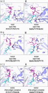
Molecular details of the impact of the K141Q substitution on crystal lattice packing at the Crystal Kappa:elbow junction. Residues in the KGP site and elbow region are colored cyan or magenta, respectively. The ASU Fab heavy chain constant (CH) and variable (VH) domains are colored light blue, whilst the packing Fab heavy chain constant domain (CH′) is colored dark blue. (a) (i) In the FabS1CE‐EPR‐1 structure (used as a representative), the side chain of residue Lys141H projects away from the crystal lattice packing site, toward the solvent. (ii) In the FabS1CE2‐EPR‐1 structure, Gln141H and Gln138H form hydrogen bonds and Van der Waals interactions with residues Thr155H′, Ser156H′, Gly157H′, Gly158H′, and Ser210H′ in the packing Fab CH domain. (b) (i) In the FabS1CE3‐EPR‐1 structure (P212121 space group structure used as a representative), the side chain of residue Lys141H is forced unfavorably away from residues in the packing Fab CH domain. Meanwhile, a small section of the packing Fab CH domain (Ser152H′ and Lys153H′) remains unresolved from the electron density (dotted line). (ii) In the FabS1CE4‐EPR‐1 structure, the side chain of Gln141H participates in hydrogen bonds and Van der Waals interactions with Gly157H′ in the packing Fab CH domain, which in turn transforms the Crystal Kappa:elbow junction into a more favorable crystal lattice packing site with an extensive interaction network, supported by interactions between residues in the packing Fab CH domain and residues in the principle Fab CH and VH domains, and elbow region (see main text for more details). (c) (i) In the FabS1CE1‐EPR‐1:EPOR‐ECD complex structure, the side chain of Lys141H is forced away from the crystal lattice packing site, and minimal intermolecular interactions form between residues in the principle Fab elbow region and packing Fab CH domain. (ii) In the FabS1CE2‐EPR‐1:EPOR‐ECD complex structure, the side chain of Gln141H forms hydrogen bond interactions with the side chain of Ser156H′ and peptide backbone amide group of Gly158H′ in the packing Fab CH domain, while elbow residue Gln138H forms interactions with Ser156H′ in the packing CH domain.
FIGURE 6
FIGURE 6
Molecular details of the incorporation of substitution E172G into the FabS1CE framework. Residues in the elbow region, FPE/FPG site in the CH domain, and “molecular socket” site (Stanfield et al., 2006) in the VH domain, are colored magenta, orange, or blue, respectively. Residues Tyr169H, Ala192H, and Tyr200H (in the CH domain) are colored blue. (a) In the FabS1CE structure, the side chain of residue Glu172H forms various hydrogen bonds and Van der Waals interactions with residues Tyr169H, Ala192H, and Tyr200H in the CH domain, which supports the position and conformation of the FPE loop region. Phe170H and Pro171H of the FPE site form weak intramolecular interactions with elbow residues, and Leu12H and Thr134H of the VH domain. (b,c) In the (b) FabS1CE1 and (c) FabS1CE2 structures, Gly172H cannot participate in the network of intramolecular interactions in an equivalent way to Glu172H. In addition, the φ and ψ dihedral angles in the peptide backbone are altered at positions 171 and 172 (see Figure S7). While a split‐Fab conformation was captured in the FabS1CE1 crystal structure, no changes to the Fab quaternary or tertiary structure occur in the FabS1CE2 crystal structure (see Figure 3a, ii and iii). Due to flexibility, the side chain of Phe136H was unresolved from the electron density in the FabS1CE1‐EPR‐1 structure.
FIGURE 7
FIGURE 7
Substitution E172G facilitate an alternative crystal lattice packing arrangement for the Fab‐EPR‐1:EPOR‐ECD complex. Crystal lattice packing arrangement (upper panel) with symmetry mates, and asymmetric unit (lower panel) of the Fab‐EPR‐1:EPOR‐ECD complex. The ASU Fab light and heavy chains are colored light blue or gray, respectively, while packing Fab light and heavy chains are colored green or dark blue, respectively. The antigen EPOR‐ECD is colored magenta. (a) In the FabS1CE:EPOR‐ECD complex structure (orthorhombic crystal system, space group P212121), one Fab:antigen complex is present in the ASU. (b) In the FabS1CE1:EPOR‐ECD complex structure (orthorhombic crystal system, space group P212121), two Fab molecules and one antigen molecule are present in the ASU. (c) In the FabS1CE2:EPOR‐ECD complex structure (orthorhombic crystal system, space group P212121), two Fab molecules and one antigen molecule are present in the ASU. For the FabS1CE1 and FabS1CE2 complex structures, the secondary Fab molecule (i.e., unbound to the antigen) domains are underscored. (d,e) The Fv:EPOR‐ECD region of the FabS1CE‐EPR‐1:EPOR‐ECD complex structure is superposed with the corresponding region in (d) the FabS1CE1‐EPR‐1:EPOR‐ECD complex structure or (e) the FabS1CE2‐EPR‐1:EPOR‐ECD complex structure. The FabS1CE1‐EPR‐1:EPOR‐ECD and FabS1CE2:EPR‐1:EPOR‐ECD complex structures are colored cyan.

References

    1. Afonine PV, Grosse‐Kunstleve RW, Echols N, Headd JJ, Moriarty NW, Mustyakimov M, et al. Towards automated crystallographic structure refinement with phenix.refine. Acta Crystallogr D Biol Crystallogr. 2012;68:352–367. - PMC - PubMed
    1. Bailey LJ, Sheehy KM, Dominik PK, Liang WG, Rui H, Clark M, et al. Locking the elbow: improved antibody fab fragments as chaperones for structure determination. J Mol Biol. 2018;430:337–347. - PMC - PubMed
    1. Barthelemy PA, Raab H, Appleton BA, Bond CJ, Wu P, Wiesmann C, et al. Comprehensive analysis of the factors contributing to the stability and solubility of autonomous human VH domains. J Biol Chem. 2008;283:3639–3654. - PubMed
    1. Berman HM, Westbrook J, Feng Z, Gilliland G, Bhat TN, Weissig H, et al. The protein data bank. 2000. Available from: http://www.rcsb.org/pdb/status.html - PMC - PubMed
    1. Bostrom J, Haber L, Koenig P, Kelley RF, Fuh G. High affinity antigen recognition of the dual specific variants of Herceptin is entropy‐driven in spite of structural plasticity. PLoS One. 2011;6:e17887. - PMC - PubMed

Substances

LinkOut - more resources