. 2024 Nov 15;210(10):1252-1266.
doi: 10.1164/rccm.202401-0078OC.

Single-Cell Analysis Reveals Novel Immune Perturbations in Fibrotic Hypersensitivity Pneumonitis

Affiliations

Single-Cell Analysis Reveals Novel Immune Perturbations in Fibrotic Hypersensitivity Pneumonitis

Amy Y Zhao et al. Am J Respir Crit Care Med. .

Abstract

Rationale: Fibrotic hypersensitivity pneumonitis (FHP) is a debilitating interstitial lung disease driven by incompletely understood immune mechanisms. Objectives: To elucidate immune aberrations in FHP in single-cell resolution. Methods: Single-cell 5' RNA sequencing was conducted on peripheral blood mononuclear cells and BAL cells obtained from 45 patients with FHP, 63 patients with idiopathic pulmonary fibrosis (IPF), 4 patients with nonfibrotic hypersensitivity pneumonitis, and 36 healthy control subjects in the United States and Mexico. Analyses included differential gene expression (Seurat), TF (transcription factor) activity imputation (DoRothEA-VIPER), and trajectory analyses (Monocle3 and Velocyto-scVelo-CellRank). Measurements and Main Results: Overall, 501,534 peripheral blood mononuclear cells from 110 patients and control subjects and 88,336 BAL cells from 19 patients were profiled. Compared with control samples, FHP has elevated classical monocytes (adjusted-P = 2.5 × 10⁻³) and is enriched in CCL3hi/CCL4hi and S100Ahi classical monocytes (adjusted-P < 2.2 × 10⁻¹⁶). Trajectory analyses demonstrate that S100Ahi classical monocytes differentiate into SPP1hi lung macrophages associated with fibrosis. Compared with both control subjects and IPF, cells from patients with FHP are significantly enriched in GZMhi cytotoxic T cells. These cells exhibit TF activities indicative of TGFβ and TNFα and NFκB pathways. These results are publicly available at http://ildimmunecellatlas.com. Conclusions: Single-cell transcriptomics of patients with FHP uncovered novel immune perturbations, including previously undescribed increases in GZMhi cytotoxic CD4+ and CD8+ T cells-reflecting this disease's unique inflammatory T cell-driven nature-as well as increased S100Ahi and CCL3hi/CCL4hi classical monocytes also observed in IPF. Both cell populations may guide the development of new biomarkers and therapeutic interventions.

Keywords: fibrotic hypersensitivity pneumonitis; idiopathic pulmonary fibrosis; interstitial lung disease; single-cell RNA sequencing; usual interstitial pneumonia.


Figures
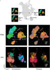
Figure 1.
Figure 1.
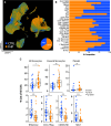
Data overview. (A) Geographic overview of the study samples, with Uniform Manifold Approximation and Projection (UMAP) representations of cells from patients from each city (New Haven, CT; Chicago, IL; Mexico City, Mexico). UMAP representation of 501,534 PBMCs, labeled by (B) all major cell types and (C) conditions, and of 88,336 BAL cells, also labeled by (D) all major cell types and (E) conditions. BAL = bronchoalveolar lavage cells; CTRL = control; FHP = fibrotic hypersensitivity pneumonitis; IPF = idiopathic pulmonary fibrosis; mDC = myeloid dendritic cell; NFHP = nonfibrotic hypersensitivity pneumonitis; NK = natural killer; pDC = plasmacytoid dendritic cell; PBMCs = peripheral blood mononuclear cells.
Figure 2.
Figure 2.
Cell type compositional comparisons of fibrotic hypersensitivity pneumonitis (FHP) versus control samples. (A) Uniform Manifold Approximation and Projection (UMAP) of PBMC samples from only patients with FHP and control subjects. (B) Cell type composition by FHP or healthy controls. (C) Percentage of cell types for each sample, grouped by disease subtype. Only significant cell type comparisons are shown. *P < 0.05, **P < 0.01, and ***P < 0.001. CTRL = control; PBMCs = peripheral blood mononuclear cells; CTL = cytotoxic lymphocyte; gdT = gamma delta T; Inter. = intermediate; ILC = innate lymphoid cell; mDC = myeloid dendritic cell; MPC = monocyte-platelet complex; MAIT = mucosal-associated invariant T cell; NK = natural killer; pDC = plasmacytoid dendritic cell; TEM = T effector memory; TReg = regulatory T cell.
Figure 3.
Figure 3.
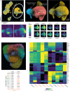
S100Ahi and CCL3hi/CCL4hi monocyte subpopulations enriched in fibrotic hypersensitivity pneumonitis (FHP) versus control. (A) Monocytes (circled) were subset from the overall data and reclustered. A Uniform Manifold Approximation and Projection (UMAP) representation of these reclustered cells is shown by (B) monocyte subtypes and (C) condition. Density graphs of monocytes for (D) controls and (E) FHP. (F) Gene expression of S100Ahi classical monocytes: S100A8, S100A9, S100A12, and CTSD. (G) Gene expression of CCL3hi/CCL4hi classical monocytes: CCL3, CCL4, CXCL3, NFKBIA, ICAM1, and IL1B. (H) UMAP of monocytes clustered by DoRothEA-VIPER transcription factor (TF) activity scores. (I) Breakdown of the DoRothEA-based clusters by percentage represented in control subjects and FHP. Significantly different clusters in FHP are marked in orange. (J) Heatmap demonstrating TFs with top variable imputed activities across monocyte DoRothEA clusters, with hierarchically clustered rows and columns. CTRL = control.
Figure 4.
Figure 4.
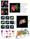
Fibrosis-associated monocytes transition into profibrotic SPP1hi macrophages. (A) Peripheral blood mononuclear cell (PBMC) monocytes and (B) BAL macrophages were integrated from the original data. (C) Uniform Manifold Approximation and Projection (UMAP) representation of integrated myeloid cells by cell type. (D) Localization of CCL3hi/CCL4hi monocytes (CCL3, CCL4), S100Ahi monocytes (S100A8, S100A9, TLR4), and profibrotic SPP1hi macrophages (SPP1, MERTK, LGMN, PLA2G7). (E) Pseudotime trajectories across all integrated PBMC and BAL myeloid cells. (F) Gene expression changes over pseudotime between S100Ahi classical monocytes to profibrotic SPP1hi macrophages, colored by pseudotime. (G) RNA velocity vector fields projected onto the UMAP embedding, demonstrating velocity vectors pointing from S100Ahi monocytes to SPP1hi macrophages. CTRL = control.
Figure 5.
Figure 5.
Cell type compositional comparisons of fibrotic hypersensitivity pneumonitis (FHP) and idiopathic pulmonary fibrosis (IPF). (A) Uniform Manifold Approximation and Projection (UMAP) of peripheral blood mononuclear cell samples from only FHP and IPF. (B) Cell type composition by FHP or IPF. CTL = cytotoxic lymphocyte; gdT = gamma delta T; Inter. = intermediate; ILC = innate lymphoid cell; mDC = myeloid dendritic cell; MPC = monocyte-platelet complex; MAIT = mucosal-associated invariant T cell; NK = natural killer; pDC = plasmacytoid dendritic cell; TEM = T effector memory; TReg = regulatory T cell.
Figure 6.
Figure 6.
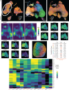
Fibrotic hypersensitivity pneumonitis (FHP) harbors distinct CD8+ T cell phenotype compared with idiopathic pulmonary fibrosis (IPF) and controls. (A) T cells (circled) were subset from the overall data and reclustered. A Uniform Manifold Approximation and Projection (UMAP) representation of these cells is shown by (B) T cell subtypes and (C) condition. Density graphs of T cells for (D) controls, (E) FHP, and (F) IPF. (G) Gene expression of cytotoxic CD4 T cells enriched in FHP: HLA-DRB1, KLRB1, TGFB1, S100A10, PLEK, and VAMP. (H) Gene expression of GZMHhi CD8+ T cells enriched in FHP: FGFBP2, GZMH, CCL5, CTSW, KLRD1, and IL1B. (I) UMAP of CD8+ T cells clustered by DoRothEA-VIPER TF (transcription factor) activity scores. (J) Breakdown of the DoRothEA-based clusters by control, FHP, and IPF. Significantly enriched clusters in FHP are marked in orange. (K) Heatmap demonstrating TFs with top variable imputed activities across CD8+ T cell DoRothEA clusters, with hierarchically clustered rows and columns. CTRL = control.

Comment in

References

    1. Alberti ML, Rincon-Alvarez E, Buendia-Roldan I, Selman M. Hypersensitivity pneumonitis: diagnostic and therapeutic challenges. Front Med (Lausanne) . 2021;8:718299. - PMC - PubMed
    1. Buendía-Roldán I, Santiago-Ruiz L, Pérez-Rubio G, Mejía M, Rojas-Serrano J, Ambrocio-Ortiz E, et al. A major genetic determinant of autoimmune diseases is associated with the presence of autoantibodies in hypersensitivity pneumonitis. Eur Respir J . 2020;56:1901380. - PMC - PubMed
    1. Costabel U, Miyazaki Y, Pardo A, Koschel D, Bonella F, Spagnolo P, et al. Hypersensitivity pneumonitis. Nat Rev Dis Primers . 2020;6:65. - PubMed
    1. Raghu G, Remy-Jardin M, Richeldi L, Thomson CC, Inoue Y, Johkoh T, et al. Idiopathic pulmonary fibrosis (an update) and progressive pulmonary fibrosis in adults: an official ATS/ERS/JRS/ALAT clinical practice guideline. Am J Respir Crit Care Med . 2022;205:e18–e47. - PMC - PubMed
    1. Selman M, Pardo A, King TE., Jr Hypersensitivity pneumonitis: insights in diagnosis and pathobiology. Am J Respir Crit Care Med . 2012;186:314–324. - PubMed