. 2024 Jul 25;84(14):2601-2617.e12.
doi: 10.1016/j.molcel.2024.05.031. Epub 2024 Jun 25.

Structure of the Hir histone chaperone complex

Affiliations

Structure of the Hir histone chaperone complex

Hee Jong Kim et al. Mol Cell. .

Abstract

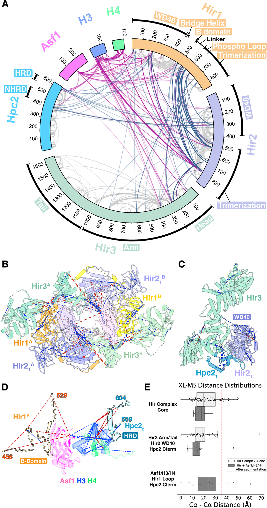
The evolutionarily conserved HIRA/Hir histone chaperone complex and ASF1a/Asf1 co-chaperone cooperate to deposit histone (H3/H4)2 tetramers on DNA for replication-independent chromatin assembly. The molecular architecture of the HIRA/Hir complex and its mode of histone deposition have remained unknown. Here, we report the cryo-EM structure of the S. cerevisiae Hir complex with Asf1/H3/H4 at 2.9-6.8 Å resolution. We find that the Hir complex forms an arc-shaped dimer with a Hir1/Hir2/Hir3/Hpc2 stoichiometry of 2/4/2/4. The core of the complex containing two Hir1/Hir2/Hir2 trimers and N-terminal segments of Hir3 forms a central cavity containing two copies of Hpc2, with one engaged by Asf1/H3/H4, in a suitable position to accommodate a histone (H3/H4)2 tetramer, while the C-terminal segments of Hir3 harbor nucleic acid binding activity to wrap DNA around the Hpc2-assisted histone tetramer. The structure suggests a model for how the Hir/Asf1 complex promotes the formation of histone tetramers for their subsequent deposition onto DNA.

Keywords: Asf1; H3/H4; HIRA; Hir; chromatin; crosslinking mass spectrometry; cryo-electron microscopy; histone chaperone; nucleosome; transcription.


Conflict of interest statement

Declaration of interests The authors declare no competing interests.

Figures

Figure 1
Figure 1. Overall cryo-EM structure of the Hir complex.
(A) Domain architecture of each subunit of the Hir complex. (B) Overall side view (left) and top view (right) of the Hir complex. The Hir complex adopts an arc-shaped roughly 2-fold symmetric dimer (copy A and copy B on the left and right, respectively). Each copy consists of one copy each of Hir1 (orange) and Hir3 (green), and two copies each of Hir2 (dark purple and light purple) and Hpc2 (indigo blue and cyan). In the right panel, two copies of the heterotrimer core are highlighted. (C and D) Overall composite density map without Asf1/H3/H4 (C) and schematic with Asf1/H3/H4 (D) in the same orientations as in (B). (E-H) Structure and location of Hir3 (E), Hir1 (F), Hir21 (G), and Hir22 (H), of the Hir complex in copy A.
Figure 2
Figure 2. Crosslinking mass spectrometry (XL-MS) of the Hir complex.
(A) Circular plot of unique cross-linked residue pairs for the Hir complex alone (steel blue) and the Hir complex-Asf1/H3/H4 (violet red). Intra-subunit unique residue pairs are indicated in gray. (B-D) Cross-links are mapped on the central core (B), Hir3 (Tail)-Hir2(WD40)-Hpc2 (C), and Asf1/H3/H4 (blue, green, magenta) complexed with the Hir1 B-domain (light orange) and the Hpc2 HRD domain (indigo blue) (D). Blue and red dashed lines indicate cross-links with Cα-Cα distances of less than 35 Å and greater than 35 Å, respectively. (E) Distance distribution plots for cross-links mapped in (B), (C), and (D) with vertical dotted line indicating a distance cutoff of 35 Å. Corresponding Cα–Cα distances were calculated to copy A and copy B for each XL, and only a shorter Cα–Cα distance was used to validate our cryo-EM model. Hir complex alone and Hir complex-Asf1/H3/H4 datasets are separately plotted.
Figure 3
Figure 3. Structure of the central core of the Hir complex at 2.96 Å resolution.
(A) Top view of the central core of the Hir complex. Boxed areas are enlarged in (C), (D), and (E), respectively. (B) Same as (A) but viewed from side. Boxed area is enlarged in (F). (C-F) Enlarged views of inter-subunit interactions of Hir22 (copy B)-Hir3 (copy B) (C), Hir1 (copy B)-Hir3 (copy B) (D), Hir22 (copy A)-Hir21(copy A)-Hir3 (copy B) (E), and Hir1 (copy A)-Hir3 (copy B) (F). Side chains involved in the inter-subunit interactions are shown. Conserved residues are labeled in bold.
Figure 4
Figure 4. Structures of the Hir3(Arm and Tail)-Hir21(WD40)-Hpc2 subcomplex.
(A) Overall side view of the Hir complex, overlaid with focused-refined map for Hir3(C-term Arm and Tail)-Hir21(WD40)-Hpc2. (B) Enlarged area of the focused-refined map in (B) but rotated by 90° (left) relative to (A). Hir3 Arm and Tail modules are in green. The Hir21 WD40 domain is in light purple. The Hpc2 is in indigo blue. (C) Enlarged view of the interface between Hir21 WD40 (light purple), Hir3 Tail (light green), Hpc2 NHRD (indigo blue) and Hpc2 HRD (cyan). Viewed roughly in the same orientation as (B). (D) Secondary structure schematics for (C).
Figure 5
Figure 5. Structure of Hir3 and conserved basic regions.
(A) Schematic diagram of the secondary structure of yeast Hir3. Canonical TPR motifs (TPR1–8) were identified by TPRpred. The Head, Arm, and Tail domains are made up of α1-α9, α10-α41, and α42-α70, respectively. The Arm and Tail domains are further divided into N-terminal and C-terminal subdomains, respectively. (B) Structure of yeast Hir3 with secondary structure annotations. Seen in two side views rotated 180° with respect to each other. (C) The basic channel and the DNA-binding patch are indicated with electrostatic potential overlaid. Boxed area is enlarged in (D) while oval area is enlarged in (E). (D) Enlarged view of the basic channel in (C) with side chains of conserved residues (W1280, Y1284, K1288, K1292, Y1328, K1336, and R1340). (E) Enlarged view of the putative DNA-binding patch in (C) with side chains of conserved residues (K1482, K1483, R1486, R1485, K1558, and K1565).
Figure 6
Figure 6. Cryptic-transcription reporter assay using Hir3 putative nucleic acid binding mutants.
(A) Schematic of cryptic promoter reporter assay. (B) Cryptic transcription phenotypes of Hir3 nucleic acid binding mutant alleles. Mutant alleles were spotted in a 10-fold dilution series on the indicated medium for three days. A low copy number plasmid pRSII315 carrying hir3 mutant alleles was transformed into the hir3Δ strain. As a control, the empty vector was transformed into the parent strain (Background) or the hir3Δ strain (Knockout). From top to bottom, 1) Background, empty vector; 2) Wildtype, HIR3 wild type; 3) ssDNA/RNA-channel 2x mutant, K1288A, K1292A; 4) ssDNA/RNA-channel 7x mutant, W1280A, Y1284A, K1288A, K1292A, Y1328A, K1336A, R1340A; 5) DNA-patch 4x mutant, K1482A, K1483A, R1485A, R1486A; 6) DNA-patch 6x mutant, K1482A, K1483A, R1485A, R1486A, K1558A, K1565A; 7) Knockout, empty vector. (C) Pull-down assay to test complex stability of hir3 alleles. Same strains as (B) were grown in SC-Leu media, pulled down by a Flag-tag on Hir1, and analyzed by SDS-PAGE.
Figure 7
Figure 7. Asymmetric binding of Asf1/H3/H4 to the central cavity and proposed model of histone deposition by the Hir complex.
(A) Cutaway views of the Hir complex, seen from bottom showing the central cavity surrounded by two copies of Hir1 (orange and yellow). Also shown is low-resolution density (gray) corresponding to one copy of Asf1/H3/H4 (magenta, blue, and green) and two copies of Hir1 WD40 domains revealed by focused refinement. Unmodeled densities (dashed circles) bridging between the Hir1 trimerization domains and Asf1/H3/H4 are due to the Hir1 acidic stretches (residues 570–620) in copy A and copy B. (B) Zoomed view of Asf1/H3/H4 flanked by the Hir1 WD40 domains of copy A (orange) and copy B (yellow). Rotated by 90° relative to the lower panel of (A). Histone H3 (blue) is tethered to the Hir1 WD40 domain of copy B (yellow) through the Hpc2 HRD (indigo blue), while Asf1 (magenta) is tethered to the Hir1 bridge helix (residues 421–435) of copy A (orange) through the B-domain and a 40-residue linker. Unique cross-linked residue pairs identified by XL-MS are indicated by red dashed lines. (C) Table summarizing the data from surface plasmon resonance measurements of interactions between histone substrates (analytes) and immobilized Hir complex (surface ligand). For yAsf1FL/hH3.3FL/hH4FL and yAsf1FL/hH3.1FL/hH4FL, binding curves were fit using a Heterogeneous Ligand model. For yeastAsf1 alone, a 1:1 binding model was used. (D) SPR kinetic curves between histone substrates and immobilized Hir complex. See also Figure S16. (E) A proposed model of histone deposition by the Hir complex. The DNA-binding patch is indicated by a blue dashed oval.

References

    1. Kornberg RD (1974). Chromatin structure: a repeating unit of histones and DNA. Science 184, 868–871. 10.1126/science.184.4139.868. - DOI - PubMed
    1. Das C, Tyler JK, and Churchill ME (2010). The histone shuffle: histone chaperones in an energetic dance. Trends Biochem Sci 35, 476–489, 2010. S0968–0004(10)00070–8 [pii] 10.1016/j.tibs.2010.04.001. - DOI - PMC - PubMed
    1. Talbert PB, and Henikoff S (2021). Histone variants at a glance. J Cell Sci 134. 10.1242/jcs.244749. - DOI - PMC - PubMed
    1. Ferrand J, Rondinelli B, and Polo SE (2020). Histone Variants: Guardians of Genome Integrity. Cells 9. 10.3390/cells9112424. - DOI - PMC - PubMed
    1. Gurard-Levin ZA, Quivy JP, and Almouzni G (2014). Histone chaperones: assisting histone traffic and nucleosome dynamics. Annu Rev Biochem 83, 487–517. 10.1146/annurev-biochem-060713-035536. - DOI - PubMed

MeSH terms

LinkOut - more resources