. 2024 Nov;31(11):1398-1409.
doi: 10.1038/s41418-024-01333-2. Epub 2024 Jun 26.

FRMD6 determines the cell fate towards senescence: involvement of the Hippo-YAP-CCN3 axis

Affiliations

FRMD6 determines the cell fate towards senescence: involvement of the Hippo-YAP-CCN3 axis

Jung-Jin Park et al. Cell Death Differ. 2024 Nov.

Abstract

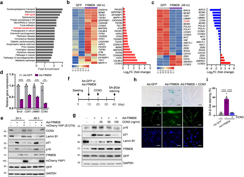
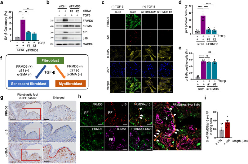
Cellular senescence, a hallmark of aging, is pathogenically linked to the development of aging-related diseases. This study demonstrates that FRMD6, an upstream component of the Hippo/YAP signaling cascade, is a key regulator of senescence. Proteomic analysis revealed that FRMD6 is upregulated in senescent IMR90 fibroblasts under various senescence-inducing conditions. Silencing FRMD6 mitigated the senescence of IMR90 cells, suggesting its requirement in senescence. Conversely, the overexpression of FRMD6 alone induced senescence in cells and in lung tissue, establishing a causal link. The elevated FRMD6 levels correlated well with increased levels of the inhibitory phosphorylated YAP/TAZ. We identified cellular communication network factor 3 (CCN3), a key component of the senescence-associated secretory phenotype regulated by YAP, whose administration attenuated FRMD6-induced senescence in a dose-dependent manner. Mechanistically, FRMD6 interacted with and activated MST kinase, which led to YAP/TAZ inactivation. The expression of FRMD6 was regulated by the p53 and SMAD transcription factors in senescent cells. Accordingly, the expression of FRMD6 was upregulated by TGF-β treatment that activates those transcription factors. In TGF-β-treated IMR90 cells, FRMD6 mainly segregated with p21, a senescence marker, but rarely segregated with α-SMA, a myofibroblast marker, which suggests that FRMD6 has a role in directing cells towards senescence. Similarly, in TGF-β-enriched environments, such as fibroblastic foci (FF) from patients with idiopathic pulmonary fibrosis, FRMD6 co-localized with p16 in FF lining cells, while it was rarely detected in α-SMA-positive myofibroblasts that are abundant in FF. In sum, this study identifies FRMD6 as a novel regulator of senescence and elucidates the contribution of the FRMD6-Hippo/YAP-CCN3 axis to senescence.


Conflict of interest statement

The authors declare no competing interests.

Figures

Fig. 1
Fig. 1. Upregulation of FRMD6 in senescent cells.
ac Proteomics analysis. a Heatmap of differentially expressed proteins (p < 0.05) in control vs. siPIX-treated IMR90 cells. ANOVA test. p < 0.05. b Pathway analysis. c Volcano plot of differentially expressed proteins. A Log2 fold change of >0.5 or <−0.5 (p < 0.05) was regarded as significant. Red, upregulated proteins; blue, downregulated proteins; gray, no differentially expressed proteins. dg Immunoblotting analysis. Lysates from siPIX-treated (d) and doxorubicin (DOX)-treated (f), and replicative senescent (g) cells were subjected to immunoblotting for the indicated proteins. e Quantification of immunoblotting shown in (d). Error bars indicate means ± SEM from three independent experiments. **p < 0.01, ***p < 0.0001, one-way ANOVA. h Co-staining images for FRMD6 and p21. Control and siPIX-treated cells were subjected to immunofluorescence. i Quantification of FRMD6-and p21-positive cells shown in (h). Scale bar, 20 μm. Error bars indicate means ± SEM from three independent experiments. ****p < 0.0001, t-test.
Fig. 2
Fig. 2. Overexpression of FRMD6 induces senescence in vitro and in vivo.
a Immunoblotting of senescence markers. b Cell proliferation assay. Cells infected with Ad-GFP (GFP only) or Ad-FRMD6 expressing GFP as a reporter were counted over 5 days of culture. c SA-β-Gal staining and quantification. SA-β-Gal staining was performed (left) and quantified for SA-β-Gal positive cells (right). Scale bars, 50 μm. d Cytokine array analysis. Infected cells were cultured for 5 days and culture media were collected and incubated with the membranes for cytokine array (top). Densitometry was performed for quantification (bottom). e Immunoblotting analysis. Lysates from Ad-GFP and Ad-FRMD6-infected lungs were subjected to immunoblotting. N = 4 mice per group. f Quantification of the blot in (e). g Immunofluorescence images for p16 and p21. Scale bar, 100 μm. h Quantification of p16 or p21-positive bronchioles in GFP-positive bronchioles shown in (g). N = 11 mice per group. Error bars indicate means ± SEM. *p < 0.05, **p < 0.01, ***p < 0.001, t-test.
Fig. 3
Fig. 3. Constitutively active YAP rescues FRMD6-induced senescence.
a Immunoblotting for the senescence markers and YAP/TAZ. Cells were transfected with plasmids for control or HA-tagged FRMD6 and lysates were subjected to immunoblotting. b Quantification of the blots shown in (a). Error bars indicate means ± SEM from three independent experiments. *p < 0.05, **p < 0.01, ***p < 0.001, t-test. c Immunofluorescence images of FRMD6 (HA-tagged) and endogenous YAP or TAZ using separate antibodies for YAP or TAZ. Cells were transfected with a plasmid encoding HA-FRMD6 and were co-stained with a combination of antibodies against HA/YAP or HA/TAZ. Note that the strong FRMD6-positive cell shows negligible YAP or TAZ staining. Scale bar, 50 μm. Immunoblotting (d) and SA-β-Gal staining (e). Cells were transfected with plasmids expressing mCherry-YAP, wild-type or a constitutively active form (S127A), followed by infection with Ad-FRMD6, after which immunoblotting and SA-β-Gal staining assays were conducted. Scale bar, 50 μm. f Quantification of SA-β-Gal positive cells shown in (e). N ≥ 200 cells per group from three independent experiments. Error bars indicate means ± SEM. **p < 0.01, t-test.
Fig. 4
Fig. 4. Identification of FRMD6-YAP/TAZ targets by RNA-seq analysis.
a KEGG pathway analysis in cells overexpressing FRMD6. b Heatmap of increased senescence-related genes in cells overexpressing FRMD6 (left) and Log2 fold change for genes (right). c Heatmap of up- or down-regulated genes in the transcriptional targets of YAP (left) and Log2 fold change for genes (right). d Validation of downregulated genes by qRT-PCR. Error bars indicate means ± SEM. N = 3, **p < 0.01, ***p < 0.001, ****p < 0.0001, t-test. e Immunoblotting. f Experimental scheme for the rescue experiment. g Immunoblotting for senescence-related proteins. h SA-β-Gal staining. i Quantification of SA-β-Gal positive cells shown in (h). N ≥ 200 cells per group from three independent experiments. Error bars indicate means ± SEM. ****p < 0.0001, t-test.
Fig. 5
Fig. 5. Identification of MST kinase as a binding partner of FRMD6.
ag Immunoblotting analysis. a IMR90 cells were transfected with a plasmid expressing HA-tagged FRMD6 and incubated with XMU-MP-1 (10 μM) for 2 h prior to harvest. Lysates were subjected to immunoblotting. b Cells were co-transfected with plasmids and lysates were immunoprecipitated using anti-HA or anti-FLAG antibody-coated Agarose beads followed by immunoblotting. c Cells were transfected with the indicated plasmids and lysates were subjected to immunoprecipitation with anti-HA beads followed by immunoblotting. d Cells transfected with the indicated plasmid were stimulated with DOX for 48 h. Lysates were immunoprecipitated by anti-HA beads and immunoblotted. e Left, lysates from DOX-treated young cells (top) or young and old cells (bottom) were immunoblotted. Right, lysates from DOX-treated young cells (top) or from old cells (bottom) were immunoprecipitated by an anti-FRMD6 antibody followed by the treatment with PP2A and immunoblotting. f DOX-treated young cells (left) and old cells (right) were incubated with XMU-MP-1 (10 μM) for 2 h prior to harvest. Lysates were then immunoprecipitated by an anti-FRMD6 antibody and immunoblotted. g Cells were co-transfected with plasmids and treated with XMU-MP-1 as described above. Lysates were immunoprecipitated with anti-HA or anti-FLAG beads followed by immunoblotting. GAPDH was used as a loading control. h Model for the interaction of FRMD6 and MST kinase, which leads to the inactivation of YAP.
Fig. 6
Fig. 6. TGF-β upregulates FRMD6 expression in IMR90 fibroblasts.
a Venn diagram for activated transcription factors. b, c Immunoblotting. Cells were treated with DOX (500 nM) in the presence of control siRNAs, p53- (b) or SMAD2-specific siRNAs (c). Representative blots are shown from two independent experiments. d Quantification of SA-β-Gal staining. N ≥ 200 cells per group from three independent experiments were counted. ****p < 0.0001, t-test. eg Immunoblotting (left) and quantification (right). Dose- (e) and time-dependent (f) changes in FRMD6 along with senescence markers in response to TGF-β. g Effect of treatment with A-8301, a TGF-β receptor I inhibitor. h, i Immunoblotting. Cells were treated with TGF-β (10 ng/ml) in the presence of control siRNAs, p53- (h) or SMAD2-specific siRNAs (i). Representative blots are shown from three independent experiments. j Immunoblotting (left) and ELISA (right) of secreted TGF-β. Culture medium was collected at 2 days post Ad-GFP (control) and Ad-FRMD6 infection and subjected to immunoblotting and ELISA. As a loading control, the membrane was stained with Coomassie Brilliant Blue. N = 3, Error bars indicate means ± SEM. **p = 0.0028, t-test k ELISA. IL-6 (left) and IL-8 (right) were quantified by ELISA using the same culture medium from (j). N = 3, Error bars indicate means ± SEM. *p = 0.0285, **p = 0.0048, t-test. l Model for the positive feedback loop via upregulated FRMD6 in TGF-β-induced senescence.
Fig. 7
Fig. 7. FRMD6 mediates TGF-β-induced senescence but not myofibroblast differentiation.
ae FRMD6 was knocked down and its effect was evaluated in TGF-β-stimulated cells. a Quantification of SA-β-Gal staining. b Immunoblotting analysis. c Immunofluorescence. Triple staining for FRMD6, p21 and α-SMA was conducted in control and FRMD6 knockdown cells after stimulation with TGF-β (10 ng/ml). Quantification of p21 (d) and α-SMA (e) positive cells in control or FRMD6 siRNA-treated cells. Error bars indicate means ± SEM from three independent experiments. ****p < 0.0001, t-test. f Schematic model for a specific role of FRMD6 in TGF-β-induced senescence but not myofibroblast differentiation. g Representative immunohistochemical images. Serially sectioned lung tissues from IPF patients were subjected to immunohistochemistry with the indicated antibodies. The rectangular areas in red (left column) are enlarged in the right column. Scale bars, 50 μm (left column); 100 μm (right column). h Representative Immunofluorescence images. Human IPF lung tissues were co-stained with FRMD6, p16, and α-SMA. FF, fibroblastic foci. Dotted lines denote the border between FF and lining cells that face the cystic space. Scale bars, 50 μm. i Quantification of FRMD6 and p16 co-staining FF in lining cells (shown in h). The size of FF denotes the long diagonal length in μm. N = 9 IPF patients.

References

    1. Coppe JP, Desprez PY, Krtolica A, Campisi J. The senescence-associated secretory phenotype: the dark side of tumor suppression. Annu Rev Pathol. 2010;5:99–118. - PMC - PubMed
    1. Lopez-Otin C, Blasco MA, Partridge L, Serrano M, Kroemer G. The hallmarks of aging. Cell. 2013;153:1194–217. - PMC - PubMed
    1. Barnes PJ, Baker J, Donnelly LE. Cellular senescence as a mechanism and target in chronic lung diseases. Am J Respir Crit Care Med. 2019;200:556–64. - PubMed
    1. Edgar BA. From cell structure to transcription: hippo forges a new path. Cell. 2006;124:267–73. - PubMed
    1. Zeng Q, Hong W. The emerging role of the hippo pathway in cell contact inhibition, organ size control, and cancer development in mammals. Cancer Cell. 2008;13:188–92. - PubMed

MeSH terms

Substances

LinkOut - more resources