. 2024 Jul;631(8020):424-431.
doi: 10.1038/s41586-024-07554-2. Epub 2024 Jun 26.

An intermediate Rb-E2F activity state safeguards proliferation commitment

Affiliations

An intermediate Rb-E2F activity state safeguards proliferation commitment

Yumi Konagaya et al. Nature. 2024 Jul.

Abstract

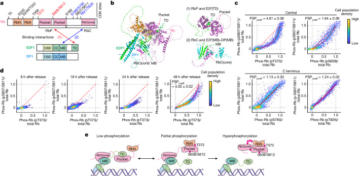
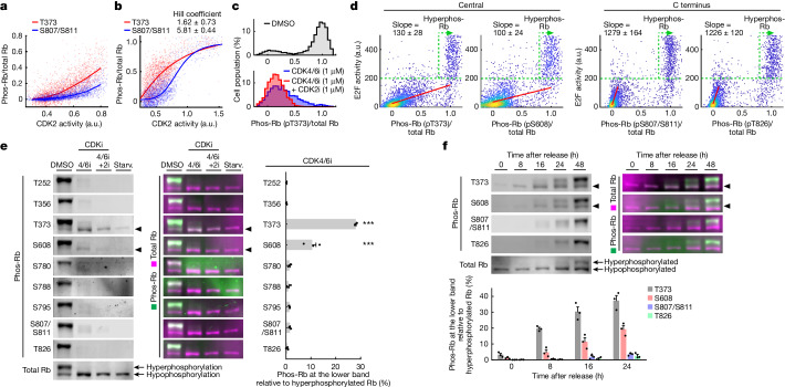
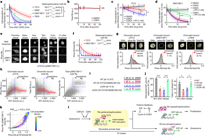
Tissue repair, immune defence and cancer progression rely on a vital cellular decision between quiescence and proliferation1,2. Mammalian cells proliferate by triggering a positive feedback mechanism3,4. The transcription factor E2F activates cyclin-dependent kinase 2 (CDK2), which in turn phosphorylates and inactivates the E2F inhibitor protein retinoblastoma (Rb). This action further increases E2F activity to express genes needed for proliferation. Given that positive feedback can inadvertently amplify small signals, understanding how cells keep this positive feedback in check remains a puzzle. Here we measured E2F and CDK2 signal changes in single cells and found that the positive feedback mechanism engages only late in G1 phase. Cells spend variable and often extended times in a reversible state of intermediate E2F activity before committing to proliferate. This intermediate E2F activity is proportional to the amount of phosphorylation of a conserved T373 residue in Rb that is mediated by CDK2 or CDK4/CDK6. Such T373-phosphorylated Rb remains bound on chromatin but dissociates from it once Rb is hyperphosphorylated at many sites, which fully activates E2F. The preferential initial phosphorylation of T373 can be explained by its relatively slower rate of dephosphorylation. Together, our study identifies a primed state of intermediate E2F activation whereby cells sense external and internal signals and decide whether to reverse and exit to quiescence or trigger the positive feedback mechanism that initiates cell proliferation.


Conflict of interest statement

The authors declare no competing interests.

Figures

Fig. 1
Fig. 1. A marked variation in the activation kinetics of E2F revealed by live cell E2F transcription analysis.
a, Model of the cell cycle decision process in G1 phase. b, Schematic of the E2F transcriptional reporter. c, Experimental design for mitogen release in MCF-10A cells. d, Top, time-course images of a cell expressing mVenus (E2F reporter) and H2B–iRFP (nucleus marker), starved for 2 days and released with growth medium. Arrows indicate cell nuclei. Scale bar, 20 μm. Bottom, mVenus intensity (in arbitrary units (a.u.)) in the cell shown in the top (1 out of 3 biological replicates). e, mVenus intensity traces in control (Con) or doxycycline-inducible HA-tagged cyclin D1-expressing cells. Doxycycline was added 5 h before release to induce cyclin D1. Cells were released with starvation medium + EGF (20 ng ml–1). Cyclin D1-expressing cells were released with DMSO or CDK4/6 inhibitor (CDK4/6i; 1 μM palbociclib). HA > 212 was used to gate cyclin D1-overexpressing cells. P  =  2.8 × 10−92 (mVenus intensity 16 h after release), calculated using two-sided, two-sample t-tests (mean ± s.e.). n = 500, 500 and 456 cells for Con + DMSO, cyclin D1 + DMSO and cyclin D1 + CDK4/6i, respectively; 1 out of 3 biological replicates. f, mVenus intensity traces after release with growth medium. Cells were released with DMSO, CDK2 inhibitor (CDK2i; 1 μM PF-07104091), CDK4/6i or CDK2i + CDK4/6i (mean ± s.e.). n = 300 cells per condition; 1 out of 4 biological replicates. g, Top, schematic of E2F activation in cells released with DMSO versus CDK4/6i. Bottom, single-cell traces of mVenus intensity after release with growth medium + DMSO or CDK4/6i.
Fig. 2
Fig. 2. E2F and CDK2 activities stay at intermediate levels and remain reversible before the positive feedback that starts S phase entry fully engages.
a, Single-cell traces of CDK2 and E2F activities. Circles, CDK2-activated or E2F-activated timing. b, Cumulative frequency of CDK2-activated, E2F-activated and S phase-entered cells. n = 5,969 cells; 1 out of 3 biological replicates. c, Cell traces were computationally aligned at S phase entry and stratified based on the variable time cells spend from E2F-active to S phase entry. CDK2 and E2F activity traces (mean per cell population). n = 244, 246, 226 and 125 cells for 5–10 h, 10–15 h, 15–20 h and 20–25 h, respectively; 1 out of 3 biological replicates. For ac, conditions were release with growth medium + CDK4/6i (1  µM). d, Data in c plotted as a phase-plane trajectory. e, Data in c analysed for the variable time cells spend from CDK2-active to CDK2 activity = 0.65 (top), and from CDK2 activity = 0.65 to S phase entry (bottom). Dashed lines indicate the median. f, Left, percentage of cells with E2F activation by 40 h after release with starvation medium + EGF (20 or 0.2 ng ml–1) ± CDK4/6i (1 µM). Right, percentage of S enter, E2F reverse and undecided cells among E2F-activated cells (mean ± s.e.). Cells were categorized based on behaviours until 40 h after release (see Methods for more detail). n = 2,655, 3,611, 1,685 and 3,042 cells for EGF 20, EGF 0.2, EGF 20 + 4/6i and EGF 0.2 + 4/6i, respectively; 3 biological replicates. g, Single-cell traces of E2F activity in cells categorized in f. h, Single-cell traces of E2F activity in S enter and E2F reverse RPE-1 cells (left), determined based on DNA content versus 5-ethynyl-2′-deoxyuridine (EdU) incorporation at the end of live-cell imaging (right). One out of 3 biological replicates. i, CDK2 and E2F activity traces (mean ± s.e.) after release with starvation medium +  EGF (20 ng ml–1) + CDK4/6i (1 µM). n = 534 (DMSO) and 476 (EGFR inhibitor (EGFRi)) cells; 1 out of 3 biological replicates. j, Data in i plotted as a phase-plane trajectory. k, Single-cell traces of CDK2 activity before and after EGFRi. G1 and S/G2 cells in i were gated based on the CRL4Cdt2 reporter signal. n = 5 cells each; 1 out of 3 biological replicates.
Fig. 3
Fig. 3. Rb is first phosphorylated at T373 before its C-terminal sites.
a, Domain architecture of Rb, E2F1 and DP1. Rb consists of a structured amino-terminal domain (RbN) and RbP. Its RbC is disordered except for a short sequence (RbC(core)) that adopts a structure after E2F binding. CC, coiled-coil domain; DBD, DNA-binding domain. b, Left, Rb–E2F1–DP1 complex prediction with AlphaFold. Rb(ΔCDK(43–928)) (CDK phosphorylation sites mutated to alanines to mimic unphosphorylated Rb), E2F1(200–437), and DP1(198–410) were used to predict the structure of the complex. Right, two interaction sites between Rb and E2F1–DP1: (1) RbP interacts with E2F(TD) and (2) RbC interacts with E2F(MB)–DP(MB). c,d, PSP plots showing single-cell correlation of Rb phosphorylation (phos-Rb) between two different sites 16 h after release with starvation medium + EGF (20 ng ml–1)  + CDK4/6i (20 nM) (c) or release after starvation medium + EGF (20 ng ml–1) + CDK4/6i (1 µM) (d). Each phosphorylation signal was normalized by the total Rb antibody signal in the same cell and each axis was adjusted to the average phosphorylation signal in S phase of 1 (when Rb is hyperphosphorylated). A red line shows fitting with a preferential relative phosphorylation–dephosphorylation rate between the two sites (PSPcoeff) (see Methods for more details). c, Rb phosphorylation at T373 (pT373), S608 (pS608), S780 (pS780) and T826 (pT826) plotted against S807/S811 (pS807/S811). n = 2,734, 2,159, 2,684 and 2203 cells for T373, S608, S780 and T826, respectively; 1 out of 3 biological replicates. d, Rb phosphorylation at T373 plotted against S807/S811. Cells were fixed 8, 16, 24 and 48 h after release. n = 2,531, 2,655, 2,713 and 2,774 cells for 8 h, 16 h, 24 h and 48 h, respectively; 1 out of 2 biological replicates for 8 h, 3 biological replicates for 16 h, 4 biological replicates for 24  and 48 h. e, Model for phosphorylation and inactivation of Rb in a two-step process. First, Rb is phosphorylated at T373 and S608/S612, which probably disrupts (1) the RbP–E2F(TD) interaction. Second, Rb phosphorylation at C-terminal sites disrupts (2) the RbC and E2F(MB)–DP(MB) interaction, leading to full release of Rb from E2F.
Fig. 4
Fig. 4. The degree of Rb phosphorylation at T373 is proportional to E2F activity during the intermediate E2F activation state.
a,b, Single-cell correlation of CDK2 activity and Rb phosphorylation under starvation medium  conditions + EGF (20 ng ml–1) + CDK4/6i (1 µM), 24 h (a) or 48 h after release (b). a, Lines indicate median Rb phosphorylation. n = 2,535 cells; 1 out of 3 biological replicates. b, Lines indicate sigmoidal fit curves. n = 1,967 (T373) and 1,867 (S807/S811) cells; 1 out of 4 biological replicates. c, Histograms of Rb phosphorylation at T373 24 h after release with starvation medium + EGF (20 ng ml–1). n = 2,416, 2,630 and 2,440 cells for DMSO, CDK4/6i and CDK4/6i + CDK2i, respectively; 1 out of 3 biological replicates. d, Single-cell correlation of Rb phosphorylation versus E2F activity 24 h after release with starvation medium + EGF (20 ng ml–1) ± CDK4/6i (1 µM). Red lines indicate Deming regression lines. Slope indicates mean ± s.e. n = 3,496, 2,999, 3,197 and 2,881 cells for T373, S608, S807/S811 and T826 respectively; 1 out of 2 technical replicates and 2 biological replicates. e,f, Western blots of phospho-RB and total Rb, and quantification of the lower Rb phosphorylation band (mean ± s.e. from 3 biological replicates). Arrowheads indicate lower Rb phosphorylation band (1 out of 3 biological replicates). e, Cells released with DMSO, CDK4/6i or CDK4/6i + CDK2i (10 μM) were assayed 16, 24 and 24 h after release with starvation (Starv.) medium + EGF (20 ng ml–1), respectively. Starved cells were assayed before release. To account for the different phospho-antibody affinities, lower Rb phosphorylation bands in the 4/6i lane were normalized by the upper Rb phosphorylation bands in the DMSO lane. P values were calculated using one-way analysis of variance (ANOVA) and Scheffé’s post hoc comparison. T373: P = 1.2 × 10−13 (vs T252), P = 1.2 × 10−13 (vs T356), P = 2.6 × 10−13 (vs S780), P = 1.9 × 10−13 (vs S788), P = 2.6 × 10−13 (vs S795), P = 2.9 × 10−13 (vs S807/S811), P = 1.5 × 10−13 (vs T826). S608: P = 1.6 × 10−6 (vs T252), P = 1.6 × 10−6 (vs T356), P = 1.0 × 10−5 (vs S780), P = 4.9 × 10−6 (vs S788), P = 1.0 × 10−5 (vs S795), P = 1.3 × 10−5 (vs S807/S811), P = 2.6 × 10−6 (vs T826). f, Cells were assayed 0, 8, 16, 24 and 48 h after release with starvation medium + EGF (20 ng ml–1) + CDK4/6i (1 µM), respectively. To account for the different phospho-antibody affinities, the lower Rb phosphorylation bands 0–24 h after release were normalized by the upper Rb phosphorylation bands 48 h after release.
Fig. 5
Fig. 5. Regulation and function of T373 phosphorylated Rb.
ad, Exponential decay fitting (mean ± s.e. from 3 biological replicates). Cells were treated with CDK2i (20 μM) 42 h after release with starvation medium + EGF (20 ng ml–1) + CDK4/6i (1 µM). One-way ANOVA and Scheffé’s post hoc comparison (a) or two-sided, two-sample t-tests (bd). a, P = 0.042 (vs S807/S811), P = 0.046 (vs T826). b, P = 0.91 (not significant (NS)). c,d, Cells were pre-treated with the PPi calyculin A (1 nM 60 min or 10 nM 30 min before CDK2i). T373: P = 0.039 (45 min), P = 4.5 × 10−3 (60 min). S807/S811: P = 7.1 × 10−4 (10 nM, 15 min), P = 6.3 × 10−3 (10  nM, 30 min). e,f, Asynchronously cycling cells in starvation medium + EGF (0.2 ng ml–1) were fixed 16 h after 50 ng ml–1 neocarzinostatin for 20 min. e, Images at different cell cycle stages during mitosis. Scale bar, 20 μm. f, Exponential decay fitting (mean ± s.e. from 3 biological replicates). Cells born 1–16 h after neocarzinostatin treatment were computationally aligned at anaphase. n = 1,160 cells. g, Representative images of MCF-10A cells stained for DNA (Hoechst), chromatin-bound phospho-Rb, and chromatin-bound total-Rb (top), and histograms of pre-extracted phospho-Rb/total-Rb (bottom). Soluble protein was pre-extracted 36 h after release with growth medium + CDK4/6i (1 µM). Scale bar, 20 μm. n = 14,385, 13,341 and 14,578 cells for T373, S807/S811 and T826, respectively; 1 out of 3 biological replicates. h, Single-cell correlation of E2F activity and pre-extracted pT373 (left), pre-extracted total-Rb (middle) and total (without pre-extraction) pS807/S811/total Rb (right) 36 h after release with growth medium + CDK4/6i (1 µM). Red lines indicate median values ± 25th and 75th percentiles. n = 14,385 (left and middle) and 22,615 cells (right); 1 out of 3 biological replicates. i, Schematic of doxycycline-inducible HA-tagged Rb constructs. j, E2F activity (left) and cells in S phase (right) after release with growth medium + CDK4/6i (1 µM). Mean ± s.e. from 4 biological replicates, except ΔCDK-3D (3 biological replicates). Endogenous Rb was knocked down 1 day before release and exogenous Rb was doxycycline-induced 5 h before release. Cells with 210 < HA < 211 were selected for analysis. Two-sided, two-sample t-tests. E2F activity: P = 1.8 × 10−5 (WT vs WT-3A), P = 9.2 × 10−4 (ΔCDK vs ΔCDK-3D). S phase cells: P = 0.013 (WT vs WT-3A), P = 0.036 (ΔCDK vs ΔCDK-3D). k, PSP plots of cells 16 h after release with starvation medium + EGF (20 ng ml–1) + DMSO (without  CDK4/6i). n = 3,580 cells; 1 out of 3 biological replicates. l, Model for a reversible primed G1 state.
Extended Data Fig. 1
Extended Data Fig. 1. Development of an E2F activity reporter and validation of the E2F and CDK2 live-cell reporters.
Related to Fig. 1. a, Schematic of E2F reporter development workflow. b, Top, schematic of E2F activation in cells released with DMSO vs CDK4/6i. A red dotted square shows the timing of RNA extraction for RNA-Seq. Bottom, MA plot (log fold change of gene expression in DMSO vs CDK4/6i (1 μM) plotted against mean log gene expression). Cells were assayed for RNA-Seq 12 h after release with growth media + DMSO or CDK4/6i (1 μM). Marked genes are candidate E2F target genes that were used for E2F activity reporter development (1 of n = 3 biological replicates. Reanalysis of published data). c, d, Fold change in pre-mRNA expression levels (measured by RT-qPCR) of the candidate E2F target genes as selected in b. In c, cells were released with growth media + DMSO or CDK4/6i (1 μM) for 14 h. In d, cells were released with growth media for 13 h and treated with DMSO or CDK4/6i (1 μM) for 1 h before RNA extraction. Relative pre-mRNA expression levels were calculated using EEF1A1 as a housekeeping gene. Expression levels with CDK4/6i were normalized by those with DMSO to calculate the fold change per gene per experiment (mean from n = 3 biological replicates). e, Fold change in mean mVenus intensity 15 h after release compared to 0 h after release. Cells expressing mVenus under the control of the E2F target gene promoters were released with growth media + DMSO or CDK4/6i (1 μM) for 15 h (mean from n = 2 biological replicates). f, Top, representative time-course images of MCF-10A cells expressing mVenus under the control of CDC6 promoter (the E2F reporter). Cells were asynchronously cycling in growth media. Scale bar = 20 μm. Bottom, single-cell trace of the mVenus intensity in the cell as shown with yellow arrowheads in Top. g, mVenus intensity (the E2F reporter) traces after release with growth media + CDK4/6i (1 μM). Cells were treated with si-Control or si-RB1 two days before release. A p-value for mVenus intensity 16 h after release was calculated using two-sided, two-sample t tests (mean ± SE. n = 100 cells for each condition. p-value = 1.7 × 10−8. 1 of n = 3 biological replicates). h, Left, histograms of E1A-HA intensity in control or Dox-inducible HA-tagged E1A-expressing cells. Dox was added 5 h before release to induce E1A. Cells were fixed 16 h after release with starvation media + EGF (20 ng/mL) to stain HA. Thresholds for E1Alow and E1Ahigh are HA < 26 and HA > 212, respectively. Right, mVenus intensity (the E2F reporter) in control, E1Alow and E1Ahigh cells 16 h after release. p-values were calculated using two-sided, two-sample t tests (mean ± SE. n = 4 biological replicates. n = 19649, 19983, 6668 cells in total for Con, E1Alow, and E1Ahigh, respectively. p-value (Con vs E1Alow) = 0.16 (n.s.), p-value (E1Alow vs E1Ahigh) = 1.9 × 10−4). i, mVenus intensity (the E2F reporter) traces in control or Dox-inducible HA-tagged E2F1-expressing cells. Dox was added 5 h before release to induce E2F1. Cells were released with starvation media + EGF (20 ng/mL). Similar as h, HA > 212 was used to gate E2F1 overexpressed cells. A p-value for mVenus intensity 16 h after release was calculated using two-sided, two-sample t tests (mean ± SE. n = 500, 484 cells for Con, E2F1, respectively. p-value = 1.3 × 10−23. 1 of n = 3 biological replicates). j, Histograms of HA-E2F1 intensity in control or Dox-inducible HA-tagged E2F1-expressing cells. Dox was added 5 h before release to induce E2F1. Cells were fixed 17 h after release with starvation media + EGF (20 ng/mL) to stain HA. A threshold for E2Fhigh (used in i) is HA > 212 (n = 8075, 3398 cells for Con and E2Fhigh, respectively. 1 of n = 3 biological replicates). k, Histograms of cyclin D1-HA intensity in control or Dox-inducible HA-tagged cyclin D1-expressing cells. Dox was added 5 h before release to induce cyclin D1. Cells were fixed 17 h after release with starvation media + EGF (20 ng/mL) and stained for HA. A threshold for cyclin D1high (used in Fig. 1e) is HA > 212 (n = 8075, 5621 cells for Con and Cyclin D1high, respectively. 1 of n = 3 biological replicates). l, Left, box plots of mVenus intensity (the E2F reporter) in control or Dox-inducible HA-tagged Myc-expressing cells. Dox was added 5 h before release to induce Myc. Cells were released with starvation media + EGF (20 ng/mL) + CDK4/6i (1 μM). HA > 210 was used to gate Myc overexpressed cells. Box centers are median values, box edges are the 25th and 75th percentiles, and whiskers are minimum and maximum values. A p-value for mVenus intensity 16 h after release was calculated using two-sided, two-sample t tests (n = 500 cells each. p-value = 0.23 (n.s.). 1 of n = 3 biological replicates). Right, single-cell correlation of anti-HA intensity and anti-Myc intensity (measured by immunofluorescence). Cells were released with starvation media + EGF (20 ng/mL) + CDK4/6i (1 μM) for 16 h. Dox treatment induces HA-tagged Myc overexpression (n = 500 cells each. 1 of n = 2 biological replicates). m, Single-cell traces of CDK2 activity after release with growth media for 16 h. Cells were released with DMSO or CDK2i (1 μM) (n = 15 cells each. 1 of n = 4 biological replicates). n, Dose-response curve of the suppression of Rb phosphorylation at S807/S811 by CDK4/6i. Cells were fixed 16 h after release with starvation media + EGF (20 ng/mL) + DMSO or CDK4/6i (20 nM, 100 nM, 1 μM). Percentage of cells with Rb phosphorylation levels > 0.5 are plotted as a function of CDK4/6i concentration. A sigmoidal fit was used to derive the half maximal inhibitory concentration, IC50. (n = 3 biological replicates. n = 9001, 9578, 9932, 8641 cells in total for 0, 20, 100, 1000 nM, respectively).
Extended Data Fig. 2
Extended Data Fig. 2. The E2F reporter is a global E2F transcriptional activity reporter, monitoring both activating and repressing E2Fs.
Related to Fig. 1. a, mVenus intensity (the E2F reporter) traces of MCF−10A cells asynchronously cycling in starvation media + EGF (20 ng/mL). Cells were treated with si-Control or si-E2F1/2/3 four hours before starting live-cell imaging. Cells that were born 22 h to 30 h after the start of live-cell imaging were analyzed. Cell traces were computationally aligned at anaphase. A p-value for mVenus intensity 5 h after anaphase was calculated using two-sided, two-sample t tests (mean ± SE. n = 125, 120 cells for si-Control, si-E2F1/2/3, respectively. p-value = 4.3 × 10−4. 1 of n = 3 biological replicates). b, mVenus intensity (the E2F reporter) traces after release with starvation media + EGF (20 ng/mL). Cells were treated with si-Control or si-E2F7 two days before release. A p-value for mVenus intensity 35 h after release was calculated using two-sided, two-sample t tests (mean ± SE. n = 913, 1026 cells for si-Control, si-E2F7, respectively. p-value = 9.0 × 10−20. 1 of n = 3 biological replicates). c, Cell traces in b were computationally aligned at S phase entry. A p-value for mVenus intensity 5 h after S phase entry was calculated using two-sided, two-sample t tests (mean ± SE. n = 796, 872 cells for si-Control, si-E2F7, respectively. p-value = 1.1 × 10−14. 1 of n = 3 biological replicates). d, Box plots of mRNA puncta area of E2F1, E2F2, E2F3, E2F7. Cells were treated with si-Control, si-E2F1, si-E2F2, or si-E2F3 one day before release with growth media and fixed 16 h after release. Cells were treated with si-Control or si-E2F7 for four hours, incubated in starvation media + EGF (20 ng/mL) for 14 h, and fixed. Box centers are median values, box edges are the 25th and 75th percentiles, and whiskers are minimum and maximum values. p-values were calculated using two-sided, two-sample t tests (E2F1: n = 721 cells for si-Control, 729 cells for si-E2F1, p-value = 3.8 × 10−119. E2F2: n = 721 cells for si-Control, 777 cells for si-E2F2, p-value = 1.1 × 10−66. E2F3: n = 721 cells for si-Control, 731 cells for si-E2F3, p-value = 5.6 × 10−141. E2F7: n = 1000 cells for si-Control, 1000 cells for si-E2F7, p-value = 3.0 × 10−145). e, E2F ChIP-Seq signals (blue plots) over CDC6 promoter region used in the E2F reporter (a green box), and detected peaks (magenta lines) with irreproducible discovery rate (IDR) cutoff = 0.05 (publicly available datasets from ENCODE). f, Single-cell correlation of mVenus intensity (the E2F reporter) and E2F target gene mRNA puncta area. Cells were fixed 16 h after growth media release. Red lines are median values and light red ranges are the 25th and 75th percentiles for E2F activity bins (n = 2192, 1833, 2836 cells for E2F1, CCNE2, CDC6, respectively. 1 of n = 4 biological replicates). g, Box plots of mRNA puncta area of E2F target genes. Cells were released with growth media + DMSO or CDK4/6i (1 μM palbociclib) and fixed 16 h after release. Box centers are median values, box edges are the 25th and 75th percentiles, and whiskers are minimum and maximum values. p-values were calculated using two-sided, two-sample t tests (E2F1: n = 806 cells for DMSO, 778 cells for 4/6i, p-value = 1.6 × 10−213. CCNE2: n = 656 cells for DMSO, 765 cells for 4/6i, p-value = 3.9 × 10−186. CDC6: n = 760 cells for DMSO, 768 cells for 4/6i, p-value = 2.8 × 10−240. 1 of n = 3 biological replicates). h, j, Representative images of MCF-10A cells expressing mVenus (the E2F reporter, in the nucleus) and FISH staining for CDC6 mRNA (puncta in the cytoplasm). Cells were fixed 16 h after release with growth media (h) or 36 h after release with growth media + CDK4/6i (1 μM) (j). Scale bar = 20 μm. i, k, Single-cell correlation of mVenus intensity (the E2F reporter) and CDC6 mRNA puncta area (left) or CDC6 mRNA puncta area multiplied by its intensity (right). Cells were fixed 16 h after release with growth media (i) or 36 h after release with growth media + CDK4/6i (1 μM) (k). Red lines are median values and light red ranges are the 25th and 75th percentiles for E2F activity bins (i: n = 1637 cells. 1 of n = 4 biological replicates. k: n = 2499 cells. 1 of n = 3 biological replicates).
Extended Data Fig. 3
Extended Data Fig. 3. Cyclin E induces CDK2 and E2F activation in CDK4/6 inhibited cells.
Related to Fig. 2. a, Single-cell traces of CDK2 activity, E2F activity, and CRL4Cdt2 activity reporter intensity after release with growth media + DMSO for 48 h (n = 25 cells each. 1 of n = 3 biological replicates). b, Cumulative frequency of CDK2-activated, E2F-activated, and S phase-entered cells after release with growth media + DMSO. A time gap between E2F-active and S phase entry was calculated at the 50% line (n = 1941 cells. 1 of n = 3 biological replicates). c, d, Left, E2F activity traces in cells treated with DMSO and CDK2i (1 μM) 14 h after release with starvation media + EGF (20 ng/mL). Right, Representative CRL4Cdt2 reporter traces in cells treated with DMSO 14 h after release with starvation media + EGF (20 ng/mL) to show examples of computational gating. Cells that were in G1 phase (c) or S phase (d) upon the drug treatment were computationally gated based on the CRL4Cdt2 reporter signal. p-values for E2F activity 6 h after the drug treatment (20 h after release) were calculated using two-sided, two-sample t tests (E2F in c: mean ± SE. n = 465, 454 cells for DMSO, CDK2i, respectively. p-value = 9.2 × 10−10. E2F in d: mean ± SE. n = 66, 88 cells for DMSO, CDK2i, respectively. p-value = 0.014. CRL4Cdt2: single-cell traces. n = 15 cells each. 1 of n = 3 biological replicates). e, f, CDK2 (left) and E2F (right) activity traces in control or Dox-inducible HA-tagged cyclin E1-expressing cells. Dox was added 5 h before release to induce cyclin E1. Cells were released with growth media + CDK4/6i (1 μM) (e: single-cell traces. n = 5 cells each. f: mean ± SE. n = 205, 363 cells for Con, Cyclin E1, respectively. 1 of n = 3 biological replicates). g, Same data in f was gated for cells entered S phase and plotted as a phase-plane trajectory. Intervals between arrows are one hour. Blue circles mark the time points for S phase entry, which were determined by the CRL4Cdt2 reporter signal (n = 6, 175 cells for Con, Cyclin E1, respectively. 1 of n = 3 biological replicates). h, Left, percentage of cells entered S phase by 40 h after release in control or Dox-inducible HA-tagged cyclin E1-expressing cells. Dox was added 5 h before release to induce cyclin E1. Cells were released with growth media + CDK4/6i (1 μM). A p-value was calculated using two-sided, two-sample t tests (mean ± SE. n = 3 biological replicates. n = 703, 834 cells in total for Con, Cyclin E1, respectively. p-value = 7.2 × 10−5). Right, histograms of cyclin E1-HA intensity in control or Dox-inducible HA-tagged cyclin E1-expressing cells. Cells were fixed 48 h after release with growth media + CDK4/6i (1 μM) to stain HA and confirm cyclin E1 induction. (n = 1420, 708 cells for Con, Cyclin E1, respectively. 1 of n = 3 biological replicates). i, Same data in Fig. 2g (S enter) was shown with CDK2 activity traces. Cells were released with starvation media + EGF (20 ng/mL) + CDK4/6i (1 μM). Dashed lines mark the time points for S phase entry, which were determined by the CRL4Cdt2 reporter signal (n = 3 cells. 1 of n = 3 biological replicates). j, Same data in Fig. 2c was shown as representative single-cell traces. CDK2 (top) and E2F (bottom) activity traces after release with growth media + CDK4/6i (1 μM). Cells were stratified and color-coded based on the time cells spend from E2F-active to S phase entry. Cell traces were computationally aligned at S phase entry (n = 3 cells each. 1 of n = 3 biological replicates). k, Box plots of CDC6 mRNA puncta area multiplied by its intensity. Cells were starved for two days for measuring CDC6 mRNA levels in G0. Cells were fixed 36 h after release with growth media + CDK4/6i (1 μM) for measuring CDC6 mRNA levels in G1 and S. G1 and S cells were gated based on the CRL4Cdt2 reporter signal. Box centers are median values, box edges are the 25th and 75th percentiles, and whiskers are minimum and maximum values. A p-value was calculated using two-sided, two-sample t tests (n = 1410, 1124, 102 cells for G0, G1, S cells, respectively. p-value (G0 vs G1) = 2.5 × 10−298. 1 of n = 3 biological replicates).
Extended Data Fig. 4
Extended Data Fig. 4. Reversible intermediate E2F activation is generalizable across non-transformed cells.
Related to Fig. 2. a, Schematic of the CRL4Cdt2 reporter, a component of the Fucci(CA) reporter system. b, Single-cell traces of E2F and CRL4Cdt2 activity as an example for “S enter” and “E2F reverse” cells categorized in Fig. 2f. MCF-10A cells were released with starvation media + EGF (20 ng/mL) (1 of n = 3 biological replicates). c, RPE-1 cells expressing the E2F reporter. Left, single-cell correlation of DNA content plotted against EdU incorporation. Dots are color-coded based on E2F activity 41 h after release with growth media. Right, E2F activity traces in cells released with growth media. G1, S, G2 cells 41 h after release are gated based on DNA content and EdU incorporation (mean ± SE. n = 431, 1865, 1466 cells for G1, S, G2 cells, respectively. 1 of n = 3 biological replicates). d, RPE-1 cells expressing the E2F reporter. Left, percentage of cells with E2F activation by 41 h after release. Cells were released with growth media + DMSO or CDK4/6i (1 μM palbociclib). Right, percentage of “S enter”, “E2F reverse”, “undecided” cells among E2F-activated cells per each condition. Cells were categorized into different groups based on the behaviors by 41 h after release. “S enter” cells are cells that entered S phase, which is determined by DNA content and EdU incorporation. “E2F reverse” cells are cells with E2F activation, without S phase entry, and with E2F activity decrease more than half from the peak. “Undecided” cells are cells with E2F activation, but without S phase entry or E2F activity decrease. (mean ± SE. n = 3 biological replicates. n = 11492, 18886 cells in total for DMSO, CDK4/6i, respectively). e, RPE-1 cells expressing the E2F reporter. Single-cell traces of E2F activity as an example for “S enter”, “E2F reverse”, “undecided” cells categorized in d. Cells were released with growth media + DMSO.
Extended Data Fig. 5
Extended Data Fig. 5. CDK2 and E2F are activated via EGFR signaling in CDK4/6 inhibited cells.
Related to Fig. 2. a, Experimental design for b and c. Cells were treated with DMSO or EGFRi (5 μM gefitinib) 42 h after release with starvation media + EGF (20 ng/mL) + CDK4/6i. Cells were lysed 4 h after DMSO or EGFRi treatment for western blots (Supplementary Fig. 1). b, c, Western blots of phospho-EGFR (p-Y1045) and total-EGFR (b) and quantification (c). A p-value was calculated using two-sided, two-sample t tests (b: 1 of n = 3 biological replicates. c: mean ± SE. n = 3 biological replicates. p-value (DMSO vs EGFRi) = 6.3 × 10−4). d, Experimental design for e. Cells were treated with DMSO or EGFRi (5 μM gefitinib) 42 h after release with starvation media + EGF (20 ng/mL) + CDK4/6i. Cells were fixed 4 h after DMSO or EGFRi treatment for immunofluorescence (mean ± SE). e, Box plots of p21 or cyclin E protein levels. Box centers are median values, box edges are the 25th and 75th percentiles, and whiskers are minimum and maximum values. p-values were calculated using two-sided, two-sample t tests (p21: n = 100 cells for DMSO, 100 cells for EGFRi, p-value = 2.5 × 10−10. Cyclin E: n = 100 cells for DMSO, 100 cells for EGFRi, p-value = 0.39 (n.s.). 1 of n = 2 biological replicates). f, Experimental design for g. Cells were treated with DMSO or EGFRi (5 μM gefitinib) 42 h after release with starvation media + EGF (20 ng/mL) + CDK4/6i. Cells were fixed 18 h after DMSO or EGFRi treatment for immunofluorescence (mean ± SE). g, Box plots of p21 or cyclin E protein levels. Box centers are median values, box edges are the 25th and 75th percentiles, and whiskers are minimum and maximum values. p-values were calculated using two-sided, two-sample t tests (p21: n = 100 cells for DMSO, 100 cells for EGFRi, p-value = 1.2 × 10−14. Cyclin E: n = 100 cells for DMSO, 100 cells for EGFRi, p-value = 1.6 × 10−30. 1 of n = 3 biological replicates). h, Experimental design for i and j. Cells were treated with DMSO or CDK2i (1 μM) 42 h after release with starvation media + EGF (20 ng/mL) + CDK4/6i for live-cell imaging. i, j, Left, E2F activity traces in cells treated with DMSO and CDK2i (1 μM). Right, Representative CRL4Cdt2 reporter traces in cells treated with DMSO to show examples of computational gating. Cells that were in G1 phase (i) or S phase (j) upon the drug treatment were computationally gated based on the CRL4Cdt2 reporter signal. p-values for E2F activity 12 h (i) or 8 h (j) after the drug treatment was calculated using two-sided, two-sample t tests (E2F in i: mean ± SE. n = 892, 876 cells for DMSO, CDK2i, respectively. p-value = 1.1 × 10−38. E2F in j: mean ± SE. n = 128, 145 cells for DMSO, CDK2i, respectively. p-value = 1.3 × 10−22. CRL4Cdt2: single-cell traces. n = 15 cells each. 1 of n = 3 biological replicates). k, Experimental design for i. Cells were treated with DMSO or CDK2i or CDK2i + CDK1i (10 μM RO-3306) 42 h after release with starvation media + EGF (20 ng/mL) + CDK4/6i. Cells were fixed 18 h after the drug treatment for immunofluorescence. l, Histograms of Rb phosphorylation at T373 (n = 1825, 1492, 1725 cells for DMSO, CDK2i, CDK2i + CDK1i, respectively. 1 of n = 3 biological replicates). m, Model for the effect of EGFRi (gefitinib) on cell cycle regulators in cells released with EGF and CDK4/6i. During the first 4 h after EGFRi, E2F is inactivated whereas CDK2 is temporarily activated due to p21 downregulation. In contrast, 4 to 18 h after EGFRi, both E2F and CDK2 are inactivated because cyclin E is also downregulated.
Extended Data Fig. 6
Extended Data Fig. 6. Validation of the antibodies and the iterative indirect immunofluorescence imaging (4i).
Related to Fig. 3. a, b, Single-cell correlation of anti-HA intensity and anti-phospho-Rb (p-T373 for a, p-S608 for b) intensity in cells expressing the Dox-inducible HA-tagged Rb constructs (measured by immunofluorescence). Cells were treated with si-RB1 one day before release to knockdown endogenous Rb, treated with Dox 5 h before release to induce Rb constructs, and fixed 36 h after release with growth media. Cells with the efficient RB1 knockdown were gated based on the correlation between anti-HA and anti-total-Rb. In a, Rb-WT-T373A and ΔCDK-T373 were compared to Rb-WT and ΔCDK, respectively and red reference lines are y = 0.8x + 200. In b, Rb-WT-T373A-S608A and ΔCDK-T373-S608 were compared to Rb-WT and ΔCDK, respectively and red reference lines are y = 0.15x + 200 (n = 3803, 3463, 2857, 3607 cells for Rb-WT, Rb-WT-T373A, ΔCDK-T373, ΔCDK, respectively (a). n = 3863, 2687, 3518, 3499 cells for Rb-WT, Rb-WT-T373A-S608A, ΔCDK-T373-S608, ΔCDK, respectively (b)). c, Workflow of the iterative indirect immunofluorescence imaging (4i) protocol. Representative images and histograms of Rb phosphorylation at T373 and S807/S811. Cells were fixed 14 h after release with starvation media + EGF (20 ng/mL). To validate the signal in the second round of 4i is due to anti-phospho-Rb (p-S807/S811) antibody, 4i was performed with (top) or without (bottom) phospho-Rb (p-S807/S811) staining, while keeping the rest same. The same cells across two rounds of 4i are shown for each protocol (top or bottom). Scale bar = 20 μm (Histograms: n = 2960, 2765 cells for with or without phospho-Rb (p-S807/S811) staining, respectively). d, Phosphorylation Site Preference (PSP) plots showing single-cell correlation of Rb phosphorylation at T373 plotted against S807/S811 or vice versa. Cells were fixed 16 h after release with starvation media + EGF (20 ng/mL) + CDK4/6i (20 nM). Left, cells were stained with anti-phospho-Rb (p-T373) antibody in the first round and anti-phospho-Rb (p-S807/S811) antibody in the second round of 4i. Right, cells were stained with anti-phospho-Rb (p-S807/S811) antibody in the first round and anti-phospho-Rb (p-T373) antibody in the second round of 4i. Each phosphorylation signal was normalized by the total Rb antibody signal in the same cell and each axis was adjusted to the average phosphorylation signal in S phase of 1. Color indicates relative cell population density. A red line shows fitting with a preferential relative phosphorylation or dephosphorylation rate between the two sites (PSPcoeff) (see more details in Methods) (n = 2734, 1727 cells for left and right, respectively. 1 of n = 2 biological replicates).
Extended Data Fig. 7
Extended Data Fig. 7. Preferential phosphorylation of Rb at T373 is generalizable in cycling cells, and slower dephosphorylation at T373 in Rb compared to other sites creates partially phosphorylated Rb/intermediate E2F activity states in cycling cells.
Related to Figs. 3 and 5. a, Left, Rb phosphorylation at T373 after anaphase measured by immunofluorescence. Cells were asynchronously cycling in starvation media + EGF (0.2 ng/mL). Pseudo-time course immunofluorescence was measured by computationally aligning cells at anaphase based on live-cell images. Cells were color-coded based on Rb phosphorylation at T373 (green: Rb phosphorylation at T373  0.6, 0-5 h after anaphase, blue: Rb phosphorylation at T373  0.6, 5-15 h). A cyan line shows median levels of Rb phosphorylation at T373 = 1.0 (n = 3 biological replicates. n = 1719 cells in total). Right, representative single-cell traces of E2F activity categorized in Left (n = 3 cells each. 1 of n = 3 biological replicates). b, Model for three different cell fates based on Rb phosphorylation across cell generations. The background color code is matched with the categorization in a. (i) Daughter cells are born with partially phosphorylated Rb involving p-T373 (green) and dephosphorylate Rb completely to undergo quiescence (blue). (ii) Daughter cells are born with partially phosphorylated Rb involving p-T373 (green) and hyperphosphorylate Rb to enter S phase (orange). (iii) Daughter cells are born with hyperphosphorylated Rb and enter S phase quickly (magenta). c, Rb dephosphorylation kinetics after anaphase at T373 (left) and S807/S811 (right) measured by immunofluorescence. Cells were asynchronously cycling in starvation media + EGF (0.2 ng/mL). Cells were treated with or without 50 ng/mL neocarzinostatin (NCS) for 20 min, and incubated in starvation media + EGF (0.2 ng/mL) again for 16 h before fixation. Pseudo-time course immunofluorescence was measured by computationally aligning cells at anaphase based on live-cell images. Cells that were born 1 h to 16 h after NCS or mock treatment were analyzed. A cyan line shows median levels of Rb phosphorylation <0.6 and a red dotted line shows a reference line of Rb phosphorylation = 1.0 (n = 3 biological replicates. n = 1719, 1160 cells in total for NCS (−), NCS (+), respectively). d, Phosphorylation Site Preference (PSP) plots showing single-cell correlation of Rb phosphorylation at T373 plotted against S807/S811. Each phosphorylation signal was normalized by the total Rb antibody signal in the same cell and each axis was adjusted to the average phosphorylation signal in S phase of 1. Color indicates relative cell population density. A red line shows fitting with a preferential relative phosphorylation or dephosphorylation rate between the two sites (PSPcoeff) (see more details in Methods). Cells asynchronously cycling in starvation media + EGF (0.2 or 20 ng/mL) were fixed 16 h after 50 ng/mL neocarzinostatin (NCS) treatment for 20 min or mock treatment (n = 3393, 1904, 3079, 2247 cells for NCS (−) in EGF (0.2 ng/mL), NCS (+) in EGF (0.2 ng/mL), NCS (−) in EGF (20 ng/mL), NCS (+) in EGF (20 ng/mL), respectively. 1 of n = 3 biological replicates). e, Box plots of 53BP1 puncta area. Box centers are median values, box edges are the 25th and 75th percentiles, and whiskers are minimum and maximum values. Cells asynchronously cycling in starvation media + EGF (0.2 or 20 ng/mL) were fixed 16 h after 50 ng/mL neocarzinostatin (NCS) treatment for 20 min or mock treatment. p-values were calculated using two-sided, two-sample t tests (n = 100 cells each, p-value (NCS (−) vs (+) in EGF (0.2 ng/mL)) = 2.0 × 10−14. p-value (NCS (−) vs (+) in EGF (20 ng/mL)) = 2.5 × 10−18. 1 of n = 2 biological replicates).
Extended Data Fig. 8
Extended Data Fig. 8. Preferential phosphorylation of Rb at T373 is generalizable across non-transformed cells, and phosphorylation of the C-terminal sites of Rb is part of the cooperative Rb hyperphosphorylation mechanism.
Related to Figs. 3 and 4. ac, Phosphorylation Site Preference (PSP) plots showing single-cell correlation of Rb phosphorylation at different sites in RPE-1 cells (a), BJ-5ta cells (b), or U2OS cells (c). Each phosphorylation signal was normalized by the total Rb antibody signal in the same cell and each axis was adjusted to the average phosphorylation signal in S phase of 1. Color indicates relative cell population density. A red line shows fitting with a preferential relative phosphorylation or dephosphorylation rate between the two sites (PSPcoeff) (see more details in Methods). Rb phosphorylation at T373 or T826 was plotted against S807/S811. a,b, Cells were fixed 8 h after release with growth media (a: n = 5019, 4900 cells for T373, T826, respectively. b: n = 3409, 3502 cells for T373, T826, respectively. 1 of n = 3 biological replicates). c, Cells asynchronously cycling in growth media were fixed 6 h after DMSO or CDK4/6i (100 nM) treatment (DMSO: n = 704, 700 cells for T373, T826, respectively. CDK4/6i: n = 850, 878 cells for T373, T826, respectively. 1 of n = 3 biological replicates). d, Single-cell correlation of CDK2 activity and Rb phosphorylation at T373 and T826. Cells were fixed 48 h after release with starvation media + EGF (20 ng/mL) + CDK4/6i (1 μM). Rb phosphorylation levels as a function CDK2 activity were fitted by sigmoidal curves (sigmoidal curves with Hill coefficients. n = 1967, 2050 cells for T373, T826, respectively).
Extended Data Fig. 9
Extended Data Fig. 9. Functions of T373 site in Rb are tested in the Rb mutant re-expression assay.
Related to Fig. 5. a, b, Histograms of total-Rb intensity (a) and E2F activity traces (b) in cells with or without RB1 knockdown or with RB1 knockdown + exogenous Rb expression. Cells were fixed 36 h after release with growth media + CDK4/6i. Cells were treated with si-RB1 one day before release to knockdown endogenous Rb and treated with Dox 5 h before release to induce exogenous Rb. A threshold for Rbhigh is Rb > 211 (n = 435, 537, 368, 173 cells for si-Control, si-RB1, si-RB1 + Rb-O.E., si-RB1 + Rbhigh, respectively. E2F: mean ± SE). c, single-cell traces of E2F activity in cells expressing the Dox-inducible HA-tagged Rb constructs. Cells were released with growth media + CDK4/6i. Cells were treated with si-RB1 one day before release to knockdown endogenous Rb and treated with Dox 5 h before release to induce exogenous Rb. To compare the same expression levels of Rb constructs, cells with 210 < HA < 211 were gated for analysis (black lines denote the example single-cell traces of 20 cells from one representative experiment. red lines denote the mean of a whole population. n = 323, 288, 383, 246 cells for Rb-WT, Rb-WT-3A, Rb-∆CDK, Rb-∆CDK-3D, respectively. n = 4 biological replicates for Rb-WT, Rb-WT-3A, Rb-∆CDK, and 3 biological replicates for Rb-∆CDK-3D).
Extended Data Fig. 10
Extended Data Fig. 10. Both CDK4/6 and CDK2 induce Rb phosphorylation at T373 before Rb hyperphosphorylation, and Rb T373 is evolutionarily conserved.
Related to Fig. 5. a, b, Phosphorylation Site Preference (PSP) plots showing single-cell correlation of Rb phosphorylation at different sites. Each phosphorylation signal was normalized by the total Rb antibody signal in the same cell and each axis was adjusted to the average phosphorylation signal in S phase of 1. Color indicates relative cell population density. A red line shows fitting with a preferential relative phosphorylation or dephosphorylation rate between the two sites (PSPcoeff) (see more details in Methods). a, Rb phosphorylation at S608, S780, T826 were plotted against S807/S811. Cells were fixed 16 h after release with starvation media + EGF (20 ng/mL) + DMSO (n = 1795, 2162, 1897 cells for S608, S780, T826, respectively. 1 of n = 3 biological replicates). b, Rb phosphorylation at T373 plotted against S807/S811. Cells were fixed 8 h after release with growth media + DMSO or CDK2i (1 μM) (n = 2167, 2080 cells for DMSO, CDK2i, respectively. 1 of n = 3 biological replicates). c, Histograms of Rb phosphorylation at T373 (left) and S807/S811 (right). Cells were fixed 8 h after release with growth media + DMSO or CDK2i (1 μM) (n = 2167, 2080 cells for DMSO, CDK2i, respectively. 1 of n = 3 biological replicates). d, A multiple sequence alignment (MSA) of T373 in Rb across vertebrates (Homo sapiens, Mus musculus, Gallus gallus, Xenopus tropicalis, Danio rerio) and invertebrate (Strongylocentrotus purpuratus) (* = fully conserved residues,: = residues of strongly similar properties - roughly equivalent to scoring > 0.5 in the Gonnet PAM 250 matrix,. = residues of weakly similar properties - roughly equivalent to scoring = 0 in the Gonnet PAM 250 matrix. See more details in Methods).

References

    1. Xia, H. et al. Tissue repair and regeneration with endogenous stem cells. Nat. Rev. Mater.3, 174–193 (2018).10.1038/s41578-018-0027-6 - DOI
    1. Álvarez-Fernández, M. & Malumbres, M. Mechanisms of sensitivity and resistance to CDK4/6 inhibition. Cancer Cell37, 514–529 (2020). 10.1016/j.ccell.2020.03.010 - DOI - PubMed
    1. Zetterberg, A., Larsson, O. & Wiman, K. G. What is the restriction point? Curr. Opin. Cell Biol.7, 835–842 (1995). 10.1016/0955-0674(95)80067-0 - DOI - PubMed
    1. Weinberg, R. A. The retinoblastoma protein and cell cycle control. Cell81, 323–330 (1995). 10.1016/0092-8674(95)90385-2 - DOI - PubMed
    1. Purvis, J. E. & Lahav, G. Encoding and decoding cellular information through signaling dynamics. Cell152, 945–956 (2013). 10.1016/j.cell.2013.02.005 - DOI - PMC - PubMed

MeSH terms

LinkOut - more resources