. 2024 Jun 26;14(1):14710.
doi: 10.1038/s41598-024-65608-x.

Tracing the invisible mutant ADNP protein in Helsmoortel-Van der Aa syndrome patients

Affiliations

Tracing the invisible mutant ADNP protein in Helsmoortel-Van der Aa syndrome patients

Claudio Peter D'Incal et al. Sci Rep. .

Abstract

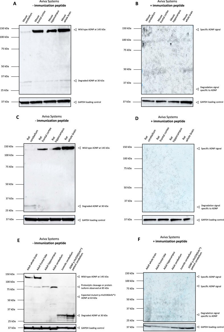
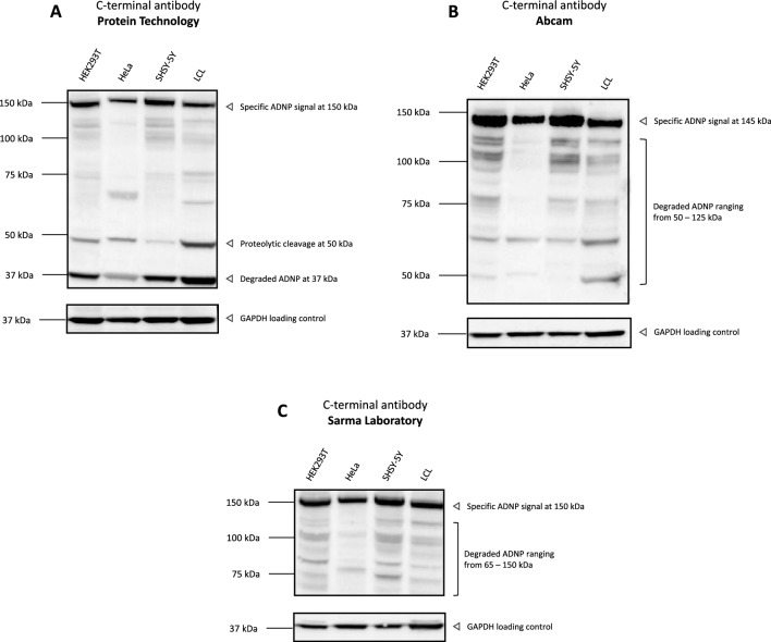
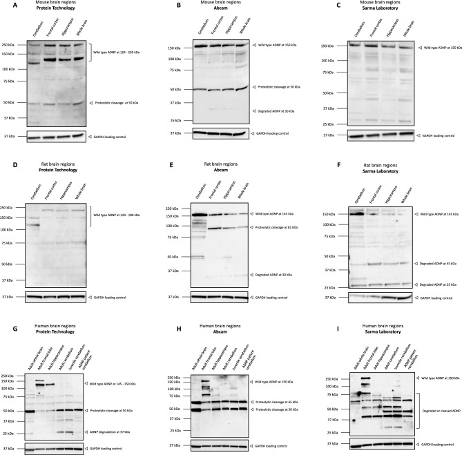
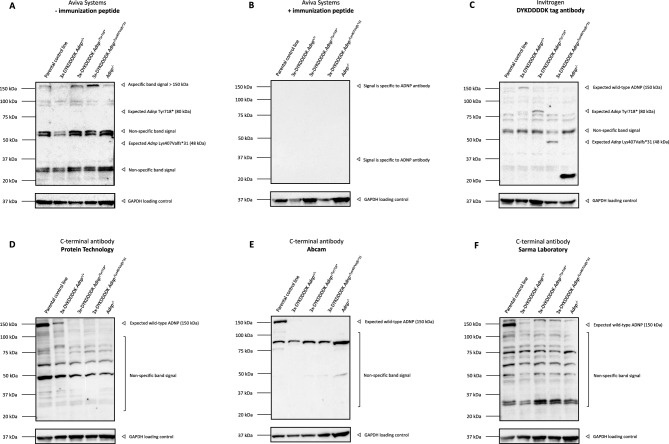
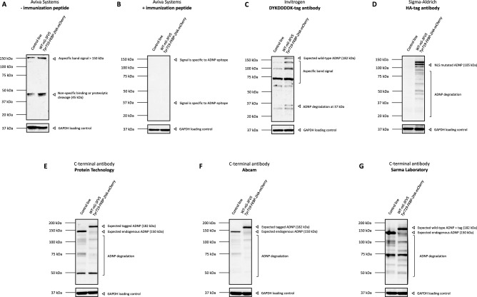
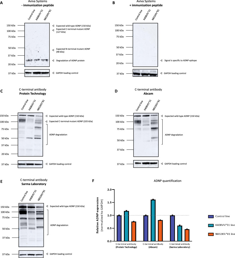
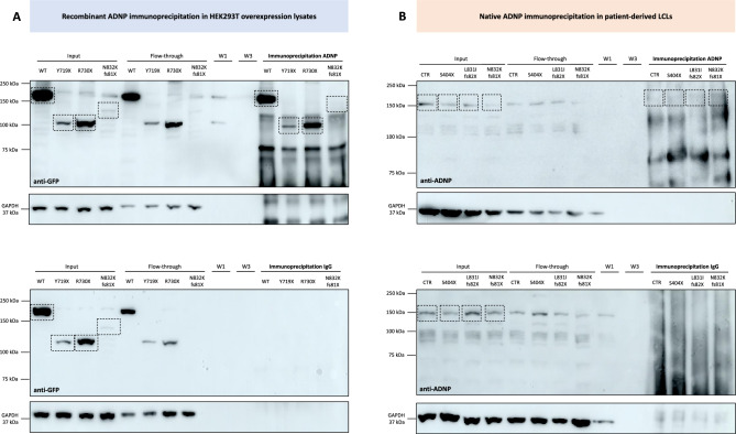
Heterozygous de novo mutations in the Activity-Dependent Neuroprotective Homeobox (ADNP) gene underlie Helsmoortel-Van der Aa syndrome (HVDAS). Most of these mutations are situated in the last exon and we previously demonstrated escape from nonsense-mediated decay by detecting mutant ADNP mRNA in patient blood. In this study, wild-type and ADNP mutants are investigated at the protein level and therefore optimal detection of the protein is required. Detection of ADNP by means of western blotting has been ambiguous with reported antibodies resulting in non-specific bands without unique ADNP signal. Validation of an N-terminal ADNP antibody (Aviva Systems) using a blocking peptide competition assay allowed to differentiate between specific and non-specific signals in different sample materials, resulting in a unique band signal around 150 kDa for ADNP, above its theoretical molecular weight of 124 kDa. Detection with different C-terminal antibodies confirmed the signals at an observed molecular weight of 150 kDa. Our antibody panel was subsequently tested by immunoblotting, comparing parental and homozygous CRISPR/Cas9 endonuclease-mediated Adnp knockout cell lines and showed disappearance of the 150 kDa signal, indicative for intact ADNP. By means of both a GFPSpark and Flag-tag N-terminally fused to a human ADNP expression vector, we detected wild-type ADNP together with mutant forms after introduction of patient mutations in E. coli expression systems by site-directed mutagenesis. Furthermore, we were also able to visualize endogenous ADNP with our C-terminal antibody panel in heterozygous cell lines carrying ADNP patient mutations, while the truncated ADNP mutants could only be detected with epitope-tag-specific antibodies, suggesting that addition of an epitope-tag possibly helps stabilizing the protein. However, western blotting of patient-derived hiPSCs, immortalized lymphoblastoid cell lines and post-mortem patient brain material failed to detect a native mutant ADNP protein. In addition, an N-terminal immunoprecipitation-competent ADNP antibody enriched truncating mutants in overexpression lysates, whereas implementation of the same method failed to enrich a possible native mutant protein in immortalized patient-derived lymphoblastoid cell lines. This study aims to shape awareness for critical assessment of mutant ADNP protein analysis in Helsmoortel-Van der Aa syndrome.

Keywords: Adnp knock-out cell line; Activity-dependent neuroprotective protein (ADNP); Antibody validation; Blocking peptide competition assay; Helsmoortel-Van der Aa syndrome (HVDAS); Immunoprecipitation.


Conflict of interest statement

The authors declare no competing interests.

Figures

Figure 1
Figure 1
Common commercially available ADNP antibodies give rise to non-specific binding. HEK293T, HeLa, SHSY-5Y and a lymphoblastoid control cell line (LCL) were lysed in RIPA buffer and used as protein samples for the assessment of the published ADNP antibodies. Samples were blocked and incubated in 5% blocking-grade non-fat dry milk/TBST with the optimized dilution listed in Table 3. The predicted molecular weight of ADNP is 124 kDa. However, only non-specific signals were detectable. GAPDH was used as a loading control. The datasheet of the tested antibodies indicated that whole or nuclear extracts from HeLa cells should be used as a positive control, which fails to raise a reliable ADNP signal in all tested antibody conditions.
Figure 2
Figure 2
Verification of the specificity of an N-terminal ADNP antibody (Aviva Systems) by performing a blocking peptide competition assay. (A) HEK293T, HeLa, SHSY-5Y and a control lymphoblastoid cell line (LCL) were lysed in RIPA buffer and used as protein samples for the assessment of N-terminal antibody of Aviva systems in a 1:1000 dilution. GAPDH was used as a loading control. The predicted molecular weight of ADNP is 124 kDa. The antibody recognizes ADNP specifically at 150 kDa in HEK293T, HeLa and SHSY-5Y cell lines, but a faint signal ranging from 75 to 150 kDa in the control LCL. (B) Western blot analysis of the blocking peptide competition assay. Supplementation of the immunization peptide in a 5 × excess to antibody concentration reduced the signal detected at 75- 150 kDa in all tested cell lines. Non-specific binding was detected after use of the immunization peptide presenting as a faint signal below the 37 kDa marker.
Figure 3
Figure 3
A polyclonal N-terminal ADNP antibody from Aviva Systems detects ADNP specifically in murine and rat tissues and suggests proteolytic processing of the protein in the human brain. Cerebellum, frontal cortex or lobe, hippocampus and whole brains of control mice, rats and humans were lysed in RIPA buffer and used as protein samples for the assessment of N-terminal antibody of Aviva systems. (AC) The predicted molecular weight of ADNP is 124 kDa. The antibody recognizes ADNP in a range of 145 kDa with (E) additional lower mass signal of 85 kDa in all human brain regions. (BDF) Western blot analysis of the blocking peptide competition assay. Supplementation of the immunization peptide in a 5 × excess to antibody concentration reduced the signal observed at 145 kDa in all tested cell lines. Importantly, the 85 kDa band suggestive for proteolytic cleavage as well as degraded ADNP signal disappeared completely after immunization peptide supplementation. GAPDH was used as a loading control.
Figure 4
Figure 4
Three independent commercially available C-terminal polyclonal ADNP antibodies detect ADNP specifically in different in vitro sample materials and show clear instability of the protein. HEK293T, HeLa, SHSY-5Y and a lymphoblastoid cell line (LCL) were lysed in RIPA buffer and used as protein samples for three different C-terminal ADNP antibodies. GAPDH was used as a loading control. The predicted molecular weight of ADNP is 124 kDa. All the tested antibodies recognized ADNP with a molecular weight of 150 kDa. Samples were blocked and incubated in 5% blocking-grade non-fat dry milk/TBST with the optimized dilution listed in Table 3.
Figure 5
Figure 5
Different C-terminal ADNP antibodies detect ADNP in the range of 150 kDa and suggest proteolytic processing of the protein in the brain. Cerebellum, frontal cortex or lobe, hippocampus and whole brains of control mice, rats, and humans were lysed in RIPA buffer and used as protein samples for the assessment with three C-terminal antibodies with the optimized dilutions listed in Table 3. GAPDH was used as a loading control. The predicted molecular weight of ADNP is 124 kDa. (A)C) Murine samples indicate detection of ADNP in the range of 150 kDa with bands suggesting proteolytic processing at 50 kDa. (DF) Rat samples indicate detection of ADNP in the range of 150 kDa with bands indicating proteolytic processing at 82 kDa after incubation with the C-terminal Abcam antibody. (GI) Human brain samples indicate detection of ADNP at different molecular weights of 124 – 150 kDa in the adult frontal lobe and hippocampus and highlight the antibody differences in detection of ADNP. The three tested antibodies showed strong band signals at lower molecular weights, which could indicate proteolytic cleavage or degradation of the protein.
Figure 6
Figure 6
Unambiguous detection of ADNP using homozygous CRISPR/Cas9 endonuclease-mediated Adnp knockout cell lines. mESCs containing either wild-type, homozygous mutants, or complete Adnp knockout were lysed in RIPA buffer and used as protein samples for the assessment with an N-terminal ADNP, 3x-DYKDDDDK, and C-terminal ADNP antibodies with the optimized dilutions listed in Table 1. GAPDH was used as a loading control. The predicted molecular weight of ADNP is 124 kDa. (A) The N-terminal antibody (Aviva Systems) recognizes ADNP in a range above its observed 150 kDa molecular weight with additional lower mass signal of 37—65 kDa in Adnp homozygous and parental control mESCs. (B) Supplementation of the immunization peptide in a 5 × excess to antibody concentration reduced all signals observed mESC lines, indicating that the N-terminal antibody does not bind ADNP specifically in mESCs. (C) Detection of wild-type and homozygous Adnp mutants by means of a C-terminal 3x-DYKDDDDK (Flag) epitope tag. Wild-type ADNP was detected in at 150 kDa in the C-terminal 3x-DYKDDDDK CRISPR/Cas9 engineered mESC line using a DYKDDDDK antibody. Truncated ADNP mutants, p.Tyr718* and p.Lys407Valfs*31, were detected at a lower molecular weight of 80 kDa, respectively 48 kDa. (DF) Wild-type ADNP detection by means of three different C-terminal antibodies in mESC lines. Wild-type ADNP was detected with a strong signal at 150 kDa in the parental control line with a rather decreased signal in the C-terminal 3x-DYKDDDDK CRISPR/Cas9 engineered mESC line. Disappearance of the 150 kDa band was observed in the mESC line with complete Adnp homozygosity, indicating a reliable molecular weight of 150 kDa for ADNP.
Figure 7
Figure 7
Unambiguous detection of ADNP using an N-terminal GFPSpark and N-DYKDDDDK (Flag) tag expression vector. (A) Western blot analysis of HEK293T cell lysates overexpressing wild-type ADNP-GFPSpark and mutated constructs using an anti-GFP antibody. (B) Western blot analysis of HEK293T cell lysates overexpressing wild-type ADNP-GFPSpark and mutated constructs using the N-terminal ADNP antibody (Aviva Systems). (C) Western blot analysis of HEK293T cell lysates overexpressing wild-type ADNP-DYKDDDDK (Flag) and mutated constructs using an anti-DYKDDDDK antibody. (D) Western blot analysis of HEK293T cell lysates overexpressing wild-type ADNP-DYKDDDDK and mutant constructs using the N-terminal ADNP antibody (Aviva Systems). The observed molecular weight of wild-type ADNP-GFPSpark is 175 kDa (including 25 kDa GFPSpark tag), respectively ADNP-DYKDDDDK 150 kDa, with each of their mutants showing a lower molecular weight as a consequence of the truncating mutations. Detection with antibodies for GFP, DYKDDDDK (Flag), and ADNP gave comparable results. GAPDH was used as a loading control in all experiments.
Figure 8
Figure 8
Western blotting of ADNP in a HCT116 colon cancer cell line, carrying the prevalent heterozygous p.Tyr719* mutation. HCT116 cells containing a wild-type and p.Tyr719* mutant allele were lysed in RIPA buffer and used as protein samples for the assessment with an N-terminal antibody, 3x-DYKDDDDK, HA-tag, and C-terminal ADNP antibodies with the optimized dilutions listed in Table 1. GAPDH was used as a loading control in all experiment. The predicted molecular weight of ADNP is 124 kDa. (A) The N-terminal antibody (Aviva Systems) recognizes ADNP in a range above its observed 150 kDa molecular weight an additional signal of 45 kDa, indicating proteolytic cleavage or non-specific binding. (B) Administration of the immunization peptide in a 5 × excess to antibody concentration reduced all signals, indicating that the N-terminal antibody does not bind ADNP specifically in HCT116 cells. (C) Detection of wild-type ADNP by means of the 3x-DYKDDDDK (Flag) epitope tag. Wild-type ADNP was detected in at 182 kDa in the 3xFlag-V5-loxP-neonGreen/3xHA-loxP-mCherry engineered line using a DYKDDDDK antibody, 32 kDa by tag insertion. (D) Detection of mutant ADNP by means of the HA-epitope tag. A truncated mutant p.Tyr719 ADNP protein was detected in at 105 kDa in the 3xFlag-V5-loxP-neonGreen/3xHA-loxP-mCherry engineered line using a HA-antibody, 25 kDa above its predicted molecular weight by tag insertion. Instability of the truncated protein was observed by a degrading smear. (E–G) Wild-type ADNP detection by means of three different C-terminal antibodies. Non-processed ADNP was detected with a strong signal at 150 kDa in the control line and at a molecular weight of 182 kDa in the genome-edited cell line. In both cases, a degrading smear was observed, indicating instability of the wild-type protein.
Figure 9
Figure 9
Western blotting of ADNP in human induced pluripotent stem cells (hiPSCs), carrying distinct heterozygous ADNP mutations mediated by CRIPSR/Cas9. (A, B) hiPSCs were lysed in RIPA buffer and analyzed by western blotting with the N-terminal antibody (Aviva Systems) with application of our blocking peptide competition assay. Here, no reliable ADNP signal was detected. The molecular weight of the ADNP mutant lines is expected to decrease to 127 kDa for the Asn832Lysfs*81, respectively to 48 kDa for the lys408Valfs*31 line. However, no signal is observed at the predicted weight for the mutations. (CE) The C-terminal antibodies of Protein Technology, Abcam, and the Sarma Laboratory were able to visualize wild-type ADNP at 150 kDa. Possessing the desired epitope for mutant ADNP detection, the C-terminal antibody of Protein technology was not able to capture the predicted truncated protein. GAPDH was used as a loading control. (F) The ADNP signal was quantified determining the ratio of the wild-type protein in mutant to control cell lines. Here, the relative ADNP expression decreased in the Asn832Lysfs*81 cell line compared to the control, whereas mutant-to-wild-type expression ratio showed a higher signal with the antibodies of Protein Technology and Abcam in the lys408Valfs*31 cell line.
Figure 10
Figure 10
Absence of a mutant ADNP protein after immunoblotting of different lymphoblastoid cell lines from Helsmoortel-Van der Aa syndrome patients. (A) LCLs of four control subjects and six patients were lysed in RIPA buffer and analyzed by western blotting with the N-terminal antibody (Aviva Systems). The expected wild-type ADNP signal presented at 150 kDa together with two non-specific bands at 50 kDa and 75 kDa with no difference in expression (p = 0.42; ns) of the wild-type protein. However, the ADNP mutants at a lower molecular weight of 127 kDa for the Asn832Lysfs*81 and Leu831Ilefs*82 mutations, respectively to 45 kDa for the Ser404* mutation, and to 10 kDa for the cell line carrying the Gln40* mutation could not be visualized. (B) Administration of the immunization peptide in a 5 × excess to antibody concentration reduced all signals, indicating that the N-terminal antibody recognized ADNP specifically in LCLs alongside non-specific band signals. (C-E) C-terminal antibodies detected wild-type ADNP at a molecular weight of 150 kDa. No mutant ADNP was observed with the antibody of Protein Technology which is capable to recognize a part of the truncated Asn832Lysfs*81 and Leu831Ilefs*82 mutations. (F) All C-terminal antibodies visualized wild-type ADNP at 150 kDa, with only the Abcam (p = 0.04; *) and Sarma Laboratory (p = 0.02; *) antibodies showing the expected reduction of ADNP in LCLs of Helsmoortel-Van der Aa syndrome patients. GAPDH was used as a loading control.
Figure 11
Figure 11
Wild-type and mutant ADNP enrichment through immunoprecipitation. The N-terminal sc-F5 ADNP IP-competent antibody was crosslinked to agarose beads and sequentially eluted in fractions (input; flow-through; three consecutive washes, W1-W3; and the immunoprecipitated fracted. IgG non-reactive beads were used as a negative control. In each lane, 20 μg of protein was separated by SDS-PAGE electrophoresis. GAPDH has been used as loading control for all western blots, and critical assessment of the accuracy of the IP method. (A) Immunoprecipitation assay of recombinant wild-type (WT) ADNP and truncating mutants (p.Tyr719*; p.Arg730*; p.Asn832Lysfs*81) in HEK293T overexpression lysates. (B) Immunoprecipitation assay of native wild-type (WT) ADNP and truncating mutants in protein extracts of LCLs derived from a control subject (CTR) and patients with the p.Ser404*, p.Leu831Ilefs*82, or p.Asn832Lysfs*81 ADNP mutation.

References

    1. Mandel S, Gozes I. Activity-dependent neuroprotective protein constitutes a novel element in the SWI/SNF chromatin remodeling complex. J. Biol. Chem. 2007;282:34448–34456. doi: 10.1074/jbc.M704756200. - DOI - PubMed
    1. Ostapcuk V, et al. Activity-dependent neuroprotective protein recruits HP1 and CHD4 to control lineage-specifying genes. Nature. 2018;557:739–743. doi: 10.1038/s41586-018-0153-8. - DOI - PubMed
    1. Sun X, Peng X, Cao Y, Zhou Y, Sun Y. ADNP promotes neural differentiation by modulating Wnt/β-catenin signaling. Nat. Commun. 2020;11:2984. doi: 10.1038/s41467-020-16799-0. - DOI - PMC - PubMed
    1. D’Incal CP, et al. Chromatin remodeler Activity-Dependent Neuroprotective Protein (ADNP) contributes to syndromic autism. Clin. Epigenet. 2023;15:45. doi: 10.1186/s13148-023-01450-8. - DOI - PMC - PubMed
    1. Pinhasov A, et al. Activity-dependent neuroprotective protein: A novel gene essential for brain formation. Brain Res. Dev. Brain Res. 2003;144:83–90. doi: 10.1016/S0165-3806(03)00162-7. - DOI - PubMed