. 2024 May 21;14(6):606.
doi: 10.3390/biom14060606.

Activation of Peroxisome Proliferator-Activated Receptor-β/δ (PPARβ/δ) in Keratinocytes by Endogenous Fatty Acids

Affiliations

Activation of Peroxisome Proliferator-Activated Receptor-β/δ (PPARβ/δ) in Keratinocytes by Endogenous Fatty Acids

Bokai Zhu et al. Biomolecules. .

Abstract

Nuclear hormone receptors exist in dynamic equilibrium between transcriptionally active and inactive complexes dependent on interactions with ligands, proteins, and chromatin. The present studies examined the hypothesis that endogenous ligands activate peroxisome proliferator-activated receptor-β/δ (PPARβ/δ) in keratinocytes. The phorbol ester treatment or HRAS infection of primary keratinocytes increased fatty acids that were associated with enhanced PPARβ/δ activity. Fatty acids caused PPARβ/δ-dependent increases in chromatin occupancy and the expression of angiopoietin-like protein 4 (Angptl4) mRNA. Analyses demonstrated that stearoyl Co-A desaturase 1 (Scd1) mediates an increase in intracellular monounsaturated fatty acids in keratinocytes that act as PPARβ/δ ligands. The activation of PPARβ/δ with palmitoleic or oleic acid causes arrest at the G2/M phase of the cell cycle of HRAS-expressing keratinocytes that is not found in similarly treated HRAS-expressing Pparb/d-null keratinocytes. HRAS-expressing Scd1-null mouse keratinocytes exhibit enhanced cell proliferation, an effect that is mitigated by treatment with palmitoleic or oleic acid. Consistent with these findings, the ligand activation of PPARβ/δ with GW0742 or oleic acid prevented UVB-induced non-melanoma skin carcinogenesis, an effect that required PPARβ/δ. The results from these studies demonstrate that PPARβ/δ has endogenous roles in keratinocytes and can be activated by lipids found in diet and cellular components.

Keywords: UVB-induced non-melanoma skin cancer; cell cycle; fatty acids; ligands; peroxisome proliferator-activated receptor.


Conflict of interest statement

The authors declare no conflicts of interest.

Figures

Figure 1
Figure 1
Phorbol ester (TPA) increases PPARβ/δ transcriptional activity in keratinocytes. (A) A reporter assay was used to detect PPARβ/δ activity in cytosolic isolates obtained from mouse primary keratinocytes after fractionation using HPLC. (B) Fatty acids known to be released by TPA treatment in wild-type keratinocytes increase the PPARβ/δ-dependent expression of Angptl4 mRNA, and this effect is absent in similarly treated Pparb/d-null keratinocytes. n = 5 independent biological replicates. (C) A ChIP assay showing increased promoter occupancy on known Angptl4 PPRE by palmitoleic acid in wild-type keratinocytes (+/+), an effect lacking in similarly treated Pparb/d-null keratinocytes (–/–), n = 5 independent biological replicates. Values represent the mean ± SD. Values with different letters are significantly different, p ≤ 0.05. * Significantly different than control, p ≤ 0.05.
Figure 2
Figure 2
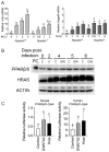
Activated HRAS increases PPARβ/δ transcriptional activity. (A) qPCR of Hras (left panel) or Angptl4 (right panel) in mock-infected or HRAS-infected Pparb/d wild-type and Pparb/d-null keratinocytes with increasing M.O.I. n = 6 independent biological replicates. (B) Representative Western blot analysis of PPARβ/δ expression in HRAS-infected wild-type keratinocytes without GW0742 (C) or treated with GW0742 (GW). PC: positive control (COS cell lysate). (C) Luciferase assay in primary keratinocytes transiently transfected with either mouse (left panel) or human (right panel) PPARβ/δ-GAL4 fusion protein and UAS-luciferase plasmids. n = 6 independent biological replicates. Values represent mean ± SD. Values with different letters are significantly different, p ≤ 0.05.
Figure 3
Figure 3
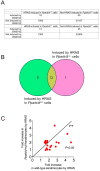
HRAS activation and GW0742 treatment induces common gene expression. (A) Overlap between the mRNAs that are induced by HRAS, and mRNAs are induced by the PPARβ/δ ligand GW0742 in primary keratinocytes. Only genes that are induced by ≥1.5 folds by either HRAS or GW0742 are included. n = 3 independent biological replicates. (B) A Venn diagram showing that 6 of 19 genes induced by GW0742 in wild-type keratinocytes are also induced by HRAS only in wild-type keratinocytes. In contrast, there was overlap in 12 genes between genotypes and treatments. (C) A plot of the fold change in genes in B induced by HRAS either in Pparb/d wild-type (x-axis) or Pparb/d-null keratinocytes (y-axis). Note that these genes are induced to a much higher fold by HRAS in wild-type keratinocytes compared to Pparb/d-null keratinocytes.
Figure 4
Figure 4
Activated HRAS increases expression of fatty acid synthesis genes. (A) qPCR of key fatty acid synthesis mRNAs in Pparb/d wild-type and Pparb/d-null keratinocytes treated with GW0742 or HRAS infection. n = 3–5 independent biological replicates. (B) Representative quantitative Western blot analysis of key fatty acid synthesis genes in Pparb/d wild-type and Pparb/d-null keratinocytes treated with GW0742 or HRAS infection. n = 4 independent biological replicates. (C) Diagram showing de novo fatty acid synthesis pathway influenced in these studies. Values represent mean ± SD. Values with different letters are significantly different, p ≤ 0.05. * Significantly different than control, p ≤ 0.05.
Figure 5
Figure 5
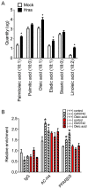
HRAS-expressing keratinocytes have higher palmitoleic and oleic acid levels, which are PPARβ/δ ligands. (A) Quantification of levels of key fatty acid in mock-infected or HRAS-infected Pparb/d wild-type keratinocytes by GC-MS. Data are compiled from three independent experiments. n = 5–6 independent biological replicates. (B) ChIP-qPCR analysis of Ac-H4 and PPARβ/δ binding to mouse Angptl4 gene promoter in response to GW0742 or oleic acid treatment in Pparb/d wild-type or Pparb/d-null keratinocytes. n = 3 independent biological replicates. Values represent mean ± SD. * Significantly different than control, p ≤ 0.05.
Figure 6
Figure 6
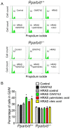
Palmitoleic and oleic acids induce G2/M arrest in HRAS-expressing keratinocytes through PPARβ/δ-dependent mechanism. Representative DNA histograms (A) and quantification of percentage of cells in G2/M phase, n = 3 independent biological replicates, (B) of mock-infected or HRAS-expressing Pparb/d wild-type or Pparb/d-null keratinocytes treated with GW0742, palmitoleic acid or oleic acid or 72 h. Representative peak of cells in G2/M phase shown above bar in each panel in (A). n = 3 independent biological replicates. Values in (B) represent mean ± SD. * Significantly different than control, p ≤ 0.05.
Figure 7
Figure 7
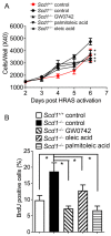
Palmitoleic and oleic acids rescue enhanced cell proliferation of HRAS-expressing keratinocytes resulting from Scd1 ablation. (A) Cell counts of HRAS-expressing keratinocytes isolated from wild-type (Scd1+/+), Scd1 knock-out (Scd1−/−), and Scd1−/− treated with GW0742, palmitoleic acid, or oleic acid for 6 days post-HRAS expression. n = 3–6 independent biological replicates. (B) BrdU-positive cells were identified by immunofluorescence after 6 days of HRAS expression. n = 4–6 independent biological replicates. Values represent mean ± SD. * Significantly different than control, p ≤ 0.05.
Figure 8
Figure 8
Palmitoleic and oleic acids rescue enhanced cell proliferation and hyperplasia resulting from Scd1 ablation. (A) H&E staining of mouse skin from wild-type and Scd1–/– mice treated topically once with TPA and with or without GW0742, palmitoleic acid, or oleic acid for 48 h. n = 3–10 independent biological replicates. The 100 µm scale in (A) is the same for images in (B). (B) Representative immunofluorescence of PCNA (red), keratin 5 (green), and DAPI (blue), and (C) quantification of percentage of PCNA-positive cells in skin of wild-type and Scd1−/− mice treated with TPA and with or without GW0742, palmitoleic acid, or oleic acid for 48 h. Higher percentage of PCNA-positive cells shown with white arrows. n = 3–10 independent biological replicates. Values represent mean ± SD. * Significantly different than control, p ≤ 0.05.
Figure 9
Figure 9
Ligand activation of PPARβ/δ with oleic acid or GW0742 inhibits UVB-induced skin cancer. SKH mice were crossed with Pparb/d wild-type or Pparb/d-null mice and irradiated with or without topical application of PPARβ/δ ligands after irradiation. Onset of tumor formation (A), number of tumors per mouse (B), and average tumor size (C) were measured weekly. n = 7–11 independent biological replicates. Each point represents mean ± S.D. * p < 0.05 compared with control.

References

    1. Green S., Wahli W. Peroxisome proliferator-activated receptors: Finding the orphan a home. Mol. Cell Endocrinol. 1994;100:149–153. doi: 10.1016/0303-7207(94)90294-1. - DOI - PubMed
    1. Berger J., Moller D.E. The mechanisms of action of PPARs. Annu. Rev. Med. 2002;53:409–435. doi: 10.1146/annurev.med.53.082901.104018. - DOI - PubMed
    1. Biddie S.C., Hager G.L. Glucocorticoid receptor dynamics and gene regulation. Stress. 2009;12:193–205. doi: 10.1080/10253890802506409. - DOI - PMC - PubMed
    1. Biddie S.C., John S., Hager G.L. Genome-wide mechanisms of nuclear receptor action. Trends Endocrinol. Metab. 2010;21:3–9. doi: 10.1016/j.tem.2009.08.006. - DOI - PMC - PubMed
    1. Hager G.L., Varticovski L. Chromatin in time and space. Biochim. Biophys. Acta. 2012;1819:631. doi: 10.1016/j.bbagrm.2012.05.002. - DOI - PMC - PubMed

MeSH terms