Review
. 2024 Jun 4;14(6):656.
doi: 10.3390/biom14060656.

Molecular Mechanisms in Tumorigenesis of Hepatocellular Carcinoma and in Target Treatments-An Overview

Affiliations
Review

Molecular Mechanisms in Tumorigenesis of Hepatocellular Carcinoma and in Target Treatments-An Overview

Raluca-Margit Szilveszter et al. Biomolecules. .

Abstract

Hepatocellular carcinoma is the most common primary malignancy of the liver, with hepatocellular differentiation. It is ranked sixth among the most common cancers worldwide and is the third leading cause of cancer-related deaths. The most important etiological factors discussed here are viral infection (HBV, HCV), exposure to aflatoxin B1, metabolic syndrome, and obesity (as an independent factor). Directly or indirectly, they induce chromosomal aberrations, mutations, and epigenetic changes in specific genes involved in intracellular signaling pathways, responsible for synthesis of growth factors, cell proliferation, differentiation, survival, the metastasis process (including the epithelial-mesenchymal transition and the expression of adhesion molecules), and angiogenesis. All these disrupted molecular mechanisms contribute to hepatocarcinogenesis. Furthermore, equally important is the interaction between tumor cells and the components of the tumor microenvironment: inflammatory cells and macrophages-predominantly with a pro-tumoral role-hepatic stellate cells, tumor-associated fibroblasts, cancer stem cells, extracellular vesicles, and the extracellular matrix. In this paper, we reviewed the molecular biology of hepatocellular carcinoma and the intricate mechanisms involved in hepatocarcinogenesis, and we highlighted how certain signaling pathways can be pharmacologically influenced at various levels with specific molecules. Additionally, we mentioned several examples of recent clinical trials and briefly described the current treatment protocol according to the NCCN guidelines.

Keywords: TERT promoter mutation; WNT/β-catenin; exosomes; hepatocellular carcinoma; histone methylation; immune checkpoint inhibitors; p53; telomere shortening; tumoral microenvironment.


Conflict of interest statement

The authors declare no conflicts of interest.

Figures

Figure 1
Figure 1
Signaling pathways activated by growth factors: the interaction of the growth factors with their specific receptors triggers the molecular cascade of PI3K/AKT/mTOR and RAS/RAF/MERK/ERK, which results in the transcription (arrow) of genes responsible for maintaining cell survival and stimulating cell proliferation, migration, and angiogenesis. TGF-β—transforming growth factor-β; EGF—epidermal growth factor; IGF I/II—insulin-like growth factor I/II; VEGF—vascular endothelial growth factor; FGF—fibroblast growth factor; HGF—hepatocyte growth factor; EGFR—epidermal growth factor receptor; IGFR I/II—insulin-like growth factor receptor I/II; VEGFR—vascular endothelial growth factor receptor; FGFR—fibroblast growth factor receptor; c-Met—mesenchymal–epithelial transition factor; SMAD—Sma gene and the Drosophila MAD (Mothers against decapentaplegic); PI3K—phosphoinositide 3-kinases; PIP2—phosphoinositide phosphatidylinositol 4,5-bisphosphate; PIP3—phosphatidylinositol-3,4,5-trisphosphate; AKT—serine/threonine-specific protein kinase (AKR mouse strain that develops spontaneous thymic lymphomas); mTOR—mammalian target of rapamycin; RAS—rat sarcoma virus; RAF—rapidly accelerated fibrosarcoma; MEK—mitogen-activated protein kinase; ERK—extracellular signal-regulated kinase; AP-1—activator protein-1; c-Jun—Jun proto-oncogene; c-FOS—a proto-oncogene that is the human homolog of the retroviral oncogene v-fos; Elk-1—ETS transcription factor ELK1.
Figure 2
Figure 2
Canonical activation of the WNT/β-catenin pathway. (a) In the absence of WNT/β-catenin cascade activation, there is an intracytoplasmic pool of β-catenin that is phosphorylated by a protein complex, known as the “destruction complex” (formed from the GSK-3β, Axin, APC, and CKI1α proteins) that will ubiquitinate the β-catenin, undergoing proteasomal degradation. As a result, β-catenin does not interact with the transcription factors. (b) When the Frizzled receptor binds any member of the WNT ligand family (WNT 1, 2, 3, 8a, 8b, 10a, 10b), it activates the pathway and β-catenin is translocated to the nucleus, where it interacts with the transcription factors (TCF/LEF, Pygo, Bcl-9, Gro/TLE) that in turn trans-activate (arrow) genes known to be associated with cell development, proliferation, and WNT regulation. LRP—low-density lipoprotein receptor-related protein; WNT—Wingless/Integrated; CKIα—casein kinase Iα; GSK-3β—glycogen synthase kinase-3 beta; APC—adenomatous polyposis coli; TCF/LEF—T-cell factor/lymphoid enhancer factor; Pygo—Pygopus protein; Bcl-9—B-cell lymphoma 9 protein; Gro/TLE—Drosophila Groucho/transducin-like enhancers of split; GLUL—glutamate-ammonia ligase; TBX3—T-box transcription factor 3; OAT—ornithine aminotransferase; HIF-1α—hypoxia inducible factor 1 subunit alpha; FOXO—forkhead box protein O1; SOX—SRY-related HMG-box genes; AXIN 2 gene; SP5—Sp5 transcription factor.
Figure 3
Figure 3
JAK/STAT signaling pathway: the activation of the JAK/STAT pathway is initiated by the binding of ligands (cytokines, growth factors) to specific receptors, associated with JAK proteins, which become activated following conformational changes triggered by the ligand. Once activated, JAK phosphorylates a tyrosine residue on the receptor, creating attachment sites for STAT that interacts with the receptor and undergoes phosphorylation by JAK, leading to the activation and dimerization of STAT proteins. The dimer is translocated to the nucleus, where it functions as a transcription factor for various genes that promote cellular proliferation, angiogenesis, tumor invasion, and metastasis. SOCs and SHP1/2 can block the cascade by competing with STAT proteins for their binding sites on the receptor or dephosphorylating JAK protein, respectively. JAK—Janus kinase; STAT—signal transducer and activator of transcription; SOCS—suppressors of cytokine signaling; SHP1/2—small heterodimer partner; BIRC5—baculoviral inhibitor of apoptosis repeat-containing 5; Mcl-1—myeloid cell leukemia-1; BCL-2—B-cell lymphoma 2; BCL2L1—B-cell lymphoma 2 like 1; VEGF—vascular endothelial growth factor; HIF-1—hypoxia inducible factor 1; MMP2—matrix metallopeptidase 2; MMP9—matrix metallopeptidase 9; SNAi2—snail family transcriptional repressor 2; TWIST—wist family bHLH transcription factor 1; bFGF—basic fibroblast growth factor.
Figure 4
Figure 4
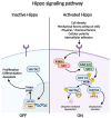
Hippo signaling pathway. When the Hippo pathway is off, YAP and TAZ proteins enter the nucleus and bind to the TEAD transcription factor, initiating the transcription of genes that promote cell survival and proliferation. Changes in the extracellular components can activate the Hippo cascade by triggering the activation of NF2 and KIBRA. These activate the kinases MST1/2, leading to the phosphorylation of the protein SAV1. The resulting complex phosphorylates the LATS1/2 and MOB by SAV1. LATS1/2 and MOB then phosphorylates YAP and TAZ. YAP/TAZ interact with the cytoplasmic protein 14-3-3, resulting in their proteasomal degradation. As a result, the transcription factor TEAD inhibits the expression of target genes. KIBRA—kidney and brain expressed protein, NF2—neurofibromatosis 2; SAV1—Salvador homolog 1; LATS1/2—large tumor suppressor homolog 1 and 2 proteins; MST1/2—monopolar spindle-one-binder proteins; YAP—Yes-associated protein; TAZ—transcriptional co-activator with PDZ-binding motif; TEAD—transcriptional enhancer activator domain; Ubiq—ubiquitin.
Figure 5
Figure 5
Hedgehog signaling pathway. In the absence of the binding of DHH, IHH, or SHH proteins to PTCH, phosphatidylinositol-4-phosphate PI(4)P binds to PTCH, thereby inhibiting the G protein-coupled SMO. SMO then interacts with SENP, resulting in the ubiquitination and degradation of the complex. When a ligand (DHH, IHH, SHH) binds to the CDO/BOC protein and the PTCH receptor, it triggers the activation of the G protein-coupled SMO receptor. This activation leads to the cleavage of Glis, releasing their active form from a protein complex in the cytoplasm. The activated protein is translocated in the nucleus, initiating the transcription of genes implicated in proliferation, migration, invasion, metastasis, and angiogenesis. DHH—Desert Hedgehog protein; IHH—Indian Hedgehog protein; SHH—Sonic Hedgehog protein; CDO/BOC—cysteine dioxygenase/brother of cysteine dioxygenase; PTCH—multitransmembrane protein patched; SMO—receptor smoothened; SENP—SUMO-specific peptidase protein; Glis—glioma-associated oncogene transcription factors.
Figure 6
Figure 6
Notch signaling pathway. Upon the binding of canonical ligands to the receptor, proteolytic cleavage is triggered by the γ—secretase complex (comprising presenilin, nicastrin, APH1, and PEN2 proteins) within the receptor’s intracellular domain (NICD). This process exposes nuclear localization sequences, enabling the NICD to translocate to the nucleus. Upon coupling with proteins from the CSL (CBF1/RBPJ—kappa/Su (H)/Lag1) family, NICD acts as a transcriptional coactivator for genes belonging to the hairy and enhancer of split 1 (HES1), HES1—related (HESR1), SOX9, P21, C—Myc, and cyclin D1 families, which are involved in cellular differentiation, proliferation, and apoptosis processes. DLK1—delta-like non-canonical Notch ligand 1; DLK2—delta-like non—canonical Notch ligand 2/EGFL9—EGF-like domain-containing protein 9; EGFL7—EGF—like domain-containing protein 7; DNER—delta/Notch-like epidermal growth factor (EGF)—related receptor; NICD—Notch intracellular domain; PEN2—presenilin 2; APH—anterior pharynx—defective; Lag1—longevity assurance gene 1; CBF1/RBPJ—kappa—C—repeat/DRE binding factor 1/recombination signal binding protein for immunoglobulin kappa J region.
Figure 7
Figure 7
Immune checkpoints inhibitors molecular mechanism. (a) When PD-1 binds with PD-L1, cells are identified as self, leading to immune tolerance. Consequently, tumor cells expressing PD-L1 evade recognition by T lymphocytes. In the same way, CTLA-4 suppresses T-cell activity by binding to CD80. (b) Antibodies against PD-1, PD-L1, and CTLA-4, known as immune checkpoint inhibitors, will attach to the ligand/receptor and stimulate the immune response against tumor cells. MHC I—major histocompatibility complex; PD-1—programmed cell death protein 1; PD-L1—programmed cell ligand 1; CTLA-4—cytotoxic T lymphocyte antigen 4; CD80—cluster of differentiation 80.

Similar articles

Cited by

References

    1. Torbenson M.S., Ng I.O.L., Park Y.N., Roncalli M., Sakarnato M. Hepatocelullar carcinoma. In: Paradis V., Fukayama M., Park Y.N., Schirmacher P., editors. WHO Classification of Tumors Editorial Board. Digestive System Tumours. 5th ed. International Agency for Research on Cancer; Lyon, France: 2019. pp. 229–232.
    1. Sung H., Ferlay J., Siegel R.L., Laversanne M., Soerjomataram I., Jemal A., Bray F. Global cancer statistics 2020: GLOBOCAN estimates of incidence and mortality worldwide for 36 cancers in 185 countries. CA Cancer J. Clin. 2021;71:209–249. doi: 10.3322/caac.21660. - DOI - PubMed
    1. Bray F., Laversanne M., Sung H., Ferlay J., Siegel R.L., Soerjomataram I., Jemal A. Global cancer statistics 2022: GLOBOCAN estimates of incidence and mortality worldwide for 36 cancers in 185 countries. CA Cancer J. Clin. 2024;74:229–263. doi: 10.3322/caac.21834. - DOI - PubMed
    1. Rizzo G.E.M., Cabibbo G., Craxì A. Hepatitis B virus-associated hepatocellular carcinoma. Viruses. 2022;14:986. doi: 10.3390/v14050986. - DOI - PMC - PubMed
    1. Wang Q., Luan W., Villanueva G.A., Rahbari N.N., Yee H.T., Manizate F., Hiotis S.P. Clinical prognostic variables in young patients (under 40 years) with hepatitis B virus-associated hepatocellular carcinoma. J. Dig. Dis. 2012;13:214–218. doi: 10.1111/j.1751-2980.2012.00577.x. - DOI - PubMed

MeSH terms