

Effect of Metaldehyde on Survival, Enzyme Activities, and Histopathology of the Apple Snail Pomacea canaliculata (Lamarck 1822)
- PMID: 38927309
- PMCID: PMC11200788
- DOI: 10.3390/biology13060428
Effect of Metaldehyde on Survival, Enzyme Activities, and Histopathology of the Apple Snail Pomacea canaliculata (Lamarck 1822)
Abstract
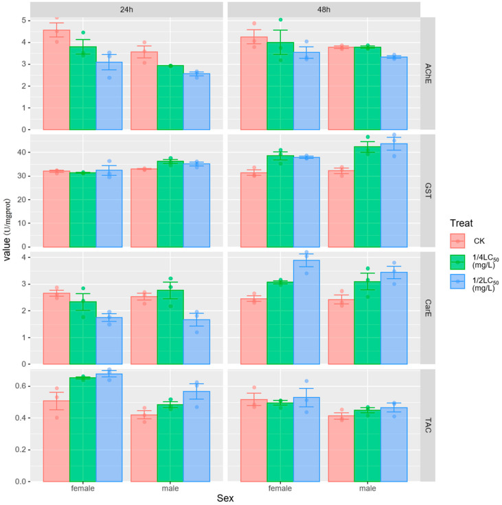
Pomacea canaliculata, as an invasive exotic species in Asia, can adversely affect crop yields, eco-environment, and human health. Application of molluscicides containing metaldehyde is one effective method for controlling P. canaliculata. In order to investigate the effects of metaldehyde on adult snails, we conducted acute toxicological experiments to investigate the changes in enzyme activities and histopathology after 24 h and 48 h of metaldehyde action. The results showed that the median lethal concentrations (LC) of metaldehyde on P. canaliculata were 3.792, 2.195, 1.833, and 1.706 mg/L at exposure times of 24, 48, 72, and 96 h, respectively. Treatment and time significantly affected acetylcholinesterase (AChE), glutathione S-transferase (GST), and total antioxidant capacity (TAC) activity, with sex significantly affecting AChE, GST, and TAC activity and time significantly affecting carboxylesterase (CarE). In addition, the interaction of treatment and time significantly affected the activity of GST, CarE and TAC. In addition, histopathological changes occurred in the digestive glands, gills and gastropods of apple snail exposed to metaldehyde. Histological examination of the digestive glands included atrophy of the digestive cells, widening of the hemolymph gap, and an increase in basophils. In treated snails, the hemolymph gap in the gills was widely dilated, the columnar cells were disorganized or even necrotic, and the columnar muscle cells in the ventral foot were loosely arranged and the muscle fibers reduced. The findings of this study can provide some references for controlling the toxicity mechanism of invasive species.
Keywords: acute toxicity; digestive gland; histopathology; molluscicide.
Conflict of interest statement
The authors declare no conflict of interest.
Figures


References
-
- Hayes K.A., Joshi R.C., Thiengo S.C., Cowie R.H. Out of South America: Multiple Origins of Non-Native Apple Snails in Asia: Invasive Ampullariids in Asia. Divers. Distrib. 2008;14:701–712. doi: 10.1111/j.1472-4642.2008.00483.x. - DOI
-
- Wada T. Strategies for Controlling the Apple Snail Pomacea canaliculata (Lamarck) (Gastropoda: Ampullariidae) in Japanese Direct-Sown Paddy Fields. Jpn. Agric. Res. Q. JARQ. 2004;38:75–80. doi: 10.6090/jarq.38.75. - DOI
-
- Boland B.B., Meerhoff M., Fosalba C., Mazzeo N., Barnes M.A., Burks R.L. Juvenile Snails, Adult Appetites: Contrasting Resource Consumption between Two Species of Applesnails (Pomacea) J. Molluscan Stud. 2007;74:47–54. doi: 10.1093/mollus/eym045. - DOI
-
- Wang W., Huang S., Liu F., Sun Y., Wang X., Yao J., Li S., Liu Y., Luo B., Zhang X., et al. Control of the Invasive Agricultural Pest Pomacea canaliculata with a Novel Molluscicide: Efficacy and Safety to Nontarget Species. J. Agric. Food Chem. 2022;70:1079–1089. doi: 10.1021/acs.jafc.1c07847. - DOI - PubMed
-
- Carlsson N.O.L., Bronmark C. Size-Dependent Effects of an Invasive Herbivorous Snail (Pomacea canaliculata) on Macrophytes and Periphyton in Asian Wetlands. Freshw. Biol. 2006;51:695–704. doi: 10.1111/j.1365-2427.2006.01523.x. - DOI
Grants and funding
LinkOut - more resources
Full Text Sources
Research Materials
Miscellaneous
