Review
. 2024 Jun 1;15(6):727.
doi: 10.3390/genes15060727.

Molecular Mechanisms Governing Sight Loss in Inherited Cone Disorders

Affiliations
Review

Molecular Mechanisms Governing Sight Loss in Inherited Cone Disorders

Chloe Brotherton et al. Genes (Basel). .

Abstract

Inherited cone disorders (ICDs) are a heterogeneous sub-group of inherited retinal disorders (IRDs), the leading cause of sight loss in children and working-age adults. ICDs result from the dysfunction of the cone photoreceptors in the macula and manifest as the loss of colour vision and reduced visual acuity. Currently, 37 genes are associated with varying forms of ICD; however, almost half of all patients receive no molecular diagnosis. This review will discuss the known ICD genes, their molecular function, and the diseases they cause, with a focus on the most common forms of ICDs, including achromatopsia, progressive cone dystrophies (CODs), and cone-rod dystrophies (CORDs). It will discuss the gene-specific therapies that have emerged in recent years in order to treat patients with some of the more common ICDs.

Keywords: CODs; CORDs; Xq28-associated disorders; achromatopsia; photoreceptors.


Conflict of interest statement

The authors declare no conflicts of interest. The funders had no role in the writing of the manuscript.

Figures

Figure 1
Figure 1
The structure of the retina. The neurosensory retina is a tri-laminar structure which sits on, and interdigitates with, the retinal pigment epithelium at the back of the eye. The optimal arrangement of the structure allows light to travel through the retina to the photoreceptors, which undergo hyperpolarization in order to initiate the visual cascade. Subsequent signalling through bipolar cells and ganglion cells (with regulation by the amacrine and horizontal interneurons) manifests with a visual impulse travelling down the optic nerve towards the brain’s occipital cortex. The information was uncovered with BioRender.com CC-BY-NC-ND.
Figure 2
Figure 2
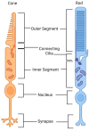
Cone and rod photoreceptors. The structures of rods and cones are similar yet distinct. Rods are much longer than cones, having more discs in their outer segment. Cones, however, are much wider. The outer segment is an extension of the photoreceptor’s specialised sensory cilium, where the ciliary membrane expands and evaginates to form disc-like formations. In rod cells, these discs fuse with and become enclosed by the overlying plasma membrane, whilst cone discs remain exposed to extracellular space. Opsins and rhodopsin (within cones and rods, respectively) are compartmentalised on the membrane of these discs, which are completely renewed every ten days via disc genesis, an actin-mediated process occurring at the connecting cilia. Discs are subsequently shed into the extracellular space at the tip of the photoreceptor for phagocytosis by the underlying RPE. The photoreceptor’s protein- and membrane-making machinery and mitochondria are located within the inner segment. This information was uncovered with BioRender.com CC-BY-NC-ND.
Figure 3
Figure 3
The prevalence of inherited cone disorders in the European population, (ACHM: achromatopsia, COD: cone dystrophy, CORD: cone–rod dystrophy, BCM: blue-cone monochromacy, BED: Bornholm eye disease, OT: oligocone trichromacy).
Figure 4
Figure 4
The Phototransduction cascade. The outer segment of a cone photoreceptor has disc-like evaginations in which opsins are compartmentalised. In the absence of light (1), transducin (formed of α, β, and γ subunits) remains bound to the opsin. cGMP, present in the outer segment, binds to and keeps open cGMP-gated membrane ion channels, allowing a steady influx of calcium and sodium ions into the cells called the dark current. This positive charge flowing into the cell leads to continued glutamate release from the synapse to the bipolar cells, maintaining the resting potential. When the light of a specific wavelength falls on and is detected by an opsin (2), 11-cis-retinal, which is bound to its opsin, undergoes a conformational change to being all-trans-retinal. This creates a conformational change in opsin, converting GDP into GTP, which binds the α subunit of transducin. This allows αtransducin to break away and bind the inhibitory γ subunits of phosphodiesterase (PDE), which in turn allows PDEα to hydrolyse cGMP to GMP. Reducing cGMP levels closes the ion channels (3), hyperpolarising the cell, reducing glutamate release, and allowing signalling to bipolar cells. This was determined using BioRender.com CC-BY-NC-ND.
Figure 5
Figure 5
Location of pathogenic mutations on the protein domains of achromatopsia-causing genes [31].
Figure 6
Figure 6
Gene prevalence in cone and cone–rod dystrophies in European populations.

References

    1. Ben-Yosef T. Inherited Retinal Diseases. Int. J. Mol. Sci. 2022;23:13467. doi: 10.3390/ijms232113467. - DOI - PMC - PubMed
    1. RetNet—Retinal Information Network. [(accessed on 14 November 2023)]. Available online: https://web.sph.uth.edu/RetNet/
    1. Mustafi D., Engel A.H., Palczewski K. Structure of Cone Photoreceptors. Prog. Retin. Eye Res. 2009;28:289–302. doi: 10.1016/j.preteyeres.2009.05.003. - DOI - PMC - PubMed
    1. Megaw R., Moye A., Zhang Z., Newton F., McPhie F., Murphy L.C., McKie L., He F., Jungnickel M.K., von Kriegsheim A., et al. Ciliary Tip Actin Dynamics Regulate Photoreceptor Outer Segment Integrity. Nat. Commun. 2024;15:4316. doi: 10.1038/s41467-024-48639-w. - DOI - PMC - PubMed
    1. Zhang Z., Moye A.R., He F., Chen M., Agosto M.A., Wensel T.G. Centriole and Transition Zone Structures in Photoreceptor Cilia Revealed by Cryo-Electron Tomography. Life Sci. Alliance. 2024;7:e202302409. doi: 10.26508/lsa.202302409. - DOI - PMC - PubMed

MeSH terms