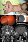
Blau Syndrome: Challenging Molecular Genetic Diagnostics of Autoinflammatory Disease
- PMID: 38927735
- PMCID: PMC11203189
- DOI: 10.3390/genes15060799
Blau Syndrome: Challenging Molecular Genetic Diagnostics of Autoinflammatory Disease
Abstract
The aim of this study was to describe the clinical and molecular genetic findings in seven individuals from three unrelated families with Blau syndrome. A complex ophthalmic and general health examination including diagnostic imaging was performed. The NOD2 mutational hot spot located in exon 4 was Sanger sequenced in all three probands. Two individuals also underwent autoinflammatory disorder gene panel screening, and in one subject, exome sequencing was performed. Blau syndrome presenting as uveitis, skin rush or arthritis was diagnosed in four cases from three families. In two individuals from one family, only camptodactyly was noted, while another member had camptodactyly in combination with non-active uveitis and angioid streaks. One proband developed two attacks of meningoencephalitis attributed to presumed neurosarcoidosis, which is a rare finding in Blau syndrome. The probands from families 1 and 2 carried pathogenic variants in NOD2 (NM_022162.3): c.1001G>A p.(Arg334Gln) and c.1000C>T p.(Arg334Trp), respectively. In family 3, two variants of unknown significance in a heterozygous state were found: c.1412G>T p.(Arg471Leu) in NOD2 and c.928C>T p.(Arg310*) in NLRC4 (NM_001199139.1). In conclusion, Blau syndrome is a phenotypically highly variable, and there is a need to raise awareness about all clinical manifestations, including neurosarcoidosis. Variants of unknown significance pose a significant challenge regarding their contribution to etiopathogenesis of autoinflammatory diseases.
Keywords: Blau syndrome; NOD2; autoinflammation; early onset sarcoidosis; neurosarcoidosis; uveitis.
Conflict of interest statement
The authors declare no conflicts of interest.
Figures


References
-
- Karacan I., Balamir A., Ugurlu S., Aydin A.K., Everest E., Zor S., Onen M.O., Dasdemir S., Ozkaya O., Sozeri B., et al. Diagnostic utility of a targeted next-generation sequencing gene panel in the clinical suspicion of systemic autoinflammatory diseases: A multi-center study. Rheumatol. Int. 2019;39:911–919. doi: 10.1007/s00296-019-04252-5. - DOI - PubMed
-
- Martin A.R., Williams E., Foulger R.E., Leigh S., Daugherty L.C., Niblock O., Leong I.U.S., Smith K.R., Gerasimenko O., Haraldsdottir E., et al. PanelApp crowdsources expert knowledge to establish consensus diagnostic gene panels. Nat. Genet. 2019;51:1560–1565. doi: 10.1038/s41588-019-0528-2. - DOI - PubMed
Publication types
MeSH terms
Substances
Supplementary concepts
LinkOut - more resources
Full Text Sources
Medical

