. 2024 May 31;11(6):557.
doi: 10.3390/bioengineering11060557.

Design and Evaluation of ScanCap: A Low-Cost, Reusable Tethered Capsule Endoscope with Blue-Green Illumination Imaging for Unsedated Screening and Early Detection of Barrett's Esophagus

Affiliations

Design and Evaluation of ScanCap: A Low-Cost, Reusable Tethered Capsule Endoscope with Blue-Green Illumination Imaging for Unsedated Screening and Early Detection of Barrett's Esophagus

Cheima Hicheri et al. Bioengineering (Basel). .

Abstract

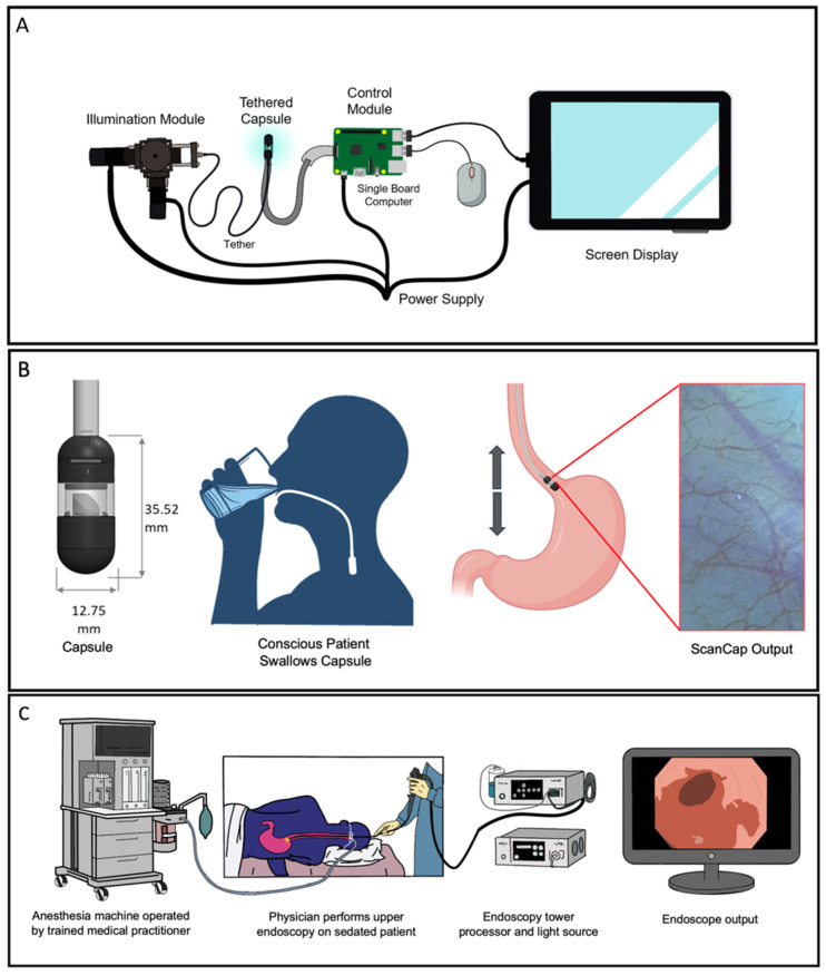
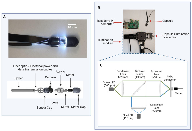
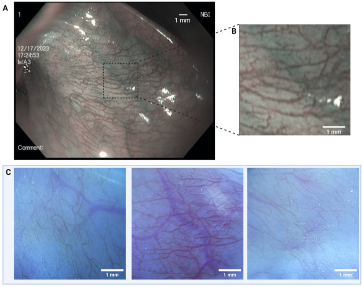
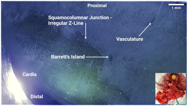
Esophageal carcinoma is the sixth-leading cause of cancer death worldwide. A precursor to esophageal adenocarcinoma (EAC) is Barrett's Esophagus (BE). Early-stage diagnosis and treatment of esophageal neoplasia (Barrett's with high-grade dysplasia/intramucosal cancer) increase the five-year survival rate from 10% to 98%. BE is a global challenge; however, current endoscopes for early BE detection are costly and require extensive infrastructure for patient examination and sedation. We describe the design and evaluation of the first prototype of ScanCap, a high-resolution optical endoscopy system with a reusable, low-cost tethered capsule, designed to provide high-definition, blue-green illumination imaging for the early detection of BE in unsedated patients. The tethered capsule (12.8 mm diameter, 35.5 mm length) contains a color camera and rotating mirror and is designed to be swallowed; images are collected as the capsule is retracted manually via the tether. The tether provides electrical power and illumination at wavelengths of 415 nm and 565 nm and transmits data from the camera to a tablet. The ScanCap prototype capsule was used to image the oral mucosa in normal volunteers and ex vivo esophageal resections; images were compared to those obtained using an Olympus CV-180 endoscope. Images of superficial capillaries in intact oral mucosa were clearly visible in ScanCap images. Diagnostically relevant features of BE, including irregular Z-lines, distorted mucosa, and dilated vasculature, were clearly visible in ScanCap images of ex vivo esophageal specimens.

Keywords: Barrett’s esophagus; cancer detection; cancer screening; capsule endoscopy; digital chromoendoscopy; esophageal cancer; global health; low-resource settings; narrow-band imaging.


Conflict of interest statement

The authors declare no conflicts of interest. The funders had no role in the design of the study; in the collection, analyses, or interpretation of data; in the writing of the manuscript; or in the decision to publish the results.

Figures

Figure 1
Figure 1
Overview of current ScanCap system and assessment versus the current standard of care. (A) The complete ScanCap system consists of a tethered capsule and a control system comprising an LED-based illumination module, a control module with a single board computer and power supply, and a screen display. (B) Proposed procedure to image the esophagus using the ScanCap endoscopy capsule. Cross-sectional overview of ScanCap; a conscious, non-sedated patient swallows the tethered capsule. Forward motion of the capsule is propelled via the natural peristaltic forces of the esophagus; the capsule is withdrawn manually using the tether. (C) Standard endoscopy procedure to image the esophagus. The patient is sedated while an endoscope is introduced into the upper gastrointestinal tract.
Figure 2
Figure 2
Schema of the ScanCap system and photographs of prototype. (A) Photograph (top) and schematic (bottom) of prototype ScanCap capsule, including tether, optical fibers that deliver blue and green light from the illumination module, 3D-printed sensor cap that holds Raspberry PiCam and lens, acrylic wall, mirror, micromotor, and 3D-printed motor cap. (B) Photograph of ScanCap capsule and control system modules, including the illumination and control modules. (C) Schematic diagram of illumination module.
Figure 3
Figure 3
Diagram of image sequence acquisition. (A) Hypothetical image capture sequence if the mirror and the camera were rotated together. (B) Actual ScanCap image sequence with rotating mirror and stationary camera. (C) Image sequence taken with ScanCap with motor rotating the mirror. In each diagram, the full ScanCap field of view (FOV) (12 × 7 mm2) and the smaller region of interest (5 × 5 mm2) are depicted. The full ScanCap FOV is utilized for mosaicking purposes and enables full coverage of the center 5 × 5 mm2 region of interest.
Figure 4
Figure 4
(A) ScanCap image of 1951 USAF resolution test target showing a 5 mm × 5 mm area of the full field of view (FOV). (B) Zoomed-in picture of the United States Air Force (USAF) standard resolution target showing that ScanCap can resolve group six, element one (7.8 µm).
Figure 5
Figure 5
(A) Representative single frames acquired while the motor rotated. Each image shows a 5 mm × 5 mm segment of the 12 × 7 mm2 field of view (FOV). (B) Stitched image of the frames captured in (A).
Figure 6
Figure 6
(A) Image of buccal mucosa (lower lip) obtained using an Olympus CV-180 endoscope with narrow-band imaging. (B) A 5 mm × 5 mm zoomed-in image from a representative area of the endoscopic capture. (C) Images of human buccal mucosa (lower lip) obtained with ScanCap. The ScanCap field of view (FOV) is cropped to show a 5 mm × 5 mm segment of the 12 × 7 mm2 FOV.
Figure 7
Figure 7
ScanCap image of resected esophageal specimen. The image shows the entire ScanCap field of view (12 mm × 7 mm). Inset, lower right: Photograph of resected specimen showing approximate location of ScanCap image acquisition (yellow rectangle). The proximal end of the esophagus specimen was on the top right, while the distal end was on the bottom left. The irregular squamocolumnar junction (Z-line) shows characteristic features of esophageal metaplasia. Barrett’s islands and dilated vasculature were also observed in the capsule images.

References

    1. Sung H., Ferlay J., Siegel R.L., Laversanne M., Soerjomataram I., Jemal A., Bray F. Global Cancer Statistics 2020: GLOBOCAN Estimates of Incidence and Mortality Worldwide for 36 Cancers in 185 Countries. CA Cancer J. Clin. 2021;71:209–249. doi: 10.3322/CAAC.21660. - DOI - PubMed
    1. José M., Arnal D., Ferrández Arenas Á., Lanas Á., María A., Arnal J.D., Ferrández Á., Lanas Arbeloa Á. Esophageal Cancer: Risk Factors, Screening and Endoscopic Treatment in Western and Eastern Countries. World J. Gastroenterol. 2015;21:7933–7943. doi: 10.3748/wjg.v21.i26.7933. - DOI - PMC - PubMed
    1. McColl K.E.L. What Is Causing the Rising Incidence of Esophageal Adenocarcinoma in the West and Will It Also Happen in the East? J. Gastroenterol. 2019;54:669–673. doi: 10.1007/S00535-019-01593-7. - DOI - PMC - PubMed
    1. Hur C., Miller M., Kong C.Y., Dowling E.C., Nattinger K.J., Dunn M., Feuer E.J. Trends in Esophageal Adenocarcinoma Incidence and Mortality. Cancer. 2013;119:1149–1158. doi: 10.1002/CNCR.27834. - DOI - PMC - PubMed
    1. Meves V., Behrens A., Pohl J. Diagnostics and Early Diagnosis of Esophageal Cancer. Viszeralmedizin. 2015;31:315–318. doi: 10.1159/000439473. - DOI - PMC - PubMed

LinkOut - more resources