. 2024 Jun 20;11(6):632.
doi: 10.3390/bioengineering11060632.

Rapid, Point-of-Care Microwave Lysis and Electrochemical Detection of Clostridioides difficile Directly from Stool Samples

Affiliations

Rapid, Point-of-Care Microwave Lysis and Electrochemical Detection of Clostridioides difficile Directly from Stool Samples

Lovleen Tina Joshi et al. Bioengineering (Basel). .

Abstract

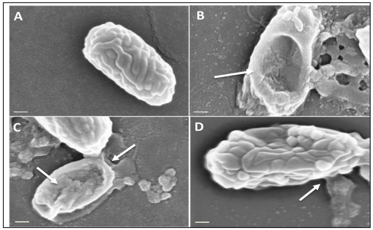
The rapid detection of the spore form of Clostridioides difficile has remained a challenge for clinicians. To address this, we have developed a novel, precise, microwave-enhanced approach for near-spontaneous release of DNA from C. difficile spores via a bespoke microwave lysis platform. C. difficile spores were microwave-irradiated for 5 s in a pulsed microwave electric field at 2.45 GHz to lyse the spore and bacteria in each sample, which was then added to a screen-printed electrode and electrochemical DNA biosensor assay system to identify presence of the pathogen's two toxin genes. The microwave lysis method released both single-stranded and double-stranded genome DNA from the bacterium at quantifiable concentrations between 0.02 μg/mL to 250 μg/mL allowing for subsequent downstream detection in the biosensor. The electrochemical bench-top system comprises of oligonucleotide probes specific to conserved regions within tcdA and tcdB toxin genes of C. difficile and was able to detect 800 spores of C. difficile within 300 µL of unprocessed human stool samples in under 10 min. These results demonstrate the feasibility of using a solid-state power generated, pulsed microwave electric field to lyse and release DNA from human stool infected with C. difficile spores. This rapid microwave lysis method enhanced the rapidity of subsequent electrochemical detection in the development of a rapid point-of-care biosensor platform for C. difficile.

Keywords: Clostridioides difficile; DNA detection; bioengineering; biosensors; electrochemistry; lysis; microwaves; point-of-care; spores.


Conflict of interest statement

The authors declare no conflicts of interest.

Figures

Figure 1
Figure 1
(a) A cylindrical aluminum cavity operating in its TM010 mode, designed to deliver 2.45 GHz of precise microwave radiation to the bacterial sample. An adjustable coupling loop is used to match the cavity to the microwave source to ensure maximum power delivery to the sample. (b) The normalized electric field distribution in the TM010 mode; the sample tube is placed in the region of the high microwave electric field, near the axis of the cavity, with the field parallel to the axis of the tube. (c) The well-known Bessel function form J0 (2.405x) for the radial dependence of electric field magnitude.
Figure 2
Figure 2
Microwave circuitry. The cavity is excited by a highly adaptable, solid-state microwave power delivery system (up to 30 W) comprising a low power source and high-power amplifier. The power sensors [5] are used to measure the incident and reflected power to ensure that maximum power transfer conditions can be attained. An additional, a wideband power sensor [6] allows the measurement of any reflected power for low duty cycles, during short pulses. The RF switch allows the microwaves to be pulsed at duty cycles ranging from 0.3% to 100% (Table S1).
Figure 3
Figure 3
Scanning Electron Microscopy of C. difficile spores before and after microwaving. Spores of strain DS1813 were imaged under SEM before and after microwaving at a peak power of 30 W rms milli-Watts at 100%, 10%, and 1% duty cycles (DC) for 5 s. A total of 40 spores per sample were imaged per DC at ×82,000 magnification, with the spores chosen here representative of spores consistently seen within the sample. White arrows indicate areas of lysis and morphological damage. (A) A control spore of DS1813 which was not exposed to microwaves. (B) DS1813 exposed to constant microwaves at 100% DC. Damage to the spore structure and debris is clearly visible on this spore. (C) DS1813 exposed to pulsed microwaves at 10% DC. Some damage to the spore structure is visible at its terminal end. (D) DS1813 exposed to pulsed microwaves at 1% DC. There is no visible damage to the spore structure. (Image scale bar = 200 nm).
Figure 4
Figure 4
Quantification of double and single-stranded DNA released from microwaved C. difficile spores. Spores of toxigenic DS1813 and non-toxigenic DS1684 strains were microwaved a peak power of 30 W rms milli-Watts at a range of duty cycles (0%, 1%, 10%, 100%) for 5 s, each at a concentration of 1.67 × 10⁷ spores/mL. The single-stranded (ss) and double-stranded (dsDNA) was quantified via Qubit Fluorometer 3.0. Each test was performed in triplicate (n = 3) (A) Concentration of ssDNA in samples of DS1813 and DS1684 (ug/mL). (B) Concentration of dsDNA in samples of DS1813 and DS1684 in ug/mL.
Figure 5
Figure 5
Electrochemical detection of toxigenic C. difficile spores suspended in sterile water following exposure to microwaves. Spores of toxigenic strain DS1813 and non-toxigenic DS1684 at a set concentration of 1.33 × 10⁴ spores/mL were exposed to duty cycles of 1%, 10%, and 100% for 5 s. Spores which were not microwaved were used at controls (0% DC). This equates to 665 spores within the 50 µL of the assay sample. The microwaved spore samples were then introduced to the VantixTM electrochemical detection system and tested for the presence of toxin genes tcdA and tcdB. The results above show voltage signals measured from toxigenic DS1813. The results from the toxin-negative DS1684 strain did not generate a measurable signal (0 mV). Each result represents the mean of two independent tests (n = 2).
Figure 6
Figure 6
Electrochemical detection of C. difficile spores at a range of concentrations in sterile water. Spores of toxigenic strain DS1813 and non-toxigenic DS1684 at concentrations ranging from 1 × 101 spores within a 50 µL sample to 1 × 10⁸ spores within a 50 µL sample were exposed to microwaves at 100% DC for 5 s. The microwaved spore samples were then introduced to the VantixTM electrochemical detection system and tested for the presence of both toxin genes tcdA and tcdB. No signal was detected for any of the DS1684 spore concentrations tested (0 mV). Each result represents the mean of two independent tests (n = 2).
Figure 7
Figure 7
Electrochemical detection of C. difficile spores at a range of concentrations in human feces. Spores of toxigenic strain DS1813 and non-toxigenic DS1684 at concentrations ranging from 1 × 101 spores within a 50 µL sample to 1 × 10⁸ spores within a 50 µL sample were exposed to microwaves at 100% DC for 5 s. The microwaved spore samples were then introduced to the VantixTM electrochemical detection system and tested for the presence of both toxin genes tcdA and tcdB. No signal was detected for any of the DS1684 spore concentrations tested (0 mV). Each result represents the mean of two independent tests (n = 2).
Figure 8
Figure 8
Presence of C. difficile in clinical stool samples. A total of 50 stool samples from patients at Public Health Wales were examined for the presence of C. difficile using selective agar culture. The data are arranged in order of bacterial number in each sample (cfu/mL). Samples 2, 3, & 4 are anomalous as these were deemed C. difficile negative by PHW. Each result represents the mean of two independent tests (n = 2).

References

    1. Guh A.Y., Mu Y., Winston L.G., Johnston H., Olson D., Farley M.M., Wilson L.E., Holzbauer S.M., Phipps E.C., Dumyati G.K., et al. Trends in US burden of Clostridioides difficile infection and outcomes. N. Engl. J. Med. 2020;382:1320–1330. doi: 10.1056/NEJMoa1910215. - DOI - PMC - PubMed
    1. Czepiel J., Dróżdż M., Pituch H., Kuijper E.J., Perucki W., Mielimonka A., Goldman S., Wultańska D., Garlicki A., Biesiada G. Clostridium difficile infection: Review. Eur. J. Clin. Microbiol. Infect. Dis. 2019;38:1211–1221. doi: 10.1007/s10096-019-03539-6. - DOI - PMC - PubMed
    1. Ahmed H., Joshi L.T. Clostridioides difficile spores tolerate disinfection with sodium hypochlorite disinfectant and remain viable within surgical scrubs and gown fabrics. Microbiology. 2023;169:001418. doi: 10.1099/mic.0.001418. - DOI - PMC - PubMed
    1. Spigaglia P. Clostridioides difficile infection (CDI) during the COVID-19 pandemic. Anaerobe. 2022;74:102518. doi: 10.1016/j.anaerobe.2022.102518. - DOI - PMC - PubMed
    1. Boyanova L., Dimitrov G., Gergova R., Hadzhiyski P., Markovska R. Clostridioides difficile resistance to antibiotics, including post-COVID-19 data. Expert Rev. Clin. Pharmacol. 2023;16:925–938. - PubMed

LinkOut - more resources