

Principles and Clinical Application of Free-Style Capillary Perforator-Based Flap for Coverage of Facial Skin Cancer Defects
- PMID: 38927912
- PMCID: PMC11201941
- DOI: 10.3390/cancers16122206
Principles and Clinical Application of Free-Style Capillary Perforator-Based Flap for Coverage of Facial Skin Cancer Defects
Abstract
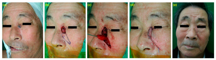
This study introduces a free-style perforator based island flap (PBIF) for the reconstruction of skin defects. From March 2012 to December 2022, a retrospective investigation was conducted on patients who underwent reconstruction for facial defects due to skin cancer. Data on the patients' gender, age, anesthesia method, diagnosis, defect location, flap size, complications, and follow-up periods were collected. There are several principles for designing the PBIF: finger-pinching method, alignment with the direction of wrinkles, the smaller width and longer length of the flap, and proximal attachment to the muscle. A total of 32 patients were included, with an average age of 63.6 years. Surgeries were performed in various regions, such as the infraorbital area, nose, cheek, philtrum, and the anterior/posterior/inferior auricular regions, with an average flap size of 7.63 cm2. There were no complications, such as venous congestion or vascular insufficiency in the skin flaps, although one case required revisional closure due to flap disruption. The PBIF is a useful and effective method for the restoration of facial defects. This method can provide simple yet aesthetically satisfying results, showing stable outcomes without complex surgeries or complications. This study indicates the potential for this method to be more widely employed in reconstructive surgeries in the future.
Keywords: basal cell carcinoma; local flap; skin cancer; squamous cell carcinoma; surgery.
Conflict of interest statement
In the interest of full disclosure, the authors declare that they have no conflicts of interest regarding the research, authorship, and/or publication of this paper. No personal relationships with other people or organizations have influenced the work presented in this manuscript.
Figures


References
-
- Helmy Ali Y., Farahat Mohamed A., Nasef M.A., Abu-Elsoud A., Dahi A., Hossni M., Ouf M.O., Zayed T., ELBatawy A., Farid M., et al. Facial skin cancer reconstructive and cosmetic outcomes: Analysis with algorithm for its management. J. Cosmet. Dermatol. 2020;19:1182–1190. doi: 10.1111/jocd.13121. - DOI - PubMed
LinkOut - more resources
Full Text Sources
