. 2024 Jun 9;25(12):6379.
doi: 10.3390/ijms25126379.

Heat Stress and Microbial Stress Induced Defensive Phenol Accumulation in Medicinal Plant Sparganium stoloniferum

Affiliations

Heat Stress and Microbial Stress Induced Defensive Phenol Accumulation in Medicinal Plant Sparganium stoloniferum

Mengru Sang et al. Int J Mol Sci. .

Abstract

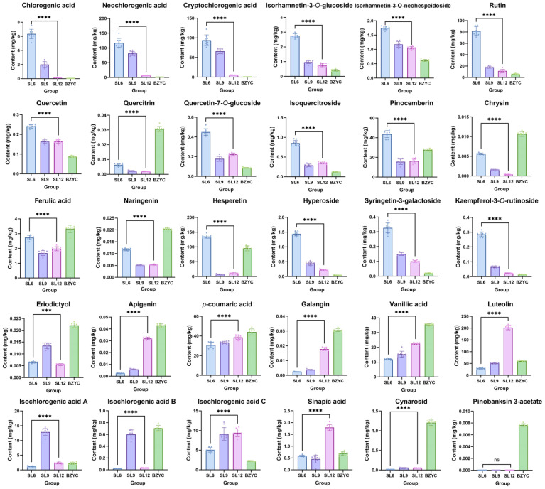
An approach based on the heat stress and microbial stress model of the medicinal plant Sparganium stoloniferum was proposed to elucidate the regulation and mechanism of bioactive phenol accumulation. This method integrates LC-MS/MS analysis, 16S rRNA sequencing, RT-qPCR, and molecular assays to investigate the regulation of phenolic metabolite biosynthesis in S. stoloniferum rhizome (SL) under stress. Previous research has shown that the metabolites and genes involved in phenol biosynthesis correlate to the upregulation of genes involved in plant-pathogen interactions. High-temperature and the presence of Pseudomonas bacteria were observed alongside SL growth. Under conditions of heat stress or Pseudomonas bacteria stress, both the metabolites and genes involved in phenol biosynthesis were upregulated. The regulation of phenol content and phenol biosynthesis gene expression suggests that phenol-based chemical defense of SL is stimulated under stress. Furthermore, the rapid accumulation of phenolic substances relied on the consumption of amino acids. Three defensive proteins, namely Ss4CL, SsC4H, and SsF3'5'H, were identified and verified to elucidate phenol biosynthesis in SL. Overall, this study enhances our understanding of the phenol-based chemical defense of SL, indicating that bioactive phenol substances result from SL's responses to the environment and providing new insights for growing the high-phenol-content medicinal herb SL.

Keywords: Sparganium stoloniferum; amino acid; chemical defense; heat stress; microbial stress; phenol.


Conflict of interest statement

The authors declare no conflicts of interest.

Figures

Figure 1
Figure 1
The contents of three kinds of chemical constituents in S. stoloniferum rhizome (SL) at different growth stages and standard SL materials (n = 6, the contents shown in (AC) are the average values). (A) The contents of amino acids in SL at different growth stages and standard SL materials. (B) The contents of phenolic acids in SL at different growth stages and standard SL materials. (C) The contents of flavonoids in SL at different growth stages and standard SL materials. SL6, SL9, and SL12 are SL samples collected in June, September, and December, respectively. BZYC is the standard SL material, set as the control sample.
Figure 2
Figure 2
Phenol contents of SL at different growth stages and standard S. stoloniferum rhizome (SL) materials (n = 6). SL6, SL9, and SL12 are SL samples collected in June, September, and December, respectively. BZYC is standard SL material, set as control sample. The ****, ***, and ns represent p < 0.0001, p < 0.001, and p > 0.05 (t-test), respectively.
Figure 3
Figure 3
Amino acids contents of SL at different growth stages and standard S. stoloniferum rhizome (SL) (n = 6). SL6, SL9, and SL12 are SL samples collected in June, September, and December, respectively. BZYC is the standard SL material, set as the control sample. The ****, **, and * represent p < 0.0001, p < 0.01, and p < 0.05 (t-test), respectively.
Figure 4
Figure 4
The bacterial communities in the S. stoloniferum rhizosphere soil from different habitats. (A) Distribution of countries growing S. stoloniferum in Aisa and the three main SL habitats in China. (B) The graph of S. stoloniferum rhizosphere soil distribution. (C) Venn diagram showing the operational taxonomic units of bacterial communities in the S. stoloniferum rhizosphere soil of three different production areas. Cartoon pictures (insets in A,B) were created with BioRender.com.
Figure 5
Figure 5
Species abundance of soil bacterial communities in rhizospheres of S. stoloniferum from different origins. (A) Heatmap of bacterial communities classified by phylum. (B) Heatmap of bacterial communities classified by class.
Figure 6
Figure 6
The contents of three kinds of chemical constituents in S. stoloniferum rhizome (SL) under microbial stress and heat stress (n = 6, the contents shown in (AC) are the average values). (A) The contents of amino acids in SL under microbial stress and heat stress. (B) The contents of phenolic acids in SL under microbial stress and heat stress. (C) The contents of flavonoids in SL under microbial stress and heat stress. The SL sample under microbial stress was named PS. The SL sample under heat stress was named HT. The negative control group was named BL.
Figure 7
Figure 7
The effects of microbial stress and heat stress on the amino acid contents of the S. stoloniferum rhizome (SL) (n = 6). The SL sample under microbial stress was named PS. The SL sample under heat stress was named HT. The negative control group was named BL. The different letters (a, b, and c) next to the error bars represent statistically significant differences at p < 0.05 (ANOVA).
Figure 8
Figure 8
The effects of microbial stress and heat stress on the phenol contents of S. stoloniferum rhizome (SL) (n = 6). The SL sample under microbial stress was named PS. The SL sample under heat stress was named HT. The negative control group was named BL. The different letters (a, b, and c) next to the error bars represent statistically significant differences at p < 0.05 (ANOVA).
Figure 9
Figure 9
Relative expression of the gene related to lignin and phenol biosynthesis in microbial stress group and negative control group of the S. stoloniferum rhizome (SL) (n = 3). Data were normalized using ACTIN3. The SL sample under heat stress was named HT. The negative control group was named BL. The ****, ***, **, and * represent p < 0.0001, p < 0.001, p < 0.01, and p < 0.05 (t-test), respectively.
Figure 10
Figure 10
Functional characterization of phenylalanine ammonia-lyase from S. stoloniferum. (A) Purification and SDS-PAGE analysis of recombinant Ss4CL produced in E. coli. Lane M, 1 and 2 represent protein molecular marker, E. coli. harboring pET28a(+)-Ss4CL construct-induced IPTG (1 mM) at 24 h and purified recombinant Ss4CL, respectively. (B) Michaelis–Menten curves for the velocity of Ss4CL. Bars indicate the standard errors of the means (n = 3). (C) LC–MS/MS analysis of p-coumaroyl-CoA produced by a 4CL reaction using recombinant Ss4CL. (C1C4) are extracted ion chromatograms of the p-coumaroyl-CoA of samples.
Figure 11
Figure 11
Functional identification of the recombinant SsC4H and SsF3′5′H. (A) LC–MS/MS analysis of p-coumaric acid produced by a C4H reaction using recombinant SsC4H. (A1A4) are extracted ion chromatograms of p-coumaric acid of samples. (B) LC–MS/MS analysis of eriodictyol produced by a F3′5′H reaction using recombinant SsF3′5′H. (B1B4) are extracted ion chromatograms of the eriodictyol of samples. (C) LC–MS/MS analysis of luteolin produced by a F3′5′H reaction using recombinant SsF3′5′H. (C1C4) are extracted ion chromatograms of the luteolin of samples.
Figure 12
Figure 12
Overall structure and molecular docking of Ss4CL, SsC4H, and SsF3′5′H from S. stoloniferum. The predicted lDDT values of each residue position of five structure rank models of Ss4CL (A), SsC4H (B), SsF3′5′H (C). (D) Predicted structure of Ss4CL and binding site of p-coumaric acid and Ss4CL. (E) Predicted structure of SsC4H and binding site of trans-cinnamic acid and SsC4H. (F) Predicted structure of SsF3′5′H and binding site of apigenin and SsF3′5′H. (G) 3D diagrams show receptor–ligand interactions between p-coumaric and Ss4CL. (H) 3D diagrams show receptor–ligand interactions between trans-cinnamic acid and SsC4H. (I) 3D diagrams show receptor–ligand interactions between apigenin and SsF3′5′H.

Similar articles

Cited by

References

    1. Salam U., Ullah S., Tang Z.-H., Elateeq A.A., Khan Y., Khan J., Khan A., Ali S. Plant Metabolomics: An Overview of the Role of Primary and Secondary Metabolites against Different Environmental Stress Factors. Life. 2023;13:706. doi: 10.3390/life13030706. - DOI - PMC - PubMed
    1. Martinez V., Mestre T.C., Rubio F., Girones-Vilaplana A., Moreno D.A., Mittler R., Rivero R.M. Accumulation of Flavonols over Hydroxycinnamic Acids Favors Oxidative Damage Protection under Abiotic Stress. Front. Plant Sci. 2016;7:838. doi: 10.3389/fpls.2016.00838. - DOI - PMC - PubMed
    1. Parida A.K., Dagaonkar V.S., Phalak M.S., Umalkar G.V., Aurangabadkar L.P. Alterations in photosynthetic pigments, protein and osmotic components in cotton genotypes subjected to short-term drought stress followed by recovery. Plant Biotechnol. Rep. 2007;1:37–48. doi: 10.1007/s11816-006-0004-1. - DOI
    1. Singh A., Sarma B.K., Upadhyay R.S., Singh H.B. Compatible rhizosphere microbes mediated alleviation of biotic stress in chickpea through enhanced antioxidant and phenylpropanoid activities. Microbiol. Res. 2013;168:33–40. doi: 10.1016/j.micres.2012.07.001. - DOI - PubMed
    1. Divekar P.A., Narayana S., Divekar B.A., Kumar R., Gadratagi B.G., Ray A., Singh A.K., Rani V., Singh V., Singh A.K., et al. Plant Secondary Metabolites as Defense Tools against Herbivores for Sustainable Crop Protection. Int. J. Mol. Sci. 2022;23:2690. doi: 10.3390/ijms23052690. - DOI - PMC - PubMed

LinkOut - more resources