Case Reports
. 2024 Jun 13;25(12):6518.
doi: 10.3390/ijms25126518.

Methylation-Based Characterization of a New IDH2 Mutation in Sinonasal Undifferentiated Carcinoma

Affiliations
Case Reports

Methylation-Based Characterization of a New IDH2 Mutation in Sinonasal Undifferentiated Carcinoma

Simon Burgermeister et al. Int J Mol Sci. .

Abstract

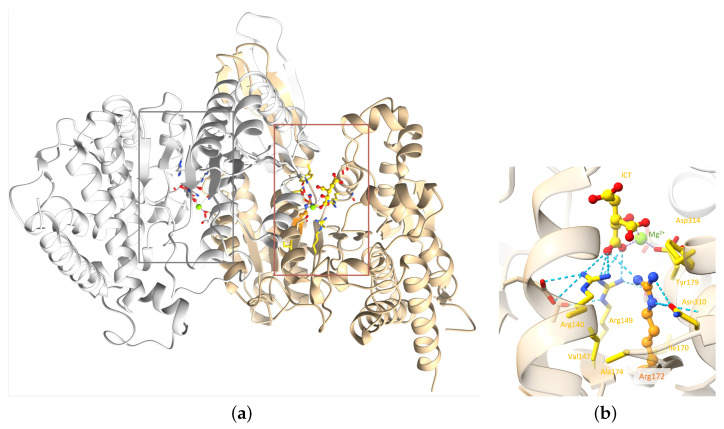
Mutations affecting codon 172 of the isocitrate dehydrogenase 2 (IDH2) gene define a subgroup of sinonasal undifferentiated carcinomas (SNUCs) with a relatively favorable prognosis and a globally hypermethylated phenotype. They are also recurrent (along with IDH1 mutations) in gliomas, acute myeloid leukemia, and intrahepatic cholangiocarcinoma. Commonly reported mutations, all associated with aberrant IDH2 enzymatic activity, include R172K, R172S, R172T, R172G, and R172M. We present a case of SNUC with a never-before-described IDH2 mutation, R172A. Our report compares the methylation pattern of our sample to other cases from the Gene Expression Omnibus database. Hierarchical clustering suggests a strong association between our sample and other IDH-mutant SNUCs and a clear distinction between sinonasal normal tissues and tumors. Principal component analysis (PCA), using 100 principal components explaining 94.5% of the variance, showed the position of our sample to be within 1.02 standard deviation of the other IDH-mutant SNUCs. A molecular modeling analysis of the IDH2 R172A versus other R172 variants provides a structural explanation to how they affect the protein active site. Our findings thus suggest that the R172A mutation in IDH2 confers a gain of function similar to other R172 mutations in IDH2, resulting in a similar hypermethylated profile.

Keywords: IDH2 mutation; methylation analysis; molecular modeling.


Conflict of interest statement

The authors declare no conflicts of interest.

Figures

Figure A1
Figure A1
Protein sequence alignment of isocitrate dehydrogenase, centered on residue 172 among a selection of vertebrate species. Sequences obtained from the Reference Sequence Database [40].
Figure 1
Figure 1
Magnetic resonance imaging showing a heterogeneous mass in the left nasal cavity. (a) T1 with Gadolinium, axial view. (b) T1 with Gadolinium, coronal view.
Figure 2
Figure 2
Histopathology of the biopsy sample. (a) H&E showing sheets of epithelioid cells without any definite lineage differentiation. The nuclei are medium- to large-sized and hyperchromatic. We note the presence of tumor necrosis and frequent mitosis. (b) H3K27me3 IHC showing an increased labeling for our sample compatible with increased histone H3K27 trimethylation.
Figure 3
Figure 3
[18F]FDG PET maximum intensity projection showing a large, irregular and intensely hypermetabolic mass in the sinonasal region. (a) lateral view. (b) frontal view.
Figure 4
Figure 4
Alignment of human and mouse protein sequences. Sequences retrieved from UniProt database [17], alignment performed with Clustal Omega v.1.2.4 [18], and sequences visualized with UCSF Chimera v.1.17.3 [19]. The color code of the alignment corresponds to the default colour code of the visualization tool. The star (*) indicates that the canonical IDH2 sequence of the organism has been used. The red rectangle indicates the position of the amino acid 172 in humans.
Figure 5
Figure 5
IDH2 3D homodimeric structure. (a) The IDH2 homodimer with the two active sites indicated by thin boxes; (b) zoom in on R172 of chain A of the complex. Chains A and B are colored tan and gray, respectively. R172 and ICT are shown as balls and sticks: Mg2+ as a green sphere and important residues as sticks. R172 is colored orange. Elements interacting directly with R172 are colored yellow. Hydrogen bonds involving the arginine 140, 149, and 172 are represented as blue dashed lines. For easier visualization, the protein is represented by transparent ribbons. (PDB ID: 5h3f [16]).
Figure 6
Figure 6
Comparison of structural models of the mutated proteins with the wild-type IDH2 active site. (a) Wild-type; (b) structural models of IDH2 p.R172G, (c) IDH2 p.R172A, (d) IDH2 p.R172S, (e) IDH2 p.R172M, and (f) IDH2 p.R172K. Only monomer A is represented for clarity. Residue 172 and ICT molecule are shown as balls and sticks: Mg2+ as a green sphere and important residues as sticks. Residue 172 is colored orange. Elements interacting directly with residue 172 are colored yellow. For easier visualization, the protein is represented by transparent ribbons. The dashed lines represent the interactions between ICT and Mg2+. Models were built based on the experimental structure with the PDB ID 5h3f [16].
Figure 6
Figure 6
Comparison of structural models of the mutated proteins with the wild-type IDH2 active site. (a) Wild-type; (b) structural models of IDH2 p.R172G, (c) IDH2 p.R172A, (d) IDH2 p.R172S, (e) IDH2 p.R172M, and (f) IDH2 p.R172K. Only monomer A is represented for clarity. Residue 172 and ICT molecule are shown as balls and sticks: Mg2+ as a green sphere and important residues as sticks. Residue 172 is colored orange. Elements interacting directly with residue 172 are colored yellow. For easier visualization, the protein is represented by transparent ribbons. The dashed lines represent the interactions between ICT and Mg2+. Models were built based on the experimental structure with the PDB ID 5h3f [16].
Figure 7
Figure 7
Heatmap with clustering using top most differentially methylated probes. Our sample (in orange) shows proximity to conventional IDH mutant SNUC (green).
Figure 8
Figure 8
UMAP representation of the M-values.

References

    1. Dogan S., Vasudevaraja V., Xu B., Serrano J., Ptashkin R.N., Jung H.J., Chiang S., Jungbluth A.A., Cohen M.A., Ganly I., et al. DNA methylation-based classification of sinonasal undifferentiated carcinoma. Mod. Pathol. 2019;32:1447–1459. doi: 10.1038/s41379-019-0285-x. - DOI - PMC - PubMed
    1. Losman J., Kaelin W.J. What a difference a hydroxyl makes: Mutant IDH, (R)-2-hydroxyglutarate, and cancer. Genes Dev. 2013;27:836–852. doi: 10.1101/gad.217406.113. - DOI - PMC - PubMed
    1. Guo J., Zhang R., Yang Z., Duan Z., Yin D., Zhou Y. Biological Roles and Therapeutic Applications of IDH2 Mutations in Human Cancer. Front. Oncol. 2021;26:644857. doi: 10.3389/fonc.2021.644857. - DOI - PMC - PubMed
    1. Showalter M.R., Hatakeyama J., Cajka T., VanderVorst K., Carraway K.L., Fiehn O., Reproducibility Project: Cancer Biology Replication Study: The common feature of leukemia-associated IDH1 and IDH2 mutations is a neomorphic enzyme activity converting alpha-ketoglutarate to 2-hydroxyglutarate. eLife. 2017;6:e26030. doi: 10.7554/eLife.26030. - DOI - PMC - PubMed
    1. Nakazawa S., Sakata K.I., Liang S., Yoshikawa K., Iizasa H., Tada M., Hamada J.I., Kashiwazaki H., Kitagawa Y., Yamazaki Y. Dominant-negative p53 mutant R248Q increases the motile and invasive activities of oral squamous cell carcinoma cells. Biomed Res. 2019;40:37–49. doi: 10.2220/biomedres.40.37. - DOI - PubMed

Publication types

Substances

Supplementary concepts

LinkOut - more resources