. 2024 Jun 17;13(6):732.
doi: 10.3390/antiox13060732.

Induction of Semaphorin 3A by Resveratrol and Pinostilbene via Activation of the AHR-NRF2 Axis in Human Keratinocytes

Affiliations

Induction of Semaphorin 3A by Resveratrol and Pinostilbene via Activation of the AHR-NRF2 Axis in Human Keratinocytes

Gaku Tsuji et al. Antioxidants (Basel). .

Abstract

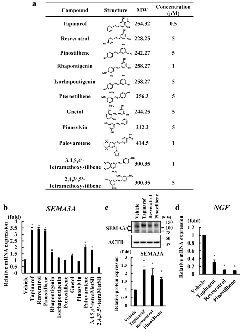
Semaphorin 3A (SEMA3A), a nerve-repellent factor produced by keratinocytes, has an inhibitory effect on nerve extension to the epidermis. Epidermal innervation is involved in pruritus in inflammatory skin diseases such as atopic dermatitis (AD) and dry skin. We previously reported that tapinarof, a stilbene molecule, upregulates SEMA3A in human keratinocytes. We also showed that this mechanism is mediated via the aryl hydrocarbon receptor (AHR), a ligand-activated transcription factor, and the nuclear factor erythroid 2-related factor 2 (NRF2) axis. Since some stilbenes activate AHR and NRF2, we attempted to identify other stilbenes that upregulate SEMA3A. We analyzed normal human epidermal keratinocytes (NHEKs) treated with 11 types of stilbenes and examined SEMA3A expression. We found that resveratrol and pinostilbene, antioxidant polyphenols, upregulated SEMA3A and increased nuclear AHR and NRF2 expression. In addition, AHR knockdown by small interfering RNA (siRNA) transfection abolished the NRF2 nuclear expression. Furthermore, AHR and NRF2 knockdown by siRNA transfection abrogated resveratrol- and pinostilbene-induced SEMA3A upregulation. Finally, we confirmed that resveratrol and pinostilbene increased SEMA3A promoter activity through NRF2 binding using ChIP-qPCR analysis. These results suggest that resveratrol and pinostilbene upregulate SEMA3A via the AHR-NRF2 axis in human keratinocytes.

Keywords: atopic dermatitis; keratinocyte; pruritus; semaphorin 3A.


Conflict of interest statement

The authors declare no conflicts of interest.

Figures

Figure 1
Figure 1
Tapinarof, resveratrol, and pinostilbene upregulated SEMA3A in NHEKs. (a) Chemical structures and concentrations of the tested stilbenes. (bd) NHEKs were treated with the indicated compounds for 72 h. mRNA (b) and protein (c) levels of SEMA3A and the NGF mRNA level (d) were analyzed. (b) MetSB, methoxystilbene. (b,d) qRT-PCR. Data are expressed as mean ± S.D.; n = 3/group. * Significant difference between the indicated compound-treated group and the vehicle group (p < 0.05). (c) Western blotting. Data are representative of triplicate experiments with similar results. SEMA3A protein levels were normalized for ACTB protein levels using ImageJ 1.48 V. Data are expressed as mean ± S.D. * Significant difference between the indicated compound-treated group and the vehicle group (p < 0.05).
Figure 2
Figure 2
Resveratrol and pinostilbene activated the AHR-NRF2 axis in NHEKs. (a,b) Western blotting. NHEKs were treated with vehicle, resveratrol, or pinostilbene for 30, 60, 120, and 180 min and then nuclear AHR and NRF2 protein levels were analyzed. Representative images from three independent experiments are shown. Protein levels of AHR and NRF2 were normalized for HDAC1 protein levels using ImageJ. Data are expressed as mean ± S.D. * Significant difference between the indicated time administration group and the pre-administration group (p < 0.05). (c,d) qRT-PCR. NHEKs were treated with vehicle, resveratrol, and pinostilbene for 72 h. Data are expressed as mean ± S.D.; n = 3/group. * p < 0.05. # Significant difference between the indicated compound-treated group and the vehicle group (p < 0.05). (e,f) Western blotting. Scrambled or AHR siRNA-transfected NHEKs were treated with vehicle, resveratrol, and pinostilbene for 30, 60, 120, and 180 min and then nuclear AHR and NRF2 protein levels were analyzed. Representative images from three independent experiments are shown. Protein levels of AHR and NRF2 were normalized for HDAC1 protein levels using ImageJ. Data are expressed as mean ± S.D. * p < 0.05. # Significant difference between si_AHR-transfected group and scrambled siRNA-transfected group (p < 0.05).
Figure 2
Figure 2
Resveratrol and pinostilbene activated the AHR-NRF2 axis in NHEKs. (a,b) Western blotting. NHEKs were treated with vehicle, resveratrol, or pinostilbene for 30, 60, 120, and 180 min and then nuclear AHR and NRF2 protein levels were analyzed. Representative images from three independent experiments are shown. Protein levels of AHR and NRF2 were normalized for HDAC1 protein levels using ImageJ. Data are expressed as mean ± S.D. * Significant difference between the indicated time administration group and the pre-administration group (p < 0.05). (c,d) qRT-PCR. NHEKs were treated with vehicle, resveratrol, and pinostilbene for 72 h. Data are expressed as mean ± S.D.; n = 3/group. * p < 0.05. # Significant difference between the indicated compound-treated group and the vehicle group (p < 0.05). (e,f) Western blotting. Scrambled or AHR siRNA-transfected NHEKs were treated with vehicle, resveratrol, and pinostilbene for 30, 60, 120, and 180 min and then nuclear AHR and NRF2 protein levels were analyzed. Representative images from three independent experiments are shown. Protein levels of AHR and NRF2 were normalized for HDAC1 protein levels using ImageJ. Data are expressed as mean ± S.D. * p < 0.05. # Significant difference between si_AHR-transfected group and scrambled siRNA-transfected group (p < 0.05).
Figure 3
Figure 3
Resveratrol and pinostilbene upregulated SEMA3A via the AHR-NRF2 axis in NHEKs. (a,b) NHEKs were transfected with siRNA targeting AHR (si_AHR) or NRF2 (si_NRF2) or scrambled control siRNA (Scram) and then treated with vehicle, tapinarof, resveratrol, or pinostilbene for 72 h. (a) qRT-PCR. SEMA3A mRNA levels. Data are expressed as mean ± S.D.; n = 3/group. * p < 0.05. # Significant difference between the indicated compound-treated group and the vehicle group (p < 0.05). (b) Western blotting. SEMA3A protein levels. Representative images of three independent experiments are shown. SEMA3A protein levels were normalized for ACTB protein levels using ImageJ. Data are expressed as mean ± S.D. * p < 0.05. # Significant difference between the indicated compound-treated group and the vehicle group (p < 0.05). N.D.: No significant difference.
Figure 4
Figure 4
Resveratrol and pinostilbene increased the promoter activity of SEMA3A via NRF2 in NHEKs. (a) Locations of AREL1 and AREL2 on the SEMA3A promoter and sequences of wild-type and mutated-type AREL1 and AREL2 are indicated. (b) Luciferase assay. NHEKs were transfected with plasmids containing the sequence of −209/+1 SEMA3A with wild-type or mutated-type AREL1 and AREL2. After transfection, cells were stimulated with resveratrol or pinostilbene for 48 h and the relative promoter activity was determined. Data are expressed as mean ± S.D.; n = 3/group. * p < 0.05. # Significant difference between the indicated compound-treated group and the vehicle group (p < 0.05). (c) ChIP-qPCR analysis using anti-NRF2 antibody. qPCR of AREL1 and AREL2 in SEMA3A promoter was performed. Data are shown as % expression compared with that of the input samples. Data are expressed as mean ± S.D.; n = 3/group. * p < 0.05. N.D.: No significant difference.

References

    1. Zeidler C., Pereira M.P., Huet F., Misery L., Steinbrink K., Ständer S. Pruritus in Autoimmune and Inflammatory Dermatoses. Front. Immunol. 2019;10:1303. doi: 10.3389/fimmu.2019.01303. - DOI - PMC - PubMed
    1. Roduit C., Frei R., Depner M., Karvonen A.M., Renz H., Braun-Fahrländer C., Schmausser-Hechfellner E., Pekkanen J., Riedler J., Dalphin J.C., et al. Phenotypes of Atopic Dermatitis Depending on the Timing of Onset and Progression in Childhood. JAMA Pediatr. 2017;171:655–662. doi: 10.1001/jamapediatrics.2017.0556. - DOI - PMC - PubMed
    1. Drucker A.M., Wang A.R., Li W.Q., Sevetson E., Block J.K., Qureshi A.A. The Burden of Atopic Dermatitis: Summary of a Report for the National Eczema Association. J. Investig. Dermatol. 2017;137:26–30. doi: 10.1016/j.jid.2016.07.012. - DOI - PubMed
    1. Weisshaar E., Bentz P., Apfelbacher C., Haufe E., Heinrich L., Heratizadeh A., Abraham S., Harder I., Kleinheinz A., Wollenberg A., et al. TREATgermany study group. Itching in Atopic Dermatitis: Patient- and Physician-reported Outcomes in the German Atopic Dermatitis Registry TREATgermany. Acta Derm. Venereol. 2023;103:adv00854. doi: 10.2340/actadv.v103.4426. - DOI - PMC - PubMed
    1. Furue K., Ulzii D., Tanaka Y., Ito T., Tsuji G., Kido-Nakahara M., Nakahara T., Furue M. Pathogenic Implication of Epidermal Scratch Injury in Psoriasis and Atopic Dermatitis. J. Dermatol. 2020;47:979–988. doi: 10.1111/1346-8138.15507. - DOI - PubMed

LinkOut - more resources