Review
. 2024 May 25;11(6):638.
doi: 10.3390/children11060638.

Congenital Lung Malformations: A Pictorial Review of Imaging Findings and a Practical Guide for Diagnosis

Affiliations
Review

Congenital Lung Malformations: A Pictorial Review of Imaging Findings and a Practical Guide for Diagnosis

Giovanna Cancemi et al. Children (Basel). .

Abstract

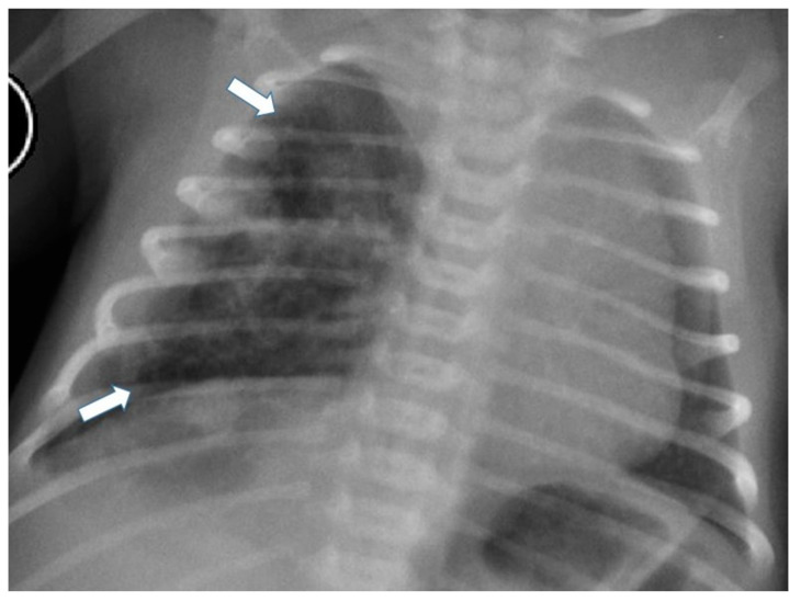
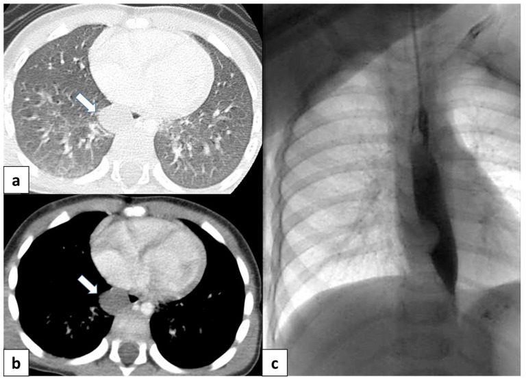
The term congenital lung malformation (CLM) is used to describe a wide range of pathological conditions with different imaging and clinical manifestations. These anomalies stem from abnormal embryological lung development, potentially occurring across various stages of prenatal life. Their natural history can be variable, presenting in a wide range of severity levels and encompassing asymptomatic individuals who remain so until adulthood, as well as those who experience respiratory distress in the neonatal period. Through the PubMed database, we performed an extensive review of the literature in the fields of congenital lung abnormalities, including their diagnostic approach and findings. From our RIS-PACS database, we have selected cases with a final diagnosis of congenital lung malformation. Different diagnostic approaches have been selected, including clinical cases studied using plain radiograph, CT scan, prenatal ultrasound, and MR images. The most encountered anomalies can be classified into three categories: bronchopulmonary anomalies (congenital pulmonary airway malformations (CPAMs), congenital lobar hyperinflation, bronchial atresia, and bronchogenic cysts), vascular anomalies (arteriovenous malformation), and combined lung and vascular anomalies (scimitar syndrome and bronchopulmonary sequestration). CLM causes significant morbidity and mortality; therefore, the recognition of these abnormalities is necessary for optimal prenatal counseling and early peri- and postnatal management. This pictorial review aims to report relevant imaging findings in order to offer some clues for differential diagnosis both for radiologists and pediatric consultants.

Keywords: MR imaging; computed tomography; congenital lung anomalies; congenital lung malformations; congenital thoracic malformations; imaging evaluation; imaging guidelines; radiography; ultrasound.


Conflict of interest statement

The authors declare no conflicts of interest.

Figures

Figure 1
Figure 1
General classification of congenital lung malformations (CLMs). CLH: congenital lobar hyperinflation; BC: bronchogenic cyst; CPAM: congenital pulmonary airway malformation; AVM: arteriovenous malformation.
Figure 2
Figure 2
Schematic representation of life-compatible types of congenital pulmonary airway malformations, which characteristics are thoroughly described in Table 1.
Figure 3
Figure 3
Chest radiograph of a newborn shows multiple large air-filled cysts in the left lower lobe (arrows), suggesting a type 1 CPAM.
Figure 4
Figure 4
Chest radiograph of a newborn shows multiple small air-filled cysts in the right upper lobe and right lower lobe (arrows), suggesting a type 2 CPAM.
Figure 5
Figure 5
A CT scan performed one month later in the same patient as in Figure 3, showing multiple large air-filled cysts involving the left lower lobe, with a maximum diameter of 60 mm, confirming the diagnostic hypothesis of type 1 CPAM.
Figure 6
Figure 6
Axial CT scan performed on a 4 month old female patient, depicting a type 1 CPAM. In image (a), it is possible to appreciate a large (>2 cm) air-filled cyst, surrounded by other multiple smaller cysts. Additionally, in image (b), a coronal CT MinIP reconstruction of the same patient is shown, which excludes communication with the tracheo-bronchial tree.
Figure 7
Figure 7
A CT scan later performed in the same patient as in Figure 4, showing multiple small air-filled cysts in the right upper lobe and in the right lower lobe, with a maximum diameter of 20 mm, confirming the diagnostic hypothesis of type 2 CPAM.
Figure 8
Figure 8
CT axial image visualized on lung window (a) of a 9 month old boy with right cystic lesions (previously seen on fetal ultrasound) shows an area of parenchymal consolidation mixed with air-filled cystic lesions, located in the right lower lobe (red circle). Contrast-enhanced acquisitions (c) demonstrate an abnormal arterial supply from systemic circulation into sequestered lung (arrow). These findings are compatible with hybrid lesion (CPAM and pulmonary sequestration). A CT scan performed after the arterial embolization of the aberrant vessel (b) shows a mild reduction in sequestered lung parenchyma (black circle).
Figure 9
Figure 9
CT image of a 4 month old girl presenting with multiple right cystic lesions <2 cm (a) compatible with CPAM type 2. Parenchymal consolidation (b) associated with the aberrant arterial (c) supply from the celiac trunk (red circle) and venous drainage into the pulmonary venous system, further evaluated with the coronal MiP reconstruction, represent a pulmonary intra-lobar sequestration. The combination of these findings supports the diagnosis of a hybrid lesion.
Figure 10
Figure 10
Incidental finding of bronchial atresia during a CT scan in a 15-year-old girl with lymphoma. In the left lower lobe, a tubular-shaped opacity (mucoid-filled bronchus) is associated with segmental hypoattenuation (arrow).
Figure 11
Figure 11
(a) CT scan in a 6 month old child shows a well-defined, rounded lesion with uniform fluid attenuation (arrow), compatible with a bronchogenic cyst; (b) after administration of intravenous contrast, bronchogenic cyst shows no enhancement or a minimally enhancing wall (arrow). (c) Dynamic esophageal fluoroscopy with oral contrast administration revealed a round, filling defect within the esophageal lumen, indicating that the cyst arises extrinsically from the bronchial tree.
Figure 12
Figure 12
A 17 year old patient with hereditary hemorrhagic telangiectasis (HHT). CT angiography with maximum intensity projection (MIP) reconstruction shows a lobulated enhancing lesion in the right middle lobe, with a feeding artery (red arrow) and a draining vein (blue arrow), compatible with pulmonary arteriovenous malformation.
Figure 13
Figure 13
Chest radiograph of a 17 year old boy shows focal lung masses within the left lower lobe with microcystic appearance due to recurrent infection, suggesting an intra-lobar sequestration (arrow).
Figure 14
Figure 14
CT angiography with maximum intensity projection (MIP) reconstruction shows an anomalous artery (red arrow) arising from left gastric artery (LGA) into sequestered lung and an anomalous vein (blue arrow) draining into inferior vena cava, in its intrahepatic tract. These findings are compatible with extra-lobar sequestration.
Figure 15
Figure 15
CT angiography with maximum intensity projection (MIP) reconstruction shows an anomalous artery (red arrow) arising from abdominal aorta and coursing toward the sequestered lung and an anomalous vein (blue arrow) draining into the pulmonary venous system. These findings are compatible with intra-lobar sequestration.
Figure 16
Figure 16
CT scans in axial and sagittal view of a 17 year old boy with suspected intra-lobar sequestration show a homogeneous soft tissue density mass within lung parenchyma (arrows).
Figure 17
Figure 17
(a) Chest radiograph of a 11 year old boy shows an anomalous vein appearing as a curvilinear opacity (arrow) in the right lower lobe, similar to a scimitar sword; note that the right lung is hypoplastic; (b) CT angiography with coronal MIP reconstruction demonstrates the entirety of the anomalous scimitar vein and its associated drainage in the inferior vena cava (arrow).

References

    1. Annunziata F., Bush A., Borgia F., Raimondi F., Montella S., Poeta M., Borrelli M., Santamaria F. Congenital Lung Malformations: Unresolved Issues and Unanswered Questions. Front. Pediatr. 2019;7:239. doi: 10.3389/fped.2019.00239. - DOI - PMC - PubMed
    1. Stocker L.J., Wellesley D.G., Stanton M.P., Parasuraman R., Howe D.T. The increasing incidence of foetal echogenic congenital lung malformations: An observational study. Prenat. Diagn. 2015;35:148–153. doi: 10.1002/pd.4507. - DOI - PubMed
    1. Biyyam D.R., Chapman T., Ferguson M.R., Deutsch G., Dighe M.K. Congenital lung abnormalities: Embryologic features, prenatal diagnosis, and postnatal radiologic-pathologic correlation. Radiographics. 2010;30:1721–1738. doi: 10.1148/rg.306105508. - DOI - PubMed
    1. Zylak C.J., Eyler W.R., Spizarny D.L., Stone C.H. Developmental lung anomalies in the adult: Radiologic-pathologic correlation. RadioGraphics. 2002;22:S25–S43. doi: 10.1148/radiographics.22.suppl_1.g02oc26s25. - DOI - PubMed
    1. Panicek D.M., Heitzman E.R., Randall P.A., Groskin S.A., Chew F.S., Lane E.J., Markarian B. The continuum of pulmonary developmental anomalies. Radiographics. 1987;7:747–772. doi: 10.1148/radiographics.7.4.3448653. - DOI - PubMed

LinkOut - more resources