Review
. 2024 Jun 13;13(12):3457.
doi: 10.3390/jcm13123457.

Peripheral Nerve Blocks for Hip Fractures

Affiliations
Review

Peripheral Nerve Blocks for Hip Fractures

Iyabo O Muse et al. J Clin Med. .

Abstract

The incidence of hip fractures has continued to increase as life expectancy increases. Hip fracture is one of the leading causes of increased morbidity and mortality in the geriatric population. Early surgical treatment (<48 h) is often recommended to reduce morbidity/mortality. In addition, adequate pain management is crucial to optimize functional recovery and early mobilization. Pain management often consists of multimodal therapy which includes non-opioids, opioids, and regional anesthesia techniques. In this review, we describe the anatomical innervation of the hip joint and summarize the commonly used peripheral nerve blocks to provide pain relief for hip fractures. We also outline literature evidence that shows each block's efficacy in providing adequate pain relief. The recent discovery of a nerve block that may provide adequate sensory blockade of the posterior capsule of the hip is also described. Finally, we report a surgeon's perspective on nerve blocks for hip fractures.

Keywords: fascia iliaca block; lateral femoral cutaneous nerve; pericapsular nerve group (PENG) block.


Conflict of interest statement

The authors declare no conflicts of interest.

Figures

Figure 1
Figure 1
Schematic diagram of the nerve supply of quadrants of the hip joint: (A) anterior and (B) posterior views. Reproduced with permission from Laumonerie et al., Pain Medicine; published by Oxford University Press, 2021 [6].
Figure 2
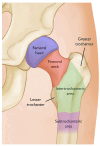
Figure 2
The areas of the femur. Most hip fractures occur in the femoral neck or intertrochanteric area. Reproduced with permission from Fischer, S and Gray, J, OrthoInfo; published by American Academy of Orthopedic Surgeons, 2020 [7].
Figure 3
Figure 3
This is an ultrasound image of a supra-inguinal fascia iliaca (SIFI) block. The white line represents the direction of the needle. The blue arrow represents the location of local anesthetic injection. IO (internal oblique muscle); AIIS (anterior inferior iliac spine); DCIA (deep circumflex iliac artery).
Figure 4
Figure 4
This is an ultrasound image of a femoral nerve block. The yellow line indicates the fascia iliaca plane. The blue line represents the needle trajectory. Local anesthetic is deposited below the nerve and above the nerve. FN (femoral nerve); FA (femoral artery); FV (femoral vein); * Nerve (ultrasound setting is in Nerve mode).
Figure 5
Figure 5
This is an ultrasound image of the lateral femoral cutaneous nerve (LFCN). The yellow arrow is pointing to the nerve. The nerve is blocked with a needle in the direction from lateral to medial.
Figure 6
Figure 6
This is an ultrasound image of the Pericapsular Nerve Group Block (PENG). Local anesthetic is injected below the psoas tendon. The needle approach is from lateral to medial. The femoral nerve (FN) and femoral artery (FA) is seen medially.
Figure 7
Figure 7
(a) Position of the patient during the posterior pericapsular deep-gluteal block. The ultrasound probe and needle are in line with the axis of the femur, with in-plane needle puncture from the posterior aspect. (b) Corresponding ultrasound image showing the gluteal muscles covering the bony landmarks: Fhead, femoral head; FN, femoral neck; GMax, gluteus maximus; GT, greater trochanter; IFL, ischiofemoral ligament as part of the posterior hip capsule; PAR, posterior acetabular rim; Pir, piriformis muscle. (c) Ultrasound image of the needle trajectory. Dashed line: needle trajectory; Blue arrows: spread of local anesthetic over the posterior acetabular rim and posterior hip capsule, deep to the piriformis muscle. Reproduced with permission from Vermeylen K et al., BJA Open; published by Elsevier, 2023 [48].

References

    1. Roberts K.C., Brox W.T., Jevsevar D.S., Sevarino K. Management of hip fractures in the elderly. J. Am. Acad. Orthop. Surg. 2015;23:131–137. doi: 10.5435/jaaos-d-14-00432. - DOI - PubMed
    1. Dixon J., Ashton F., Baker P., Charlton K., Bates C., Eardley W. Assessment and Early Management of Pain in Hip Fractures: The Impact of Paracetamol. Geriatr. Orthop. Surg. Rehabil. 2018;9:2151459318806443. doi: 10.1177/2151459318806443. - DOI - PMC - PubMed
    1. Ackermann L., Schwenk E.S., Lev Y., Weitz H. Update on medical management of acute hip fracture. Clevel. Clin. J. Med. 2021;88:237–247. doi: 10.3949/ccjm.88a.20149. - DOI - PubMed
    1. Downey C., Kelly M., Quinlan J.F. Changing trends in the mortality rate at 1-year post hip fracture—A systematic review. World J. Orthop. 2019;10:166–175. doi: 10.5312/wjo.v10.i3.166. - DOI - PMC - PubMed
    1. Guay J., Kopp S. Peripheral Nerve Blocks for Hip Fractures in Adults. Cochrane Database Syst. Rev. 2020;11:CD001159. doi: 10.1002/14651858.cd001159.pub3. - DOI - PMC - PubMed

LinkOut - more resources