Review
. 2024 Jun 13;13(12):3459.
doi: 10.3390/jcm13123459.

PET Molecular Imaging in Breast Cancer: Current Applications and Future Perspectives

Affiliations
Review

PET Molecular Imaging in Breast Cancer: Current Applications and Future Perspectives

Sanaz Katal et al. J Clin Med. .

Abstract

Positron emission tomography (PET) plays a crucial role in breast cancer management. This review addresses the role of PET imaging in breast cancer care. We focus primarily on the utility of 18F-fluorodeoxyglucose (FDG) PET in staging, recurrence detection, and treatment response evaluation. Furthermore, we delve into the growing interest in precision therapy and the development of novel radiopharmaceuticals targeting tumor biology. This includes discussing the potential of PET/MRI and artificial intelligence in breast cancer imaging, offering insights into improved diagnostic accuracy and personalized treatment approaches.

Keywords: FAPI; FDG; FDHT; FES; PET; PET/CT; PET/MRI; androgen receptor (AR); artificial intelligence (AI); breast cancer; deep learning (DL); estrogen receptor (ER); human epidermal growth factor receptor 2 (HER2); machine learning (ML); progesterone receptor (PR).


Conflict of interest statement

The authors declare no conflict of interest.

Figures

Figure 1
Figure 1
Lower FDG avidity in certain BC subtypes. Fused axial PET/CT (A), axial PET only (B), and MIP PET/CT (C) demonstrate low-grade FDG uptake in biopsy-proven left breast lobular carcinoma (arrow) in a 55-year-old patient. The lower SUVmax of invasive lobular carcinoma (ILC) primary tumors compared to invasive ductal carcinomas (IDCs) is attributed to factors such as lower tumoral cell density and reduced proliferation rates.
Figure 2
Figure 2
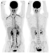
Physiologic brown fat uptake. MIP FDG PET/CT imaging (A) demonstrates extensive brown fat uptake, leading to a technically limited, non-diagnostic study in a 45-year-old patient. The patient returned for a repeat PET scan (B) on another date post-administration of beta-blockers.
Figure 3
Figure 3
FDG PET/CT in BC initial staging. PET/CT MIP images of two breast cancer patients (A,B) demonstrate intensely FDG-avid multifocal left breast cancer, with an unexpected extensive burden of regional and non-regional nodal disease as well as multifocal distant osseous and pulmonary metastases.
Figure 4
Figure 4
FDG PET/CT in BC nodal staging. The PET/CT MIP (A) and axial fused PET/CT (B,C) images demonstrate a hypermetabolic right breast primary (red arrow) with multiple hypermetabolic ipsilateral right level I/II axillary (white arrow) and internal mammary (green arrow) nodal metastases.
Figure 5
Figure 5
FDG PET/CT in BC nodal staging. The PET/CT MIP (A) and axial fused PET/CT (B,C) images demonstrate high FDG uptake within the left breast primary (red arrow) with solitary ipsilateral internal mammary nodal disease (white arrow) in a 55-year-old patient referred for staging of a newly diagnosed TNBC. Internal mammary node positivity is more common in centrally or medially located BCs.
Figure 6
Figure 6
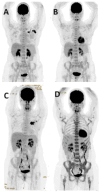
FDG PET/CT in response assessment. Baseline PET/CT MIP images (A,C) show FDG-avid left BC and ipsilateral axillary nodal metastases in two patients before neoadjuvant chemotherapy (NAC). Post-NAC PET/CT MIP images (B,D) demonstrate an excellent treatment response, with significantly reduced FDG uptake in the primary and axillary nodal metastases following neoadjuvant chemotherapy.

References

    1. Sung H., Ferlay J., Siegel R.L., Laversanne M., Soerjomataram I., Jemal A., Bray F. Global Cancer Statistics 2020: GLOBOCAN Estimates of Incidence and Mortality Worldwide for 36 Cancers in 185 Countries. CA Cancer J. Clin. 2021;71:209–249. - PubMed
    1. Giaquinto A.N., Sung H., Miller K.D., Kramer J.L., Newman L.A., Minihan A., Jemal A., Siegel R.L. Breast Cancer Statistics, 2022. CA Cancer J. Clin. 2022;72:524–541. doi: 10.3322/caac.21754. - DOI - PubMed
    1. [(accessed on 20 March 2024)]; Available online: https://www.canceraustralia.gov.au/cancer-types/breast-cancer/statistics.
    1. Taori K., Dhakate S., Rathod J., Hatgaonkar A., Disawal A., Wavare P., Bakare V., Puri R.P. Evaluation of breast masses using mammography and sonography as first line investigations. Open J. Med. Imaging. 2013;3:40–49. doi: 10.4236/ojmi.2013.31006. - DOI
    1. Bevers T.B., Niell B.L., Baker J.L., Bennett D.L., Bonaccio E., Camp M.S., Stehman K. NCCN Guidelines® Insights: Breast Cancer Screening and Diagnosis, Version 1.2023. J. Natl. Compr. Cancer Netw. 2023;21:900–909. doi: 10.6004/jnccn.2023.0046. - DOI - PubMed