. 2024 Jun 19;13(12):3598.
doi: 10.3390/jcm13123598.

Normative Values for Sternoclavicular Joint and Clavicle Anatomy Based on MR Imaging: A Comprehensive Analysis of 1591 Healthy Participants

Affiliations

Normative Values for Sternoclavicular Joint and Clavicle Anatomy Based on MR Imaging: A Comprehensive Analysis of 1591 Healthy Participants

Theo Morgan Languth et al. J Clin Med. .

Abstract

Background: The clavicle remains one of the most fractured bones in the human body, despite the fact that little is known about the MR imaging of it and the adjacent sternoclavicular joint. This study aims to establish standardized values for the diameters of the clavicle as well as the angles of the sternoclavicular joint using whole-body MRI scans of a large and healthy population and to examine further possible correlations between diameters and angles and influencing factors like BMI, weight, height, sex, and age. Methods: This study reviewed whole-body MRI scans from the Study of Health in Pomerania (SHIP), a German population-based cross-sectional study in Mecklenburg-Western Pomerania. Descriptive statistics, as well as median-based regression models, were used to evaluate the results. Results: We could establish reference values based on a shoulder-healthy population for each clavicle parameter. Substantial differences were found for sex. Small impacts were found for height, weight, and BMI. Less to no impact was found for age. Conclusions: This study provides valuable reference values for clavicle and sternoclavicular joint-related parameters and shows the effects of epidemiological features, laying the groundwork for future studies. Further research is mandatory to determine the clinical implications of these findings.

Keywords: MRI diagnostics; SHIP; clavicular anatomy; reference values; sternoclavicular joint.


Conflict of interest statement

The authors declare no conflicts of interest. The funders had no influence in the design of the study, in the collection, analysis, or interpretation of data, in the writing of the manuscript, or in the decision to publish the results.

Figures

Figure 1
Figure 1
How to measure the sternal angle (SA): Angle between the articular surfaces of the sternoclavicular joint and the sternum, utilizing tangents drawn from specific points, T1 weighting the 3D frontal layer with a slice thickness of 3 mm.
Figure 2
Figure 2
How to measure clavicular angle (CA): angle representing the inclination between the articular surface of the SCJ and the clavicle, derived from tangents and forming the acute angle beta, T1 weighting the 3D frontal layer with a slice thickness of 3 mm.
Figure 3
Figure 3
How to measure the clavicular—sternal angle (CSB): The vertical axis of the sternum, deriving from a line drawn from the middle point of the cranial end of the manubrium to the xiphoid process, and the lengthwise axis of the clavicle, drawn from the middle of the lateral clavicle to the middle of the medial clavicle, produced the obtuse angle gamma. T1 weights the 3D frontal layer with a slice thickness of 3 mm.
Figure 4
Figure 4
Schematic figure of the superior view of the clavicle bone illustrates the measurement areas and examples of measurement for A = maximal lateral diameter, B = minimal diameter, and C = maximal medial diameter.
Figure 5
Figure 5
How to measure the max. medial diameter (MAXMCL): Measured at the medial end of the clavicle from the furthest ventral to dorsal points with an orthogonal line to the clavicle axis, T1 weighting has a slice thickness of 3 mm (C).
Figure 6
Figure 6
How to measure the min. diameter (MINCL): Measured similarly to MAXMCL but focusing on the least distant ventral to dorsal points of the medial clavicle, T1 weighting has a slice thickness of 3 mm (B).
Figure 7
Figure 7
How to measure the max. lateral diameter (MAXLCL): Measured at the lateral end of the clavicle, from the furthest ventral to dorsal points, with an orthogonal line to the clavicle axis, T1 weighting has a slice thickness of 3 mm (A).
Figure 8
Figure 8
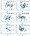
(ag) Bland–Altman plots for interreader variability: (a) Clavicular angle (CA, n = 177), (b) clavicular–sternal angle (CSB, n = 174), (c) sternal angle (SA, n = 176), (d) max. medial diameter (Max MCL, n = 189), (e) max. lateral diameter (MaxClavR, n = 104), (f) max. clavicular diameter (MaxCL, n = 196), (g) minimal clavicular diameter (MinCL, n = 174).
Figure 8
Figure 8
(ag) Bland–Altman plots for interreader variability: (a) Clavicular angle (CA, n = 177), (b) clavicular–sternal angle (CSB, n = 174), (c) sternal angle (SA, n = 176), (d) max. medial diameter (Max MCL, n = 189), (e) max. lateral diameter (MaxClavR, n = 104), (f) max. clavicular diameter (MaxCL, n = 196), (g) minimal clavicular diameter (MinCL, n = 174).
Figure 9
Figure 9
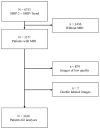
Selection of the participants (composition of analyzed MRI scans and number of rolled-out patients).
Figure 10
Figure 10
Excluded patients for the reference table due to the exclusion criteria in order to define a shoulder-healthy population.
Figure 11
Figure 11
Showing the Pearson coefficient for all measured variables by comparing the right and left sides.

Similar articles

References

    1. OECD and European Union . Availability and Use of Diagnostic Technologies. OECD; Paris, France: 2018. - DOI
    1. Guglielmi G., Cascavilla A., Scalzo G., Salaffi F., Grassi W. Imaging of sternocostoclavicular joint in spondyloarthropaties and other rheumatic conditions. [(accessed on 2 June 2024)];Clin. Exp. Rheumatol. 2009 27:402–408. Available online: http://www.ncbi.nlm.nih.gov/pubmed/19604431. - PubMed
    1. Restrepo C.S., Martinez S., Lemos D.F., Washington L., McAdams H.P., Vargas D., Lemos J.A., Carrillo J.A., Diethelm L. Imaging Appearances of the Sternum and Sternoclavicular Joints. RadioGraphics. 2009;29:839–859. doi: 10.1148/rg.293055136. - DOI - PubMed
    1. Farpour F., Phan S.J., Burns J., Tehranzadeh J. Enhanced MR imaging of the shoulder, and sternoclavicular and acromioclavicular joint arthritis in primary hemochromatosis. Rheumatol. Int. 2011;31:395–398. doi: 10.1007/s00296-009-1193-z. - DOI - PMC - PubMed
    1. Johnson M.C., Jacobson J.A., Fessell D.P., Kim S.M., Brandon C., Caoili E. The sternoclavicular joint: Can imaging differentiate infection from degenerative change? Skelet. Radiol. 2010;39:551–558. doi: 10.1007/s00256-009-0802-y. - DOI - PubMed

LinkOut - more resources