

The Chimeric LFC and DCIA Flap in Combined Mandibular and Condylar Head and Neck Reconstruction-A Case Series
- PMID: 38930140
- PMCID: PMC11204853
- DOI: 10.3390/jcm13123613
The Chimeric LFC and DCIA Flap in Combined Mandibular and Condylar Head and Neck Reconstruction-A Case Series
Abstract
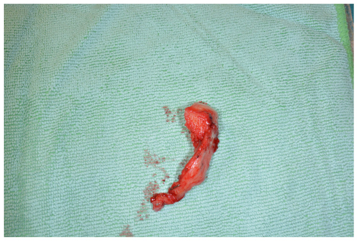
Background: Defects of the ascending ramus of the mandible, including the condylar head and neck or the whole temporomandibular joint (TMJ), are difficult to reconstruct. Reconstruction is mainly based on the use of alloplastic joint prosthesis, costochondral grafting, distraction osteogenesis of the dorsal part of the mandibular ramus, or osseous microvascular flaps of various origin. With the objective of developing a method that overcomes the restrictions of these methods, we recently introduced a sequential chimeric flap consisting of a lateral femoral condyle flap (LFC) and deep circumflex iliac artery flap (DCIA) for reconstruction of up to half of the mandible and the condylar head and neck. Methods: The chimeric flap was used in four patients with the following diagnoses: therapy-refractory osteomyelitis, extended recurrent odontogenic keratozyst, Goldenhar syndrome, and adenocarcinoma of the parotid gland. After a diagnostic workup, LFC and DCIA flaps were harvested in all patients and used in a sequential chimeric design for the reconstruction of the mandibular body and condylar head and neck. Results: Follow-up from at least 24 months up to 70 month after surgery showed a successful reconstruction in all four patients. The LFC provided a cartilaginous joint surface, allowing for a satisfactory masticatory function with a stable occlusion and unrestricted mouth opening and preserved or regained lateral and medial excursions in all patients. The DCIA allowed for a bony reconstruction anatomically resembling a non-atrophied mandibular body. No flap-related complications were observed. Conclusions: The sequential chimeric LFC and DCIA flap is an appropriate method for reconstructing up to half of the mandible and the condylar head and neck. It is suitable in cases where alloplastic joint replacement cannot be used or where other methods have failed. Due to the necessity of harvesting two flaps, the burden of care is increased, and a careful indication is required. The technique is reserved for maxillofacial surgeons who have already gained significant experience in the field of microsurgery.
Keywords: DCIA flap; TMJ reconstruction; chimera flap; lateral femoral condyle flap; mandibular reconstruction; maxillofacial surgery; plastic and reconstructive surgery.
Conflict of interest statement
The authors declare no conflicts of interest.
Figures


References
LinkOut - more resources
Full Text Sources
