

Manufacturing of 3D-Printed Hybrid Scaffolds with Polyelectrolyte Multilayer Coating in Static and Dynamic Culture Conditions
- PMID: 38930181
- PMCID: PMC11205028
- DOI: 10.3390/ma17122811
Manufacturing of 3D-Printed Hybrid Scaffolds with Polyelectrolyte Multilayer Coating in Static and Dynamic Culture Conditions
Abstract
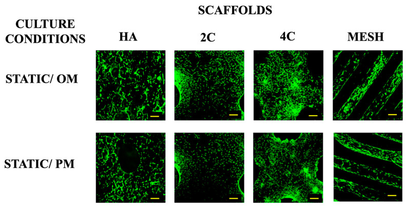
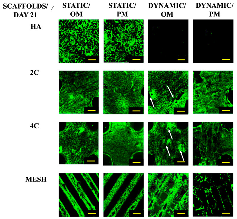
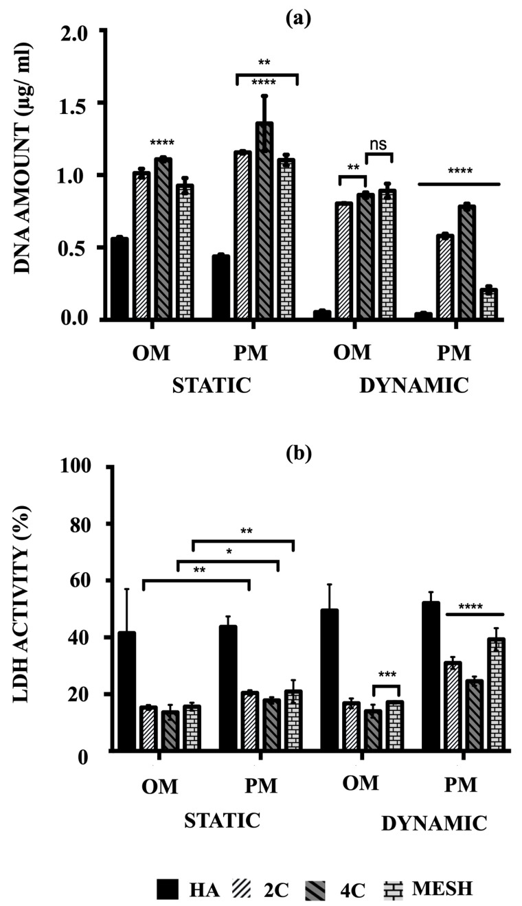
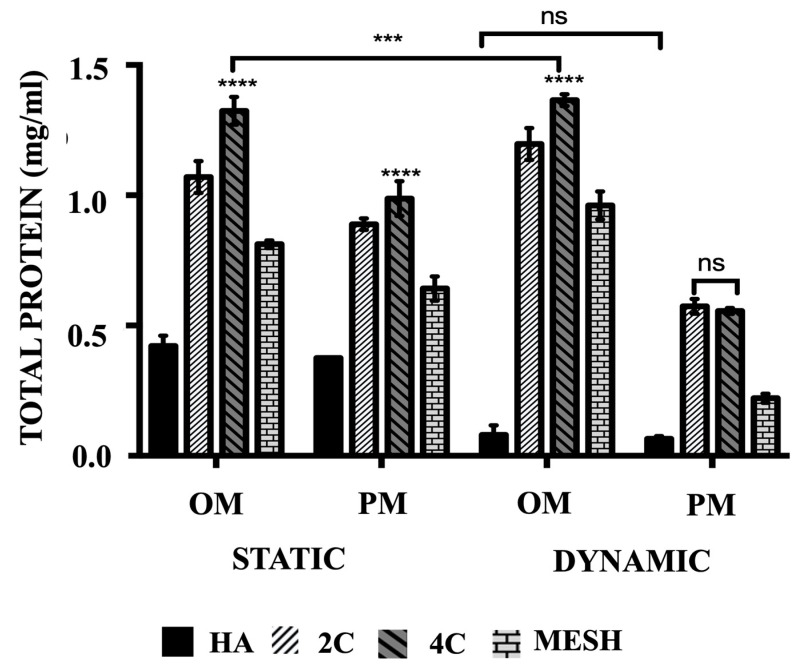
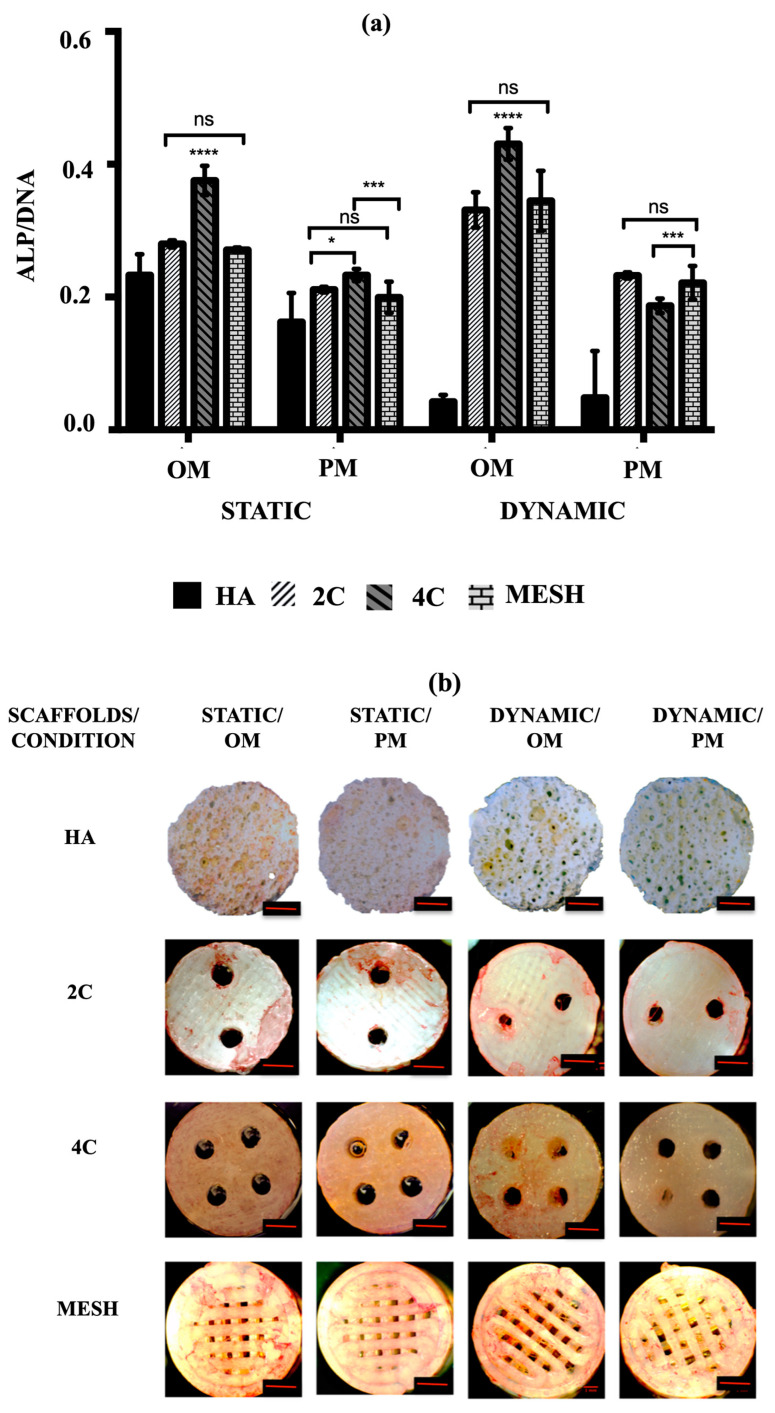
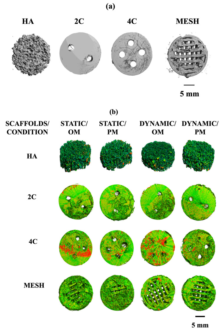
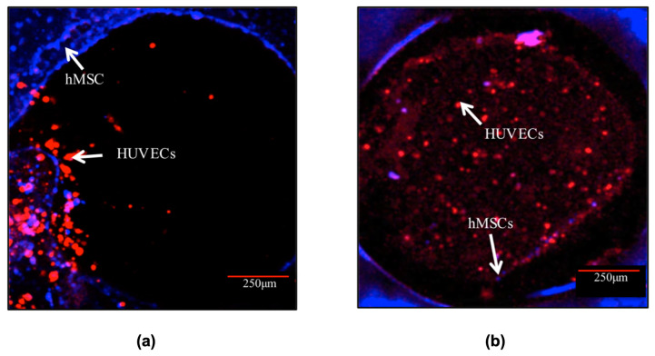
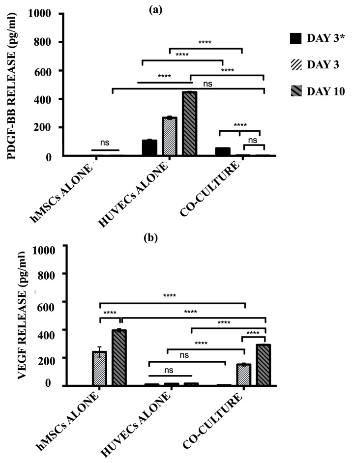
Three-dimensional printing (3DP) has emerged as a promising method for creating intricate scaffold designs. This study assessed three 3DP scaffold designs fabricated using biodegradable poly(lactic) acid (PLA) through fused deposition modelling (FDM): mesh, two channels (2C), and four channels (4C). To address the limitations of PLA, such as hydrophobic properties and poor cell attachment, a post-fabrication modification technique employing Polyelectrolyte Multilayers (PEMs) coating was implemented. The scaffolds underwent aminolysis followed by coating with SiCHA nanopowders dispersed in hyaluronic acid and collagen type I, and finally crosslinked the outermost coated layers with EDC/NHS solution to complete the hybrid scaffold production. The study employed rotating wall vessels (RWVs) to investigate how simulating microgravity affects cell proliferation and differentiation. Human mesenchymal stem cells (hMSCs) cultured on these scaffolds using proliferation medium (PM) and osteogenic media (OM), subjected to static (TCP) and dynamic (RWVs) conditions for 21 days, revealed superior performance of 4C hybrid scaffolds, particularly in OM. Compared to commercial hydroxyapatite scaffolds, these hybrid scaffolds demonstrated enhanced cell activity and survival. The pre-vascularisation concept on 4C hybrid scaffolds showed the proliferation of both HUVECs and hMSCs throughout the scaffolds, with a positive expression of osteogenic and angiogenic markers at the early stages.
Keywords: 3DP hybrid scaffolds; co-culture; fused deposition modelling; polyelectrolyte multilayers; pre-vascularisation; rotating wall vessels; stem cells.
Conflict of interest statement
The authors declare no conflicts of interest. The funders had no role in the design of the study; in the collection, analyses, or interpretation of data; in the writing of the manuscript; or in the decision to publish the results.
Figures
Similar articles
-
Polyelectrolyte multi-layers assembly of SiCHA nanopowders and collagen type I on aminolysed PLA films to enhance cell-material interactions.Colloids Surf B Biointerfaces. 2017 Nov 1;159:445-453. doi: 10.1016/j.colsurfb.2017.07.086. Epub 2017 Aug 14. Colloids Surf B Biointerfaces. 2017. PMID: 28837894
-
Development of mussel-inspired 3D-printed poly (lactic acid) scaffold grafted with bone morphogenetic protein-2 for stimulating osteogenesis.J Mater Sci Mater Med. 2019 Jun 20;30(7):78. doi: 10.1007/s10856-019-6279-x. J Mater Sci Mater Med. 2019. PMID: 31222566
-
Fused Deposition Modeling Printed PLA/Nano β-TCP Composite Bone Tissue Engineering Scaffolds for Promoting Osteogenic Induction Function.Int J Nanomedicine. 2023 Oct 17;18:5815-5830. doi: 10.2147/IJN.S416098. eCollection 2023. Int J Nanomedicine. 2023. PMID: 37869064 Free PMC article.
-
Towards resorbable 3D-printed scaffolds for craniofacial bone regeneration.Orthod Craniofac Res. 2023 Dec;26 Suppl 1:188-195. doi: 10.1111/ocr.12645. Epub 2023 Mar 13. Orthod Craniofac Res. 2023. PMID: 36866957 Review.
-
Design and fabrication of porous biodegradable scaffolds: a strategy for tissue engineering.J Biomater Sci Polym Ed. 2017 Nov;28(16):1797-1825. doi: 10.1080/09205063.2017.1354674. Epub 2017 Jul 24. J Biomater Sci Polym Ed. 2017. PMID: 28707508 Review.
References
LinkOut - more resources
Full Text Sources
