Review
. 2024 May 29;15(6):720.
doi: 10.3390/mi15060720.

Cell Migration Assays and Their Application to Wound Healing Assays-A Critical Review

Affiliations
Review

Cell Migration Assays and Their Application to Wound Healing Assays-A Critical Review

Chun Yang et al. Micromachines (Basel). .

Abstract

In recent years, cell migration assays (CMAs) have emerged as a tool to study the migration of cells along with their physiological responses under various stimuli, including both mechanical and bio-chemical properties. CMAs are a generic system in that they support various biological applications, such as wound healing assays. In this paper, we review the development of the CMA in the context of its application to wound healing assays. As such, the wound healing assay will be used to derive the requirements on CMAs. This paper will provide a comprehensive and critical review of the development of CMAs along with their application to wound healing assays. One salient feature of our methodology in this paper is the application of the so-called design thinking; namely we define the requirements of CMAs first and then take them as a benchmark for various developments of CMAs in the literature. The state-of-the-art CMAs are compared with this benchmark to derive the knowledge and technological gap with CMAs in the literature. We will also discuss future research directions for the CMA together with its application to wound healing assays.

Keywords: cell migration assay; system and design perspective; wound healing assay.


Conflict of interest statement

The authors declare no conflicts of interest in this work.

Figures

Figure 1
Figure 1
Various CMAs based on the mechanical depletion approach: (a) a pipette removing cells manually on a dish plate of cell culture [10], reproduced with the permission of [37]; (b) multiple pipettes creating eight CFZs simultaneously based on a commercially available CMA device called OrisTM, adapted with the permission of [28]; (c) stamping devices applying consistent forces on cells to improve consistency in the size of CFZs, adapted with the permission of [27]; (d) a microfluidic device creating CFZs automatically with the pneumatic actuation by deflecting a flexible membrane, adapted with the permission of [29]; (e) a microfluidic device achieving a high consistency of CFZs with various forces, adapted with the permission of [30]; and (f) a microfluidic device creating 400 CFZs at one operation in a shear stress condition, adapted with the permission of [31].
Figure 2
Figure 2
A magnetically controlled robot creating CFZs, adapted with permission from [32]: (a) cells cultured on a PDMS-based microfluidic chip; (b) the driving robot controls the scratch robot magnetically: (I) initial position of driving robot (green) and scratching robot (red), (II) the scratching robot driven by the driving robot starts moving, (III) the driving force required to move the scratch robot can be adjusted by the distance between the two robots horizontally (∆x) and vertically (∆h); (c) schematics of different geometries of CFZs (line wound, plus wound, rectangle wound, and triangle wound); and (d) images of line-shaped wounds at different time intervals. Legend white bar is equal to 400 μm.
Figure 3
Figure 3
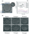
Structural design of a bio-inspired cell chamber for studying the uniformity of flow distribution, adapted with permission from [56]: (a) a cell chamber in octagonal shape; (b) cell chambers with different shapes including square, pentagon, hexagon, heptagon, nonagon, and decagon; and (c) the velocities varying in different shapes of cell chambers with the same inlet velocity. The scale bar is 300 μm.
Figure 4
Figure 4
Various signal molecules in the deformation pathway: a protein network (i.e., talin, vinculin, paxillin, focal adhesion kinase (FAK), vasodilator stimulated phosphoprotein (VASP), and other proteins) interconnected between cells and the extracellular matrix (ECM). Adapted with permission from [74].
Figure 5
Figure 5
Schematic diagram of the structural design aspects of engineered biomimetic materials in macroscale, microscale, and nanoscale: the design aspects in macroscale are related to external structural characteristics, such as overall shape and size; the design aspects in microscale include microwells, micropores, microchannels, microgels, and microfibers; and the design aspects in nanoscale include nanofibers and nanoparticles. Adapted with permission from [91].

References

    1. Bhatia S.N., Ingber D.E. Microfluidic organs-on-chips. Nat. Biotechnol. 2014;32:760–772. doi: 10.1038/nbt.2989. - DOI - PubMed
    1. Junaid A., Mashaghi A., Hankemeier T., Vulto P. An end-user perspective on Organ-on-a-Chip: Assays and usability aspects. Curr. Opin. Biomed. Eng. 2017;1:15–22. doi: 10.1016/j.cobme.2017.02.002. - DOI
    1. MacQueen L., Sun Y., Simmons C.A. Mesenchymal stem cell mechanobiology and emerging experimental platforms. J. R. Soc. Interface. 2013;10:20130179. doi: 10.1098/rsif.2013.0179. - DOI - PMC - PubMed
    1. Rothbauer M., Zirath H., Ertl P. Recent advances in microfluidic technologies for cell-to-cell interaction studies. Lab. Chip. 2018;18:249–270. doi: 10.1039/c7lc00815e. - DOI - PubMed
    1. Pijuan J., Barceló C., Moreno D.F., Maiques O., Sisó P., Marti R.M., Macià A., Panosa A. Cell Migration, Invasion, and Adhesion Assays: From Cell Imaging to Data Analysis. Front. Cell Dev. Biol. 2019;7:107. doi: 10.3389/fcell.2019.00107. - DOI - PMC - PubMed