. 2024 May 21;16(6):817.
doi: 10.3390/v16060817.

Investigating the Genetic Diversity of Hepatitis Delta Virus in Hepatocellular Carcinoma (HCC): Impact on Viral Evolution and Oncogenesis in HCC

Affiliations

Investigating the Genetic Diversity of Hepatitis Delta Virus in Hepatocellular Carcinoma (HCC): Impact on Viral Evolution and Oncogenesis in HCC

Horng-Heng Juang et al. Viruses. .

Abstract

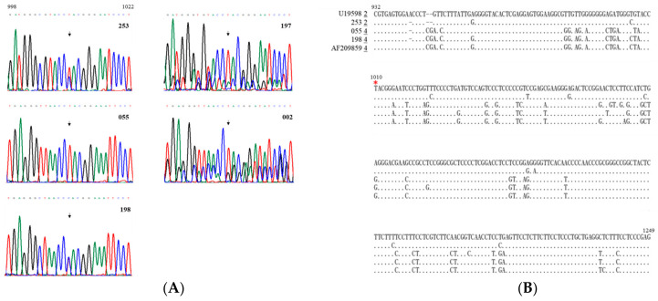
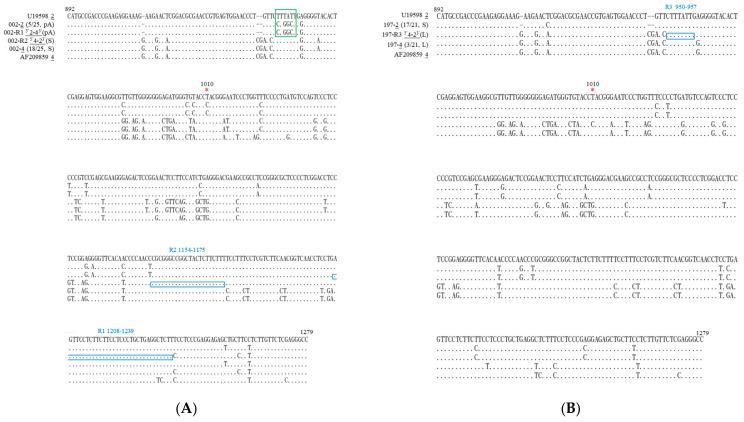
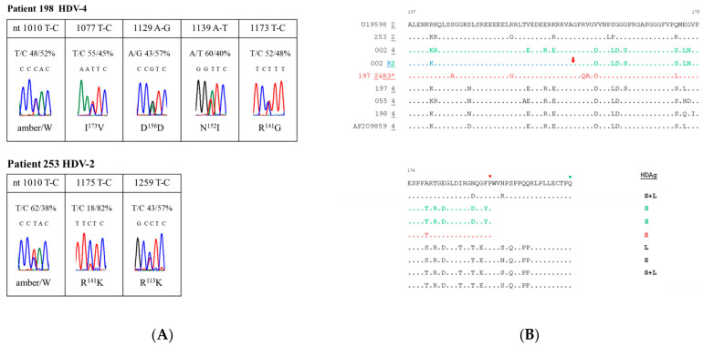
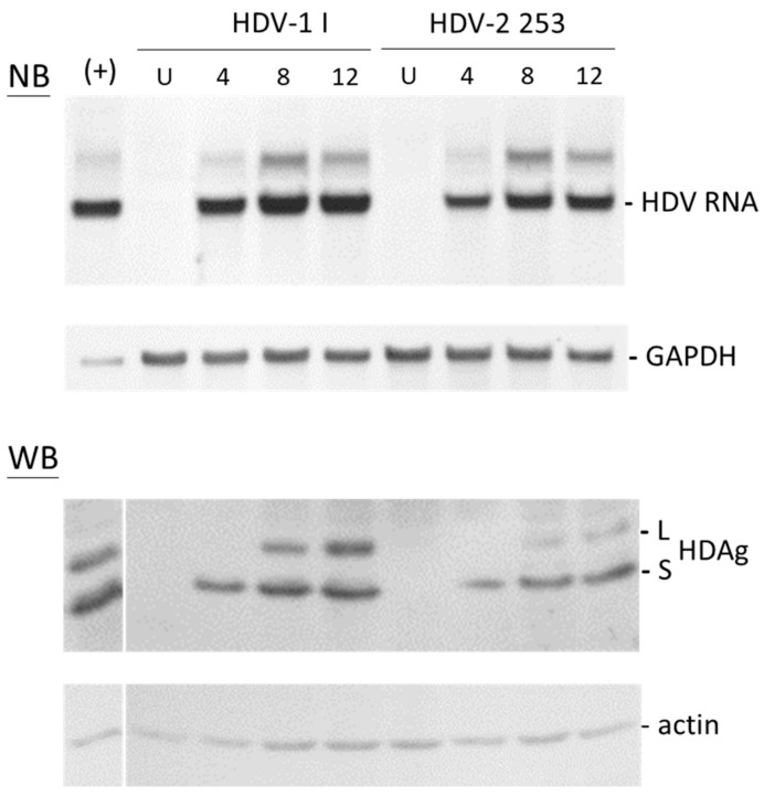
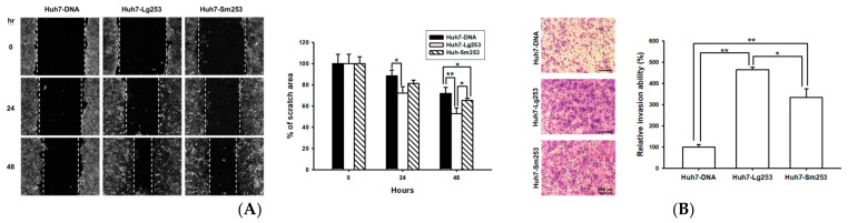
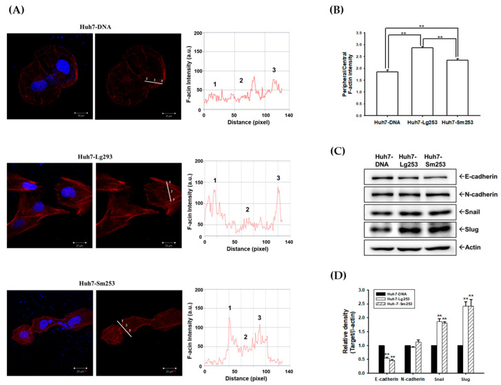
Hepatitis delta virus (HDV), an RNA virus with two forms of the delta antigen (HDAg), relies on hepatitis B virus (HBV) for envelope proteins essential for hepatocyte entry. Hepatocellular carcinoma (HCC) ranks third in global cancer deaths, yet HDV's involvement remains uncertain. Among 300 HBV-associated HCC serum samples from Taiwan's National Health Research Institutes, 2.7% (8/300) tested anti-HDV positive, with 62.7% (5/8) of these also HDV RNA positive. Genotyping revealed HDV-2 in one sample, HDV-4 in two, and two samples showed mixed HDV-2/HDV-4 infection with RNA recombination. A mixed-genotype infection revealed novel mutations at the polyadenylation signal, coinciding with the ochre termination codon for the L-HDAg. To delve deeper into the possible oncogenic properties of HDV-2, the predominant genotype in Taiwan, which was previously thought to be less associated with severe disease outcomes, an HDV-2 cDNA clone was isolated from HCC for study. It demonstrated a replication level reaching up to 74% of that observed for a widely used HDV-1 strain in transfected cultured cells. Surprisingly, both forms of HDV-2 HDAg promoted cell migration and invasion, affecting the rearrangement of actin cytoskeleton and the expression of epithelial-mesenchymal transition markers. In summary, this study underscores the prevalence of HDV-2, HDV-4, and their mixed infections in HCC, highlighting the genetic diversity in HCC as well as the potential role of both forms of the HDAg in HCC oncogenesis.

Keywords: RNA recombination; cell migration and invasion; delta antigen; hepatitis delta virus; hepatocellular carcinoma.


Conflict of interest statement

The authors declare no conflicts of interest.

Figures

Figure 1
Figure 1
HDV-related sequences in serum samples from HCC patients. (A) The sequencing chromatogram spanning nucleotides 998 to 1022. Each of the DNA nucleotides adenine (A, green), cytosine (C, blue), guanine (G, black), and thymine (T, red), have their own color on the chromatogram for visualization. The sequences are summarized above the sequencing chromatograms. RNA samples obtained from patients’ sera underwent RT-PCR, followed by direct sequencing of a region encompassing the amber/W RNA editing site, marked by arrowheads. (B) Sequences spanning nucleotides 932 to 1249, acquired through direct sequencing of PCR products from HCC patients 253, 055, and 198, with conserved nucleotides indicated by dots. Reference isolates for HDV-2 and HDV-4 are Taiwanese T3 (GenBank accession number U19598) [34] and Taiwan-IIb-1 (GenBank accession number AF209859) clones, respectively [32]. The genotype of each clone is underlined and presented preceding the sequence. The amber/W editing site is highlighted with a red star.
Figure 2
Figure 2
HDV-associated sequences found in patients 002 (A) and 197 (B) exhibiting mixed-genotype infection. Alignment of representative HDV sequences (nucleotides 892–1279) is presented, obtained via cloning and sequencing of PCR products. In patients 002 (A) and 197 (B), two (R1 and R2) and one (R3) recombinants were identified, respectively. The genotype of each clone is underlined. The crossover regions are indicated by blue rectangles, while the amber/W editing site is denoted by a red star. The polyadenylation signal and the sequence of the pA mutant of HDV-2 in patient 002 are depicted within a green rectangle.
Figure 3
Figure 3
Amino acid sequences of the HDAg in the sera of HCC patients. (A) An overview of sequence variations, recognized as double peaks in sequencing chromatograms of patients 198 and 253. Only minor peaks, accounting for over 10% of the population, were depicted in this analysis. Percentages of specific nucleotide (nt) residues and resulting amino acid changes are summarized above and below each sequencing chromatogram. (B) Alignment displaying the amino acid sequences at the C-terminus of different HDAg clones discovered in five HCC patients. Each patient’s most conserved sequences are depicted, with dots indicating amino acids identical to those of the reference strain, U19598 [34]. Red arrows highlight crossover regions. The last amino acid positions for S- and L-HDAg are marked with a red and green triangle, respectively. Summaries of the HDAg variants encoded by HDV sequences associated with HCC are presented alongside the end of the amino acid sequences. *: the amino acid sequences of HDV-2 and R3 from patient 197 are identical.
Figure 4
Figure 4
Time course of HDV-1 I and HDV-2 253 in HuH-7 cells following transfection with a genome expression plasmid. RNA and proteins were collected at specified intervals post-transfection. Analysis of HDV antigenomic RNA and HDAg was conducted by NB and WB assays, respectively. The uniform loading of RNA and protein samples was verified through hybridization with a glyceraldehyde-3-phosphate dehydrogenase (GAPDH) probe and an actin-specific antibody, respectively. U: untransfected cells.
Figure 5
Figure 5
Impact of HCC serum-derived Sm253 and Lg253 expression on cell migration and invasion in Huh7 cells. (A) Both Sm253 and Lg253 promote cell migration. Huh7 cells overexpressing either Sm253 or Lg253 were evaluated through a wound healing assay. Control cells were transfected with a vector. Images were captured at 0, 24, and 48 h, with the dotted line denoting the average leading edge of cellular migration. ImageJ software was utilized to measure the wound area, and the data are presented as mean percentages (±SE; n = 4) relative to the control cells. (B) Transwell invasion assays were conducted to assess the invasion capacities of Sm253 and Lg253. Images were acquired at 24 h and a representative image from three independent experiments was displayed. Data are depicted as mean percentages (±SE, n = 3) compared to the control cells. * indicates p < 0.05, ** indicates p < 0.01.
Figure 6
Figure 6
Sm253 and Lg253 modulate the F-actin distribution and expression of EMT markers. (A) The F-actin was stained using Texas Red X-Phalloidin, with the nuclei counterstained with DAPI (blue), and the immunofluorescence was captured using a confocal microscope. (B) The quantitative analysis of the F-actin fluorescence intensity. The intensities were measured along the line from the peripheral to the center of the cells, and the quantitative analyses of the F-actin fluorescence intensity of control cells and cells expressing Lg253 and Sm253 are shown (± SE, n = 6). ** indicates p < 0.01. (C) Immunoblot assays determined E-cadherin, N-cadherin, Snail, and Slug protein levels. (D) The quantitative analyses of the immunoblots are presented as a relative density of EMT markers. Actin protein level was determined to compare the total protein levels in each lane. ** indicates p < 0.01.

Similar articles

Cited by

References

    1. Rizzetto M., Hoyer B., Canese M.G., Shih J.W., Purcell R.H., Gerin J.L. Delta Agent: Association of delta antigen with hepatitis B surface antigen and RNA in serum of delta-infected chimpanzees. Proc. Natl. Acad. Sci. USA. 1980;77:6124–6128. doi: 10.1073/pnas.77.10.6124. - DOI - PMC - PubMed
    1. Yan H., Zhong G., Xu G., He W., Jing Z., Gao Z., Huang Y., Qi Y., Peng B., Wang H., et al. Sodium taurocholate cotransporting polypeptide is a functional receptor for human hepatitis B and D virus. eLife. 2012;1:e00049. doi: 10.7554/eLife.00049. - DOI - PMC - PubMed
    1. Chen H.Y., Shen D.T., Ji D.Z., Han P.C., Zhang W.M., Ma J.F., Chen W.S., Goyal H., Pan S., Xu H.G. Prevalence and burden of hepatitis D virus infection in the global population: A systematic review and meta-analysis. Gut. 2019;68:512–521. doi: 10.1136/gutjnl-2018-316601. - DOI - PubMed
    1. Miao Z., Zhang S., Ou X., Li S., Ma Z., Wang W., Peppelenbosch M.P., Liu J., Pan Q. Estimating the global prevalence, disease progression, and clinical outcome of hepatitis delta virus infection. J. Infect. Dis. 2020;221:1677–1687. doi: 10.1093/infdis/jiz633. - DOI - PMC - PubMed
    1. Stockdale A.J., Kreuels B., Henrion M.Y.R., Giorgi E., Kyomuhangi I., de Martel C., Hutin Y., Geretti A.M. The global prevalence of hepatitis D virus infection: Systematic review and meta-analysis. J. Hepatol. 2020;73:523–532. doi: 10.1016/j.jhep.2020.04.008. - DOI - PMC - PubMed

Publication types

Associated data