. 2024 May 24;16(6):832.
doi: 10.3390/v16060832.

Acute and Long COVID Intestinal Changes in an Experimental Model of Coronavirus in Mice

Affiliations

Acute and Long COVID Intestinal Changes in an Experimental Model of Coronavirus in Mice

Hussain Hussain et al. Viruses. .

Abstract

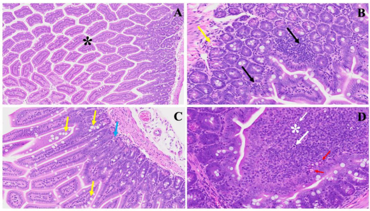
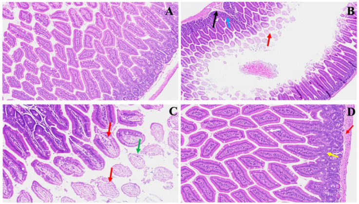
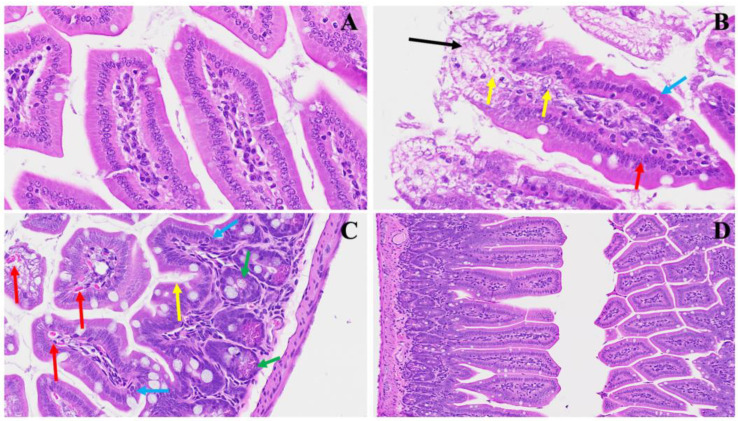
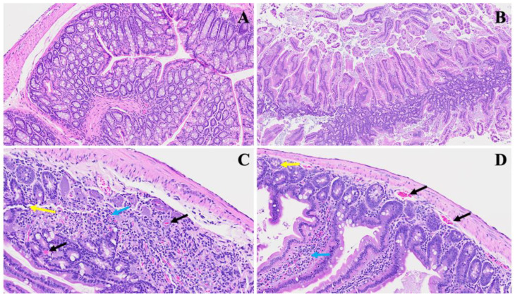
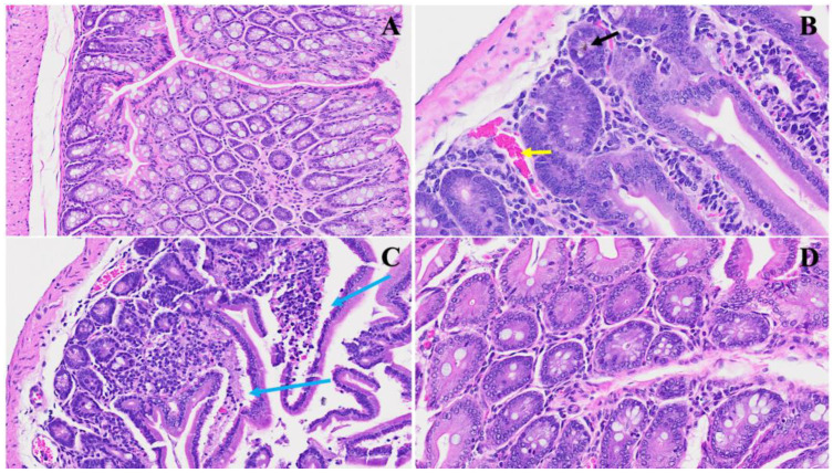
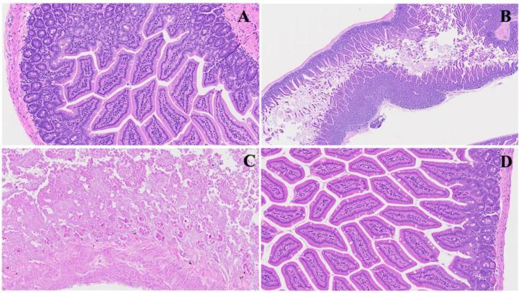
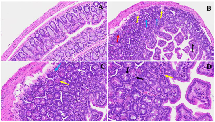
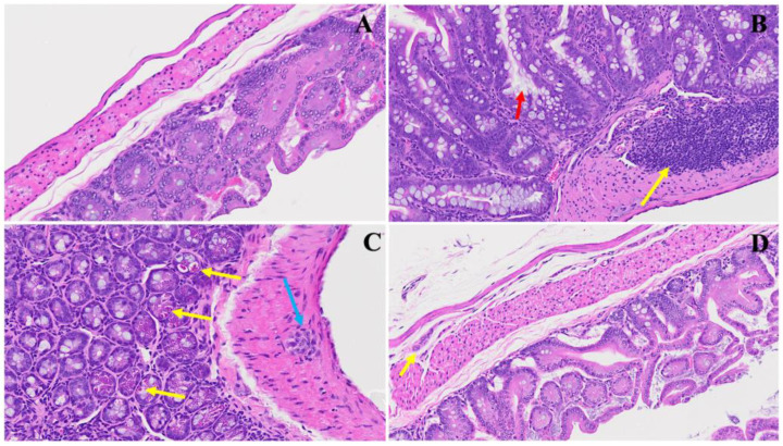
The COVID-19 pandemic, which emerged in early 2020, has had a profound and lasting impact on global health, resulting in over 7.0 million deaths and persistent challenges. In addition to acute concerns, there is growing attention being given to the long COVID health consequences for survivors of COVID-19 with documented cases of cardiovascular abnormalities, liver disturbances, lung complications, kidney issues, and noticeable cognitive deficits. Recent studies have investigated the physiological changes in various organs following prolonged exposure to murine hepatitis virus-1 (MHV-1), a coronavirus, in mouse models. One significant finding relates to the effects on the gastrointestinal tract, an area previously understudied regarding the long-lasting effects of COVID-19. This research sheds light on important observations in the intestines during both the acute and the prolonged phases following MHV-1 infection, which parallel specific changes seen in humans after exposure to SARS-CoV-2. Our study investigates the histopathological alterations in the small intestine following MHV-1 infection in murine models, revealing significant changes reminiscent of inflammatory bowel disease (IBD), celiac disease. Notable findings include mucosal inflammation, lymphoid hyperplasia, goblet cell hyperplasia, and immune cell infiltration, mirroring pathological features observed in IBD. Additionally, MHV-1 infection induces villous atrophy, altered epithelial integrity, and inflammatory responses akin to celiac disease and IBD. SPIKENET (SPK) treatment effectively mitigates intestinal damage caused by MHV-1 infection, restoring tissue architecture and ameliorating inflammatory responses. Furthermore, investigation into long COVID reveals intricate inflammatory profiles, highlighting the potential of SPK to modulate intestinal responses and restore tissue homeostasis. Understanding these histopathological alterations provides valuable insights into the pathogenesis of COVID-induced gastrointestinal complications and informs the development of targeted therapeutic strategies.

Keywords: fibrosis; goblet cell; infection; intestine; long COVID; murine hepatitis virus-1; pili.


Conflict of interest statement

Paidas is a Scientific Advisory Board Member of BioIncept, LLC, with stock options. The other authors have no conflicts of interest or competing interests to declare.

Figures

Figure 1
Figure 1
This illustrates the histopathological changes observed in acute extensive intestinal inflammation in the MHV-1 model in small and large intestines. In Panel (A), the typical architecture of the large intestine is depicted, with intact villi highlighted by a star. Panel (B) demonstrates colonic mucosal inflammation and lymphoid hyperplasia, indicated by black arrows, accompanied by microthrombi marked by yellow arrows. In Panel (C), hyperplasia of goblet cells scattered throughout the colonic mucosa is shown (yellow arrows), along with various inflammatory cells within the lamina propria (blue arrow). Panel (D) reveals diffuse proliferation of lymphoid tissue (white star), microbleeding (red arrow), and melanocytosis (white arrows) in the colonic mucosa. These findings provide insight into the pathological features associated with acute significant intestinal changes in the MHV-1 model. (H&E, original magnification 66× (AD)). MHV-1 infection alone (n = 16), healthy control (n = 7), and SPK-treated mice (n = 5).
Figure 2
Figure 2
This shows acute small and large intestinal changes induced by MHV-1 infection. In Panel (A), a histological section displays the typical regular colonic layers. Panel (B) exhibits early manifestations of disease, with evident mucosal layer sloughing (red arrow), concomitant inflammatory alterations in the muscularis mucosa layer (black arrow), and overall inflammation (blue arrow). Panel (C) depicts diverse stages of villus degeneration (red arrows) alongside the presence of crypt apoptotic bodies (green arrow). In Panel (D), regenerative responses during the acute phase of SPK treatment are evident, including the reconstruction of the muscularis mucosa layer (red arrow), normalization of goblet cells (white arrow), restoration of crypt and lamina propria anatomy (yellow arrow), and mitigation of inflammatory changes. (H&E, original magnification 66× (AD)). MHV-1 infection alone (n = 16), healthy control (n = 7), and SPK-treated mice (n = 5).
Figure 3
Figure 3
This portrays the acute small intestinal changes ensuing from MHV-1 infection. In Panel (A), a depiction of normal crypt morphology with intact goblet cells is observed. Panel (B) showcases several mitotic figures within the villi (yellow arrows) alongside sloughing villi (black arrow) and the destruction of enterocytes, representing the simple columnar epithelium (red arrow) accompanied by edema surrounding enteroendocrine cells (blue arrow). Panel (C) further elucidates the presence of edema surrounding enteroendocrine cells (blue arrow), dying Paneth cells (green arrow), increased mucus secretion (yellow arrow), and invasion of red blood cells (red arrows). Panel (D) demonstrates the restoration of regular histopathological changes following SPK administration characterized by reduced sloughing and normalization of goblet cells. These observations offer insights into the dynamic alterations occurring in the small intestine during MHV-1 infection and hint at the potential therapeutic benefits of SPK treatment. (H&E, original magnification 66× (AC) and 22× (D)). MHV-1 infection alone (n = 16), healthy control (n = 7), and SPK-treated mice (n = 5).
Figure 4
Figure 4
Representation of acute small bowel damage induced by MHV-1 infection. Panel (A) demonstrates normal small bowel histology, while Panel (B) depicts severe inflammatory changes penetrating all layers of the small intestine, including papillary necrosis, apoptosis, and an inflamed lamina propria. Panel (C) highlights the infiltration of immune cells, with neutrophils (yellow arrow) and lymphocytes (blue arrow) evident alongside observable microthrombi (black arrows). Panel (D) further illustrates microthrombi (black arrows), dying crypts (yellow arrows), and pronounced villi inflammation (blue arrows). (H&E, original magnification 22× (AD)). MHV-1 infection alone (n = 16), healthy control (n = 7), and SPK-treated mice (n = 5).
Figure 5
Figure 5
SPK restoration of intestinal architecture post MHV-1 infection. Panel (A) displays standard intestinal architecture in a control group. In Panel (B), acute inflammatory changes, microthrombi (yellow arrow), and hemosiderin deposition (black arrow) are evident in ileum. Panel (C) illustrates acute severe inflammatory changes resulting in villi destruction (blue arrows). Panel (D) demonstrates the restoration of these changes following SPK treatment. (H&E, original magnification 66× (AD)). (MHV-1 infection alone (n = 16), healthy control (n = 7), and SPK-treated mice (n = 5)).
Figure 6
Figure 6
Acute ileum changes post MHV-1 infection. Panel (A) illustrates the histology of a normal ileum. In Panel (B), acute severe hyperplasia of crypts is observed, accompanied by various stages of inflammatory changes. Panel (C) showcases the massive destruction of one part of the ileum, characterized by the blunting of crypts and villi and the loss of brush borders. Panel (D) depicts the restoration of tissue architecture following SPK treatment. (H&E, original magnification 66× (A,D) and 22× (B,C)). (MHV-1 infection alone (n = 16), healthy control (n = 7), and SPK-treated mice (n = 5)).
Figure 7
Figure 7
This illustrates the duodenum intestinal changes associated with long COVID. In Panel (A), a depiction of standard small intestinal architecture is shown. Panel (B) highlights alterations observed in long COVID, including nests of erythrocytosis indicated by yellow arrows, diffused inflammation marked by a red arrow, lymphocyte invasion denoted by blue arrows, along with an increased number of goblet cells, various apoptotic bodies, congestion, and thrombosis indicated by a black arrow. Panel (C) exhibits severe inflammatory cellular invasion (blue arrow), accompanied by an increase in Paneth cells (yellow arrows) and the presence of neutrophils (white arrow). Panel (D) demonstrates various inflammatory cell infiltrates (yellow arrow) alongside erythrocytosis, with evident apoptotic bodies (black arrows) and an increase in Paneth cells. (H&E, original magnification 66× (AD)). (4 MHV-1 infection, 4 healthy control, 4 SPK treated group).
Figure 8
Figure 8
This depicts the SPK effects observed in long COVID in the small intestine (jejunum and ileum). Panel (A) presents the standard architecture observed across all intestinal layers. In Panel (B), lymphoid hyperplasia is highlighted by the yellow arrow, alongside an increase in goblet cells with large mucus discharges indicated by the red arrow. Panel (C) illustrates Auerbach’s plexus (blue arrow), dying Paneth cells showing apoptotic bodies (yellow arrows), and diffused inflammatory cell infiltrates. Panel (D) demonstrates the restoration of regular intestinal layers, normalized goblet cell numbers, reduced inflammation, and the return of average Paneth cell counts. Additionally, the presence of the submucosal (Meissner’s) plexus and myenteric (Auerbach’s) plexus (yellow arrow) in their normal state is noted. (H&E, original magnification 66× (A,D) and 22× (B,C)). (4 MHV-1 infection, 4 healthy control, 4 SPK treated group).

References

    1. World Health Organization . Who Coronavirus (COVID-19) Dashboard. World Health Organization; Geneva, Switzerland: [(accessed on 20 July 2023)]. Available online: https://covid19.who.int/
    1. Davis H.E., McCorkell L., Vogel J.M., Topol E.J. Long COVID: Major findings, mechanisms, and recommendations. Nat. Rev. Microbiol. 2023;3:133–146. doi: 10.1038/s41579-022-00846-2. - DOI - PMC - PubMed
    1. Sullivan M.K., Lees J.S., Drake T.M., Docherty A.B., Oates G., Hardwick H.E., Russell C.D., Merson L., Dunning J., Nguyen-Van-Tam J.S., et al. Acute Kidney Injury in Patients Hospitalized With COVID-19 From the ISARIC WHO CCP-UK Study: A Prospective, Multicentre Cohort Study. Nephrol. Dial. Transplant. 2022;37:271–284. doi: 10.1093/ndt/gfab303. - DOI - PMC - PubMed
    1. Mortaz E., Tabarsi P., Jamaati H., Dalil Roofchayee N., Dezfuli N.K., Hashemian S.M., Moniri A., Marjani M., Malekmohammad M., Mansouri D., et al. Increased Serum Levels of Soluble TNF-α Receptor Is Associated with ICU Mortality in COVID-19 Patients. Front. Immunol. 2021;12:592727. doi: 10.3389/fimmu.2021.592727. - DOI - PMC - PubMed
    1. Schiffl H., Lang S.M. Long-term interplay between COVID-19 and chronic kidney disease. Int. Urol. Nephrol. 2023;55:1977–1984. doi: 10.1007/s11255-023-03528-x. - DOI - PMC - PubMed

Publication types