. 2024 May 29;16(6):872.
doi: 10.3390/v16060872.

Cationic Residues of the HIV-1 Nucleocapsid Protein Enable DNA Condensation to Maintain Viral Core Particle Stability during Reverse Transcription

Affiliations

Cationic Residues of the HIV-1 Nucleocapsid Protein Enable DNA Condensation to Maintain Viral Core Particle Stability during Reverse Transcription

Helena Gien et al. Viruses. .

Abstract

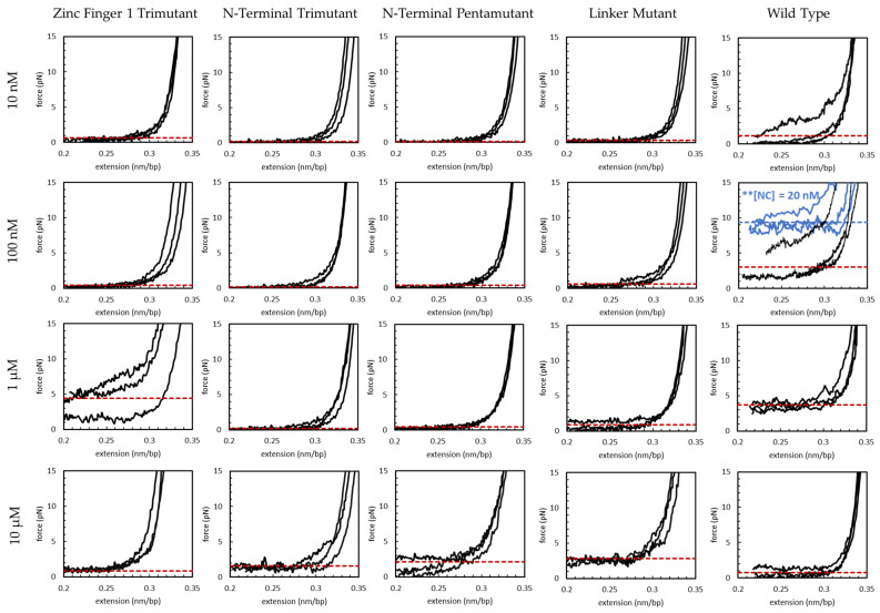
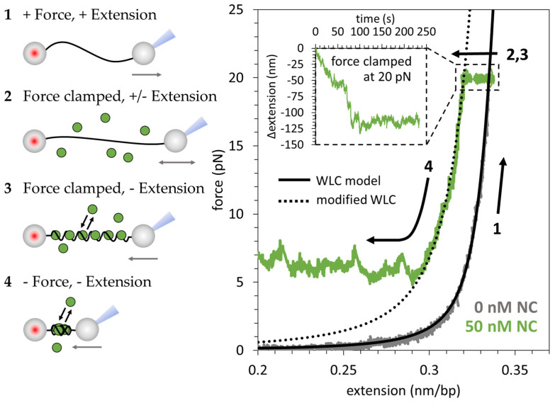
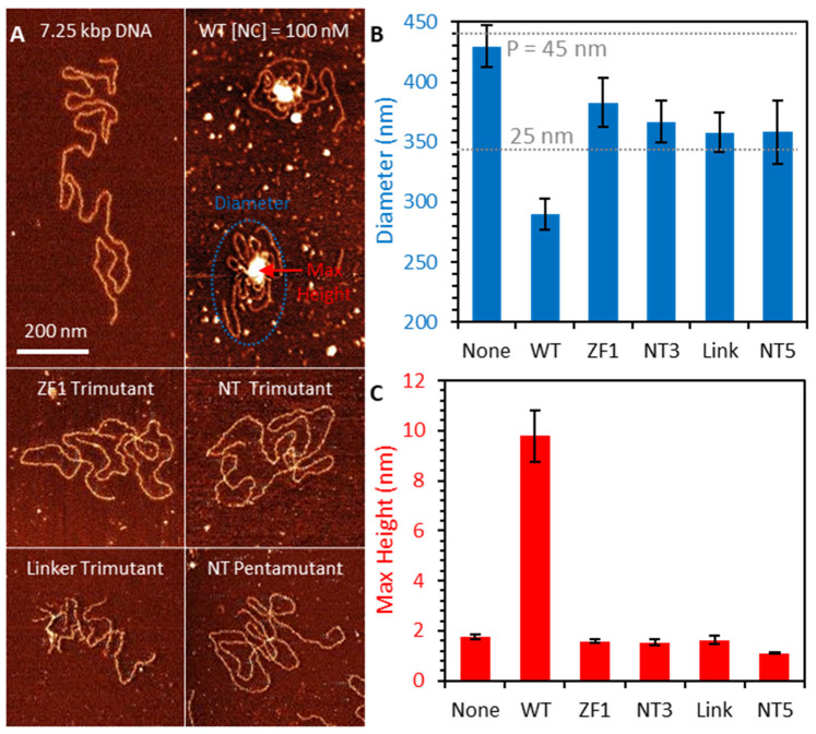
The HIV-1 nucleocapsid protein (NC) is a multifunctional viral protein necessary for HIV-1 replication. Recent studies have demonstrated that reverse transcription (RT) completes in the intact viral capsid, and the timing of RT and uncoating are correlated. How the small viral core stably contains the ~10 kbp double stranded (ds) DNA product of RT, and the role of NC in this process, are not well understood. We showed previously that NC binds and saturates dsDNA in a non-specific electrostatic binding mode that triggers uniform DNA self-attraction, condensing dsDNA into a tight globule against extending forces up to 10 pN. In this study, we use optical tweezers and atomic force microscopy to characterize the role of NC's basic residues in dsDNA condensation. Basic residue mutations of NC lead to defective interaction with the dsDNA substrate, with the constant force plateau condensation observed with wild-type (WT) NC missing or diminished. These results suggest that NC's high positive charge is essential to its dsDNA condensing activity, and electrostatic interactions involving NC's basic residues are responsible in large part for the conformation, size, and stability of the dsDNA-protein complex inside the viral core. We observe DNA re-solubilization and charge reversal in the presence of excess NC, consistent with the electrostatic nature of NC-induced DNA condensation. Previous studies of HIV-1 replication in the presence of the same cationic residue mutations in NC showed significant defects in both single- and multiple-round viral infectivity. Although NC participates in many stages of viral replication, our results are consistent with the hypothesis that cationic residue mutations inhibit genomic DNA condensation, resulting in increased premature capsid uncoating and contributing to viral replication defects.

Keywords: DNA condensation; HIV-1 nucleocapsid protein; atomic force microscopy; capsid uncoating; optical tweezers.


Conflict of interest statement

The authors declare no conflicts of interest. The funders had no role in the design of the study; in the collection, analyses, or interpretation of data; in the writing of the manuscript, or in the decision to publish the results.

Figures

Figure A1
Figure A1
n = 3 replicates are shown for return curves of varying concentrations of WT and basic residue variants of HIV-1 NC. The average condensing force (measured below 0.3 nm/bp) is shown for each case (red dashed line). Data for ** [NC] = 20 nM is included in the 100 nM wild-type panel (blue solid lines) to illustrate the maximum average condensing force (blue dashed line) observed for WT NC.
Figure 1
Figure 1
Sequence of wild-type (WT) and basic residue mutation variants of the HIV-1 nucleocapsid protein (NC). Zinc binding residues are highlighted in blue and basic residues are highlighted in red. Individual mutated residues are shown in green and indicated with an arrow. (A) WT; (B) K3A/R7A/R10A/K11A/K14A (N-terminal pentamutant); (C) R7A/R10A/K11A (N-terminal trimutant); (D) K14A/K20A/K26A (zinc finger 1 trimutant); (E) R29A/R32A/K33A/K34A (zinc finger linker mutant). The sequence shown is for the NL4-3 isolate (GenBank accession no. AF324493).
Figure 2
Figure 2
NC’s electrostatic binding mode collapses dsDNA into a dense globule along a constant force plateau. (1) Protein-free double-stranded (ds)DNA (gray) is extended until reaching 20 pN substrate tension, following the extensible worm-like chain (WLC) model (black line). The force-extension profile for bare dsDNA is consistent with the WLC polymer model. (2) A force feedback loop is applied to clamp the construct tension at 20 pN while 50 nM HIV-1 nucleocapsid protein (NC) is flowed into the cell. (3) NC binds the dsDNA (green). The NC-dsDNA complex reaches an equilibrium state within ~150 s. (4) The force clamp is removed, and the end-to-end distance is reduced in a controlled manner. At first, the force extension profile is consistent with a modified WLC model with a shortened persistence length (dotted line). Below a critical force, the force-extension profile deviates from the modified WLC model. In this low-force regime, NC maintains an approximately constant tension across the substrate as the end-to-end distance is reduced.
Figure 3
Figure 3
NC-induced overcharging of dsDNA. (A) Bare double-stranded (ds)DNA has a large persistence length (~50 nm) limiting DNA crossings and increasing end-to-end distance (i) due to dsDNA’s uniform negative charge preventing self-interaction (ii). Atomic force microscopy (AFM) imaging (iii) and force-extension measurements (iv) are consistent with the extensible worm-like chain (WLC) model with a persistence and contour length typical for B-DNA. (B) Low, sub-saturating concentrations of HIV-1 nucleocapsid protein (NC) effectively soften (reduce the rigidity of) dsDNA by inducing local bending. AFM imaging shows increased flexibility and frequent dsDNA intersections while force-extension curves lack the typical condensation force plateau (iv). The overall charge of the complex is negative, as the net charge of bound NC does not exceed the dsDNA net charge. (C) AFM images show a partial condensate surrounded by single layer dsDNA while the force-extension curves exhibit a partial condensation force plateau that eventually decays to zero force due to uncondensed DNA. The probability of phase separation occurring increases with increasing NC:bp. (D) Stoichiometrically optimized NC:bp binding (one NC per ~5 bp [30]) leads to complete dsDNA condensation into the tight globule. Mutual repulsion between mobile dsDNA-bound NC proteins might create a periodic +/− charge pattern on the dsDNA surface that arranges in counterphase (+ vs. −) against an analogous +/− charge pattern on an adjacent dsDNA, thereby leading to the mutual attraction schematically depicted in (D(ii)). A maximum density NC/dsDNA globule is observed on AFM and force-extension curve observes complete NC/dsDNA condensation along maximum condensation force plateau (Fc=~9 pN). (E) Oversaturating dsDNA with NC leads to the inversion of the complex net charge from negative to positive and de-condensation. Charge inversion prevents attachment to the positively charged AFM surface and a condensation force plateau and dsDNA softening are absent in the force-extension curves.
Figure 4
Figure 4
Basic residue mutations inhibit NC-induced dsDNA condensation. The average condensing force is plotted as a function of condensing agent concentration. A representative return trace is shown in each inset. The full set of experimental replicates can be found in Figure A1. (A) The average condensing force is plotted as a function of wild type HIV-1 nucleocapsid protein (WT NC) concentration. Error bars are standard error of the mean for n = 3 measurements. WT NC achieves a maximum average condensing force at [NC] = 20 nM. Above this critical concentration, the system becomes overcharged and condensing force decreases. (B) The zinc finger 1 (ZF1) trimutant variant achieves a maximum average condensing force at [NC] = 1 µM, ~50× higher than WT. (CE) The N-terminal and linker basic residue variants only show condensation at [NC] = 10 µM. The maximum condensing force observed for these variants is lower than those observed for the ZF1 trimutant and the WT, and occurs at a 10× higher concentration. Overcharging is not observed for these variants at the concentrations studied. (F) The maximum average condensing forces and corresponding concentrations are reported for previously studied condensing agents, as detailed in Table A1. Data points show the minimum and maximum concentrations required for condensation and the maximum condensation force and corresponding concentration. Solid lines are provided to guide the eye and do not necessarily represent the exact distribution shape. NC achieves a maximum condensing force at a >100× lower concentration than reported for previously studied condensing agents.
Figure 5
Figure 5
AFM imaging of dsDNA-NC complexes. (A) dsDNA in the absence of NC spreads out on the imaging surface, with minimal self-intersection. Incubation with 100 nM WT NC prior to deposition causes DNA to partially condense, both creating a large, multilayer globule with increased height (red) and reducing the spread of the DNA (blue). Incubation with NC mutants (also 100 nM) increases DNA flexibility, slightly reducing spread and increasing self-intersection, but does not create condensed globule. (B) Average diameter of DNA constructs incubated with each NC variant shows large reduction for WT NC, but a smaller effect consistent with a 2-fold reduction in DNA persistence length is observed for NC mutants. (C) Max height of DNA-NC complexes shows condensed globule formation for WT NC only, with NC mutants leaving DNA in single layer conformation.

References

    1. Aiken C., Rousso I. The HIV-1 capsid and reverse transcription. Retrovirology. 2021;18:29. doi: 10.1186/s12977-021-00566-0. - DOI - PMC - PubMed
    1. Briggs J.A.G., Simon M.N., Gross I., Kräusslich H.-G., Fuller S.D., Vogt V.M., Johnson M.C. The stoichiometry of Gag protein in HIV-1. Nat. Struct. Mol. Biol. 2004;11:672–675. doi: 10.1038/nsmb785. - DOI - PubMed
    1. Li S., Hill C.P., Sundquist W.I., Finch J.T. Image reconstructions of helical assemblies of the HIV-1 CA protein. Nature. 2000;407:409–413. doi: 10.1038/35030177. - DOI - PubMed
    1. Ganser B.K., Li S., Klishko V.Y., Finch J.T., Sundquist W.I. Assembly and Analysis of Conical Models for the HIV-1 Core. Science. 1999;283:80–83. doi: 10.1126/science.283.5398.80. - DOI - PubMed
    1. Perilla J.R., Schulten K. Physical properties of the HIV-1 capsid from all-atom molecular dynamics simulations. Nat. Commun. 2017;8:15959. doi: 10.1038/ncomms15959. - DOI - PMC - PubMed

Publication types

Associated data

LinkOut - more resources