. 2024 Jun 9;16(6):935.
doi: 10.3390/v16060935.

Applying Flow Virometry to Study the HIV Envelope Glycoprotein and Differences Across HIV Model Systems

Affiliations

Applying Flow Virometry to Study the HIV Envelope Glycoprotein and Differences Across HIV Model Systems

Jonathan Burnie et al. Viruses. .

Abstract

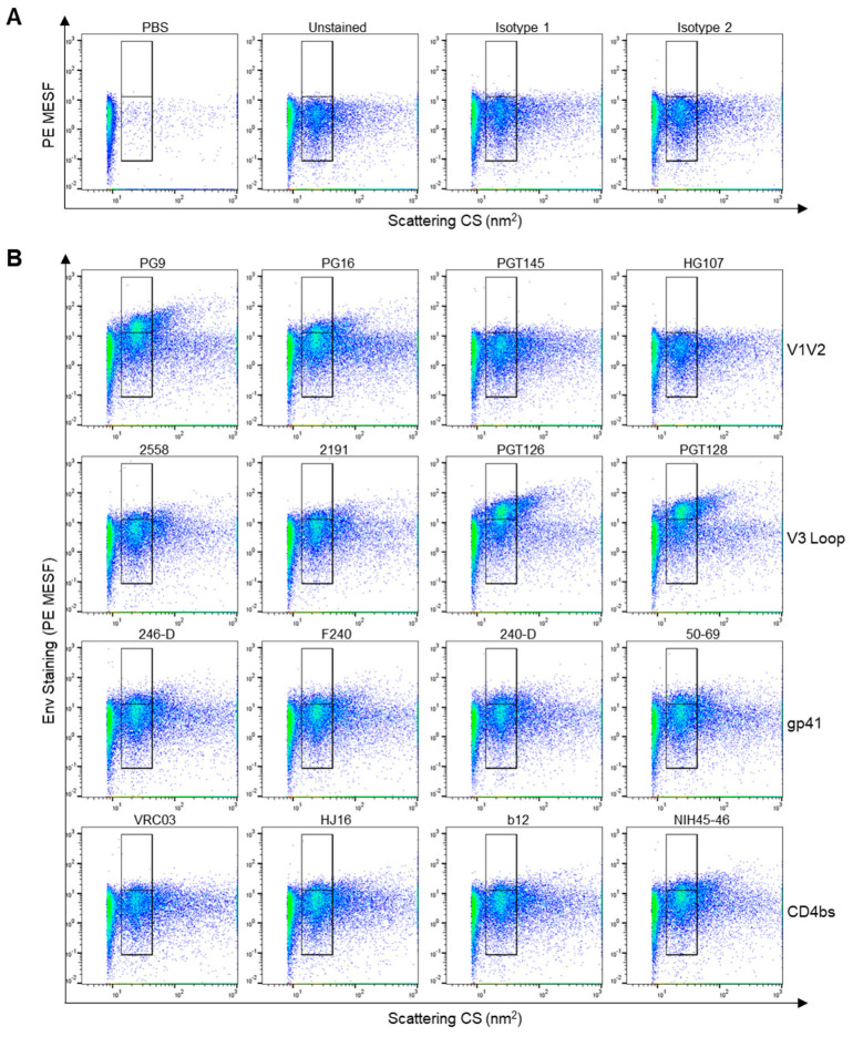
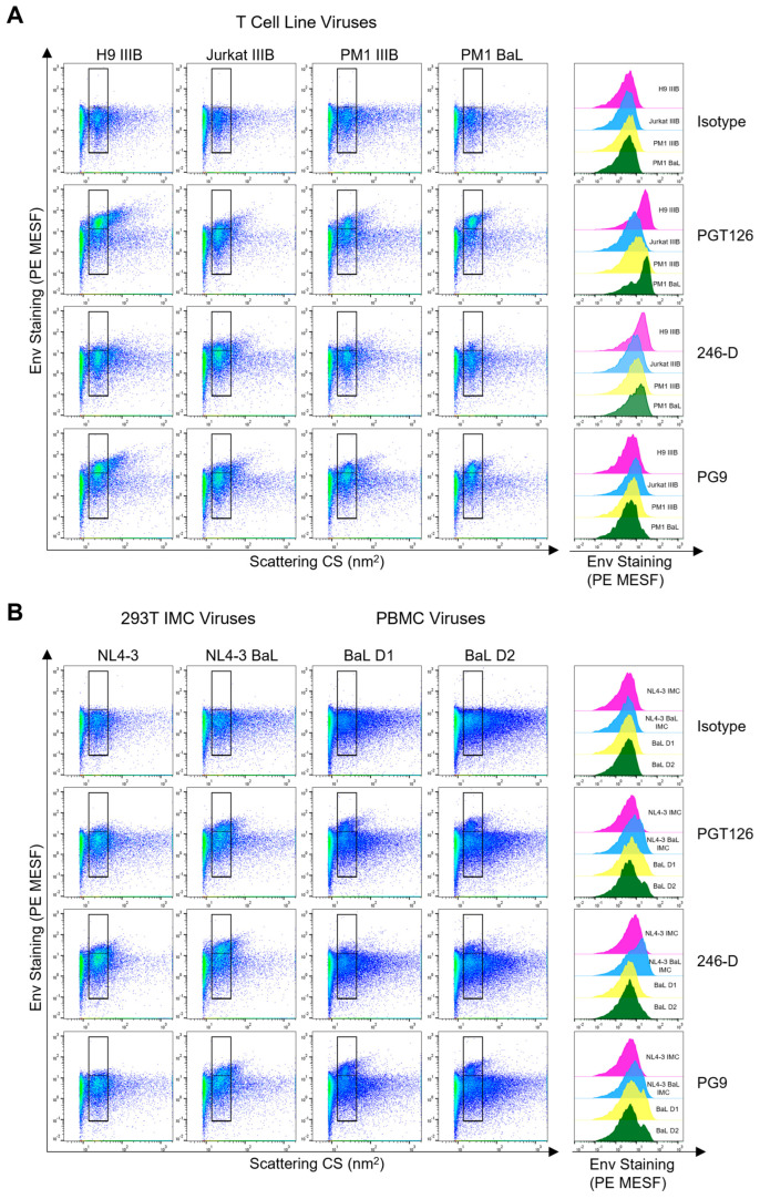
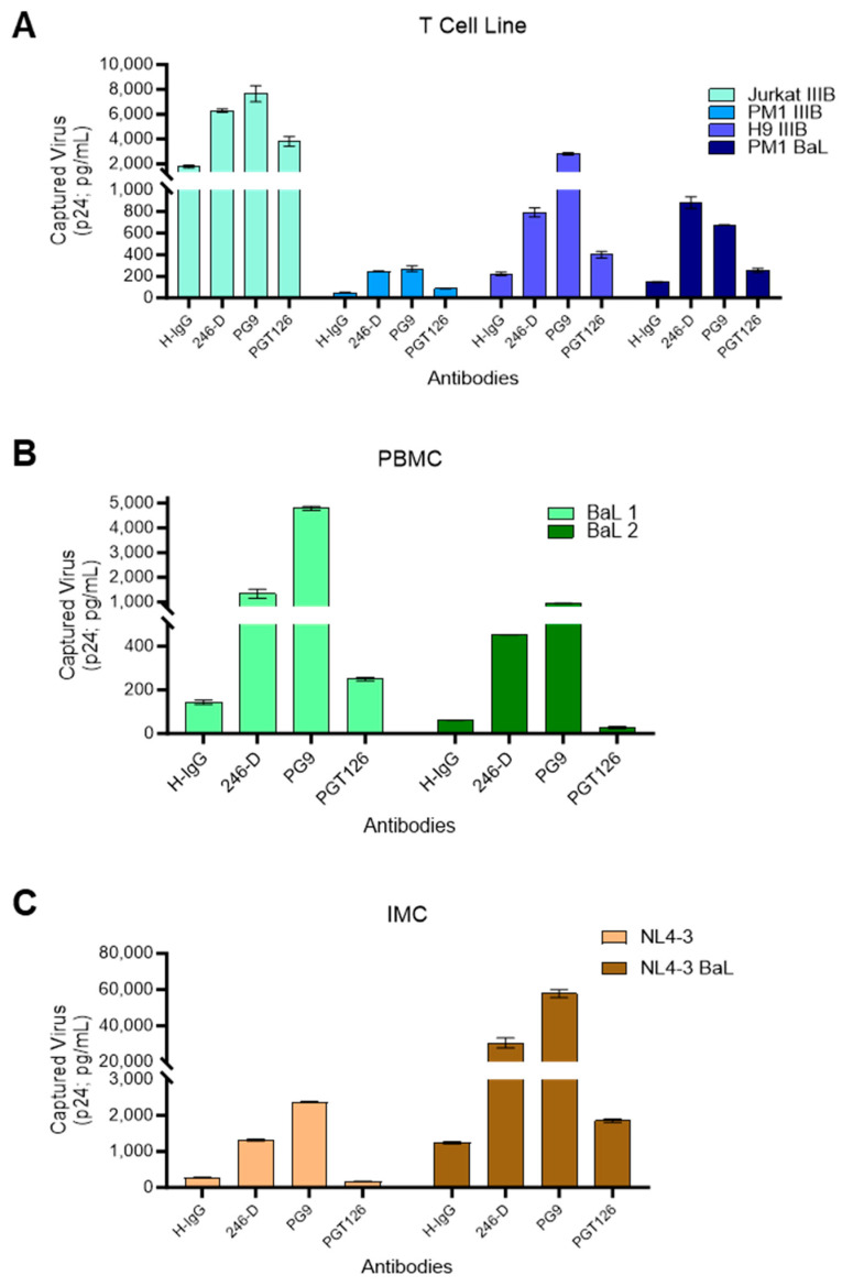
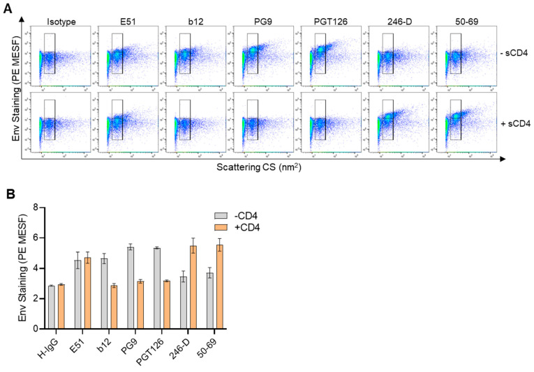
The HIV envelope glycoprotein (Env) is a trimeric protein that facilitates viral binding and fusion with target cells. As the sole viral protein on the HIV surface, Env is important both for immune responses to HIV and in vaccine designs. Targeting Env in clinical applications is challenging due to its heavy glycosylation, high genetic variability, conformational camouflage, and its low abundance on virions. Thus, there is a critical need to better understand this protein. Flow virometry (FV) is a useful methodology for phenotyping the virion surface in a high-throughput, single virion manner. To demonstrate the utility of FV to characterize Env, we stained HIV virions with a panel of 85 monoclonal antibodies targeting different regions of Env. A broad range of antibodies yielded robust staining of Env, with V3 antibodies showing the highest quantitative staining. A subset of antibodies tested in parallel on viruses produced in CD4+ T cell lines, HEK293T cells, and primary cells showed that the cellular model of virus production can impact Env detection. Finally, in addition to being able to highlight Env heterogeneity on virions, we show FV can sensitively detect differences in Env conformation when soluble CD4 is added to virions before staining.

Keywords: Env conformation; HIV Env; HIV trimer; calibrated flow virometry; gp120/gp41; human immunodeficiency virus (HIV); molecules of equivalent soluble fluorophore (MESF); nanoscale flow cytometry; neutralization; virion capture.


Conflict of interest statement

The authors declare no conflicts of interest. The funders had no role in the design of the study; in the collection, analyses, or interpretation of data; in the writing of the manuscript; or in the decision to publish the results.

Figures

Figure 1
Figure 1
Anti-Env staining on the HIVIIIB isolate produced in H9 CD4+ T cells. (A) Dot plots displaying flow virometry control stains, including PBS alone, unstained virus, and virus stained with different anti-human isotype control antibodies. Gates are set on the scattering profile (x-axis) of the HIV virus. Positive staining is shown in the upper gate, while background levels are within the lower gate, as determined with isotype controls. (B) Selected antibody staining from quantitative data reported in Table 1, with mAbs targeting the variable loops 1 and 2 (V1V2), V3 loop, gp41 or the CD4bs shown across each row. Representative plots from three independent experiments are shown.
Figure 2
Figure 2
Comparing Env staining across different cellular models of virus production. (A) Dot plots displaying anti-Env antibody staining (PGT126, 246-D, PG9) of viral isolates (IIIB or BaL, as indicated) produced in the H9, Jurkat, or PM1 cell lines. Positive staining is shown in the upper gate, while background levels as assessed by the isotype control are shown in the lower gate. Histogram overlays displaying the range of Env staining for each virus are shown to the right of the dot plots. The levels of PE-fluorescence represented on histograms are generated from the total virus staining (i.e., spanning the upper and lower gates). (B) In the left two panels, dot plots display anti-Env antibody staining of viruses (NL4-3 and NL4-3 BaL) produced through transfection of infectious molecular clones (IMC) in HEK293T cells. In the two rightmost panels, plots display antibody staining of the HIVBaL isolate produced in PBMC from two different donors (D1 and D2). Representative plots are shown from at least two independent experiments.
Figure 3
Figure 3
Comparison of virus immunocapture with anti-Env antibodies across different virus model systems. Plate-based virion immunocapture assays were performed with wells coated with the antibodies PGT126, 246-D, PG9, or an isotype control. Captured viruses were lysed and HIV-1 p24 Gag was quantified using p24 AlphaLISA as an indicator of the amount of virus capture. (A) Viruses (IIIB and BaL) produced in T cell lines (PM1, Jurkat, H9) were added to the wells at a normalized concentration of input virus (50 ng/mL of viral p24). (B) The HIVBaL isolate propagated in two different PBMC donors (BaL 1 and BaL 2), and (C) viruses produced from the transfection of infectious molecular clones (IMC; NL4-3 BaL and NL4-3) in HEK293T cells were tested at their undiluted titers. Results represent the mean ± SD of duplicate wells and are representative of three independent experiments.
Figure 4
Figure 4
Validating the neutralization profile of mAbs PGT126 and PG9 against different viruses. (A) Neutralization sensitivities of IIIB and BaL viruses grown in the PM1 and H9 CD4+ T cell lines using PG9 (blue) and PGT126 (red) mAbs. (B) Neutralization of HIV BaL produced in two different PBMC donors (D1 and D2). (C) Neutralization of virus produced through the transfection of HEK293T cells with the NL4-3-BaL infectious molecular clone. All neutralization tests were performed using the TZM-bl reporter cell assay, with the infection of control samples (in absence of mAbs) set at 100%. Data are representative of two independent experiments tested with triplicate wells.
Figure 5
Figure 5
Staining HIVIIIB in the presence or absence of soluble CD4. (A) Dot plots depicting flow virometry staining of HIVIIIB in the presence (+CD4) or absence (−sCD4) of soluble CD4. Positive staining is shown in the upper gate, while background levels fall within the lower gate. Plots shown are representative from three independent experiments. (B) Quantified staining data showing the mean ± SD of three replicates as in (A), generated from the individual median PE MESF values derived from all events within both virus gates (upper and lower) of each replicate stain.
Figure 6
Figure 6
Comparing direct and indirect staining methods in flow virometry to label HIV Env. Indirect staining of HIVIIIB propagated in H9 CD4+ T cells with unlabeled primary anti-gp120 antibodies (PGT145 and PG9), and a PE-conjugated secondary antibody (top row). Direct staining of the same HIVIIIB virus stocks using PE-labelled PGT145 and PG9 antibodies (bottom row). Positive staining is shown in the upper gate, while background levels fall within the lower gate. Median PE MESF values that were generated from all virus events within the merged upper and lower gates are enumerated on the plots.

Similar articles

Cited by

References

    1. Johnston M.I., Fauci A.S. An HIV Vaccine—Evolving Concepts. N. Engl. J. Med. 2007;356:2073–2081. doi: 10.1056/NEJMra066267. - DOI - PubMed
    1. Burton D.R., Desrosiers R.C., Doms R.W., Koff W.C., Kwong P.D., Moore J.P., Nabel G.J., Sodroski J., Wilson I.A., Wyatt R.T. HIV Vaccine Design and the Neutralizing Antibody Problem. Nat. Immunol. 2004;5:233–236. doi: 10.1038/ni0304-233. - DOI - PubMed
    1. Overbaugh J., Morris L. The Antibody Response against HIV-1. Cold Spring Harb. Perspect. Med. 2012;2:a007039. doi: 10.1101/cshperspect.a007039. - DOI - PMC - PubMed
    1. Wei X., Decker J.M., Wang S., Hui H., Kappes J.C., Wu X., Salazar-Gonzalez J.F., Salazar M.G., Kilby J.M., Saag M.S., et al. Antibody Neutralization and Escape by HIV-1. Nature. 2003;422:307–312. doi: 10.1038/nature01470. - DOI - PubMed
    1. Checkley M.A., Luttge B.G., Freed E.O. HIV-1 Envelope Glycoprotein Biosynthesis, Trafficking, and Incorporation. J. Mol. Biol. 2011;410:582–608. doi: 10.1016/j.jmb.2011.04.042. - DOI - PMC - PubMed

Publication types

MeSH terms

Substances