. 2024 Jun 1;12(6):605.
doi: 10.3390/vaccines12060605.

A Unique mRNA Vaccine Elicits Protective Efficacy against the SARS-CoV-2 Omicron Variant and SARS-CoV

Affiliations

A Unique mRNA Vaccine Elicits Protective Efficacy against the SARS-CoV-2 Omicron Variant and SARS-CoV

Xiaoqing Guan et al. Vaccines (Basel). .

Abstract

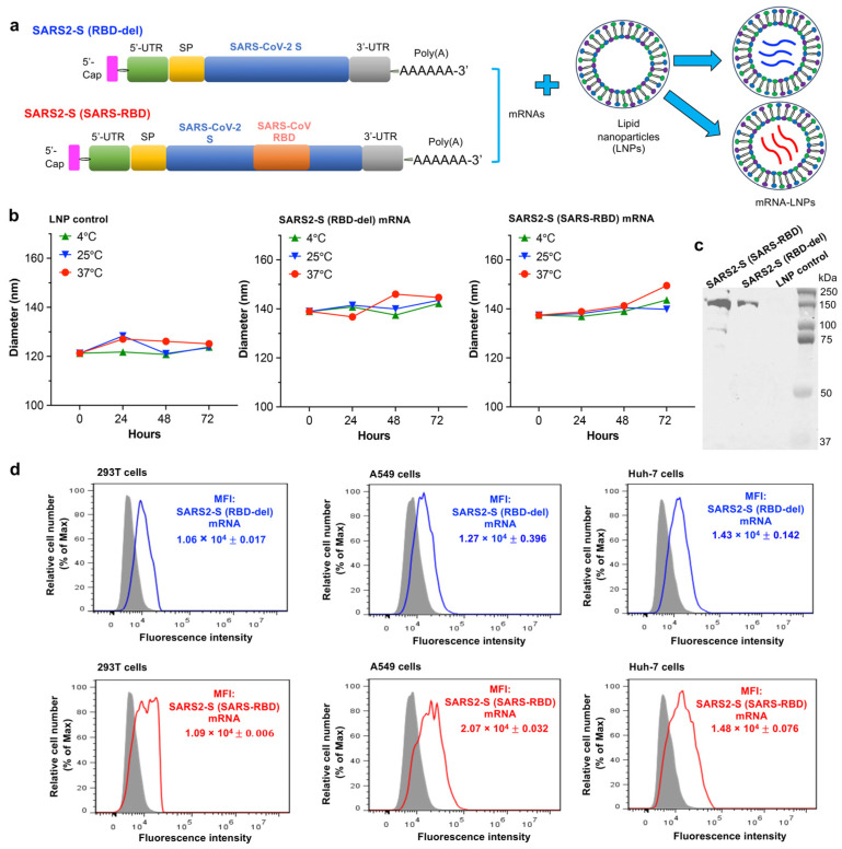
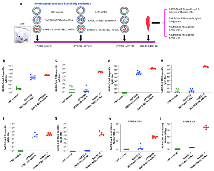
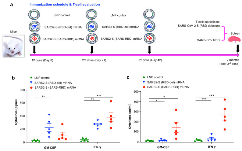
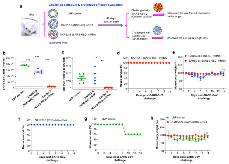
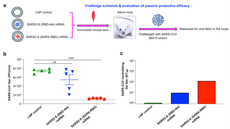
The highly pathogenic coronaviruses SARS-CoV-2 and SARS-CoV have led to the COVID-19 pandemic and SARS outbreak, respectively. The receptor-binding domain (RBD) of the spike (S) protein of SARS-CoV-2, particularly the Omicron variant, has frequent mutations, resulting in the reduced efficiency of current COVID-19 vaccines against new variants. Here, we designed two lipid nanoparticle-encapsulated mRNA vaccines by deleting the mutant RBD of the SARS-CoV-2 Omicron variant (SARS2-S (RBD-del)) or by replacing this mutant RBD with the conserved and potent RBD of SARS-CoV (SARS2-S (SARS-RBD)). Both mRNA vaccines were stable at various temperatures for different time periods. Unlike SARS2-S (RBD-del) mRNA, SARS2-S (SARS-RBD) mRNA elicited effective T-cell responses and potent antibodies specific to both SARS-CoV-2 S and SARS-CoV RBD proteins. It induced strong neutralizing antibodies against pseudotyped SARS-CoV-2 and SARS-CoV infections and protected immunized mice from the challenge of the SARS-CoV-2 Omicron variant and SARS-CoV by significantly reducing the viral titers in the lungs after Omicron challenge and by completely preventing SARS-CoV-induced weight loss and death. SARS2-S (SARS-RBD)-immunized serum antibodies protected naïve mice from the SARS-CoV challenge, with its protective efficacy positively correlating with the neutralizing antibody titers. These findings indicate that this mRNA vaccine has the potential for development as an effective vaccine against current and future SARS-CoV-2 variants and SARS-CoV.

Keywords: COVID-19; SARS-CoV; SARS-CoV-2; coronavirus; receptor-binding domain; spike protein; unique mRNA vaccine.


Conflict of interest statement

The authors declare no conflicts of interest.

Figures

Figure 1
Figure 1
Construction and characterization of mRNA vaccines. (a) Construction of nucleoside-modified SARS2-S (SARS-RBD) and SARS2-S (RBD-del) mRNAs. Each of the synthesized mRNAs encodes tissue plasminogen activator (tPA) signal peptide (SP) and SARS-CoV-2 Omicron spike (S) protein with a deleted receptor-binding domain (RBD) or with the inserted SARS-CoV RBD, and contains a 5′ cap, 5′ untranslated region (UTR), 3′-UTR, and 3′ poly(A) tail. The synthesized mRNAs were formulated with lipid nanoparticles (LNPs) for vaccine delivery. (b) Detection of the stability of LNP-formulated mRNA vaccines or LNP control under different conditions. mRNA vaccines or control were stored at 4 °C, 25 °C, and 37 °C for 0, 24, 48, and 72 h, respectively, followed by measurement of particle sizes (hydrodynamic diameter) by DLS. (c) Western blot for detection of expression of mRNA-encoding protein. Each mRNA-LNP was incubated with 293T cells for 72 h at 37 °C and the culture supernature was tested by SARS2-S (SARS-RBD)-immunized mouse sera. kDa, protein molecular weight marker. (d) Representative figures of flow cytometry analysis of expression of mRNA-encoding protein. Each mRNA-LNP was incubated with 293T, A549, and Huh-7 cells, respectively, for 48 h at 37 °C, which were then stained with FITC-labeled mouse-anti-His antibody and measured for fluorescence intensity by flow cytometry. The grey-shaded region represents LNP-incubated cell controls. MFI indicates the Median Fluorescence Intensity. The data are expressed as mean ± standard deviation of the mean (s.e.m.) of triplicate wells. One experimental repeat was performed and similar results were obtained.
Figure 2
Figure 2
Evaluation of antibody responses and neutralizing antibodies induced by mRNA vaccines against infection of SARS-CoV-2 and SARS-CoV. (a) BALB/c mice were vaccinated with each mRNA vaccine, or LNP control, for a total of 3 times at 3-week intervals and bled 10 days post-last vaccination to test, by ELISA, for serum IgG antibodies targeting the RBD-deleted SARS-CoV-2 S (b) or SARS-CoV RBD (c) protein. These sera were also evaluated, by ELISA, for serum IgG1 subtype antibodies targeting the aforementioned SARS-CoV-2 S (d) or SARS-CoV RBD (e) protein, as well as for serum IgG2a subtype antibodies targeting the aforementioned SARS-CoV-2 S (f) or SARS-CoV RBD (g) protein. The SARS-CoV-2 S or SARS-CoV RBD protein was pre-coated to the ELISA plates and the respective antibody (Ab) titer was reported as mean ± s.e.m. (from five mice per group). The aforementioned mouse sera were also detected for neutralizing antibodies against pseudoviruses expressing the respective S proteins of SARS-CoV-2 ancestral strain (h) and the ancestral strain (Tor2) of SARS-CoV (i). The neutralizing antibody titer (NT50: 50% neutralizing Ab titer) is expressed as mean ± s.e.m. (from five mice per group). One experimental repeat was performed and similar results were obtained.
Figure 3
Figure 3
Evaluation of mRNA vaccine-induced T-cell responses. (a) BALB/c mice were immunized with each mRNA vaccine or LNP control for 3 times at 3-week intervals and splenocytes collected 2 months post-last vaccine dose were tested for cytokine expression by Multiplex assay. Isolated splenocytes were stimulated with RBD-deleted SARS-CoV-2 S (b) or SARS-CoV RBD (c) protein and the expressed cytokines (pg/mL) were measured in the supernatant. Significant differences among different groups are shown as * (p < 0.05), ** (p < 0.01), and *** (p < 0.001). The experiments were repeated once, resulting in similar results.
Figure 4
Figure 4
Evaluation of mRNA vaccine-induced protective efficacy against SARS-CoV-2 and SARS-CoV. (a) At a time of 40 days after the last immunization, BALB/c mice were i.n.-challenged with the SARS-CoV-2 Omicron variant (BA.1, 105 PFU/mL) and lungs were collected two days later to measure viral titers by plaque assay and viral replication by qRT-PCR. Evaluation of viral titers (b) and viral replication (c) in the lungs. The viral titers were reported as the PFU/mL of lung tissues. The viral (nucleocapsid gene) replication was normalized to glyceraldehyde-3-phosphate dehydrogenase (GAPDH). The data are presented as mean ± s.e.m. (from five mice per group). One experimental repeat was performed and similar results were obtained. (dh) In a separate experiment, vaccinated mice were challenged (i.n.) with the MA15 strain of SARS-CoV (500 PFU/mL) and investigated for survival and weight loss for 14 days after the virus challenge. The data (in (e) and (h)) are expressed as mean ± s.e.m. of three (for surviving mice in the LNP control group from day 9) to five mice (for mRNA vaccine groups and LNP control group by day 8) in each group. Significant differences among different groups are shown as * (p < 0.05), ** (p < 0.01), and *** (p < 0.001).
Figure 5
Figure 5
Evaluation of passive protective efficacy of mRNA vaccine-induced mouse serum antibodies. (a) Experimental procedure and challenge schedule. Naïve BALB/c mice were i.p.-injected with the pooled sera of mice receiving vaccines (SARS2-S (SARS-RBD) mRNA or SARS2-S (RBD-del) mRNA) or LNP control, i.n.-challenged with the heterologous SARS-CoV (MA15 strain, 400 PFU/mL) 12 h later, and measured for viral titers in the lungs by plaque assay two days post-challenge. (b) Evaluation of viral titers after serum transfer. The viral titers were expressed as the PFU/mL of lung tissues. The data are presented as mean ± s.e.m. (from five mice per group). ** (p < 0.01) and *** (p < 0.001) indicate significant differences among different groups. (c) Plaque reduction neutralization assay was tested for the aforementioned pooled mouse sera against infection of the heterologous authentic MA15 strain of SARS-CoV. The 50% neutralizing antibody (Ab) titer (NT50) was calculated and the data are expressed as mean ± s.e.m. (from duplicate wells of pooled sera per group). One experimental repeat was performed and similar results were obtained.

Similar articles

References

    1. Zhou P., Yang X.L., Wang X.G., Hu B., Zhang L., Zhang W., Si H.R., Zhu Y., Li B., Huang C.L., et al. A pneumonia outbreak associated with a new coronavirus of probable bat origin. Nature. 2020;579:270–273. doi: 10.1038/s41586-020-2012-7. - DOI - PMC - PubMed
    1. World Health Organization Coronavirus (COVID-19) Dashboard. 2023. [(accessed on 28 April 2024)]. Available online: https://covid19.who.int/
    1. Stadler K., Masignani V., Eickmann M., Becker S., Abrignani S., Klenk H.D., Rappuoli R. SARS—beginning to understand a new virus. Nat. Rev. Microbiol. 2003;1:209–218. doi: 10.1038/nrmicro775. - DOI - PMC - PubMed
    1. Fronteira I., Sidat M., Magalhães J.P., de Barros F.P.C., Delgado A.P., Correia T., Daniel-Ribeiro C.T., Ferrinho P. The SARS-CoV-2 pandemic: A syndemic perspective. One Health. 2021;12:100228. doi: 10.1016/j.onehlt.2021.100228. - DOI - PMC - PubMed
    1. Ksiazek T.G., Erdman D., Goldsmith C.S., Zaki S.R., Peret T., Emery S., Tong S., Urbani C., Comer J.A., Lim W., et al. A novel coronavirus associated with severe acute respiratory syndrome. N. Engl. J. Med. 2003;348:1953–1966. doi: 10.1056/NEJMoa030781. - DOI - PubMed

LinkOut - more resources