. 2024 Jun 18;12(6):676.
doi: 10.3390/vaccines12060676.

S2 Peptide-Conjugated SARS-CoV-2 Virus-like Particles Provide Broad Protection against SARS-CoV-2 Variants of Concern

Affiliations

S2 Peptide-Conjugated SARS-CoV-2 Virus-like Particles Provide Broad Protection against SARS-CoV-2 Variants of Concern

Chang-Kyu Heo et al. Vaccines (Basel). .

Abstract

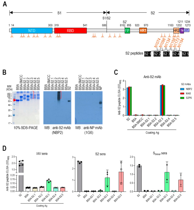
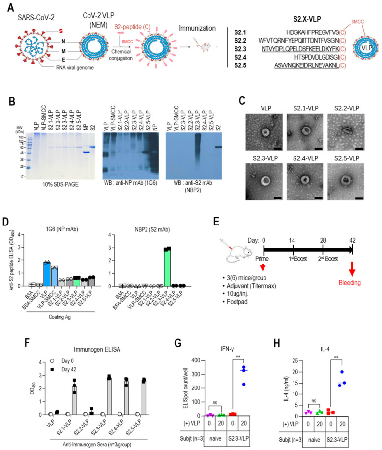
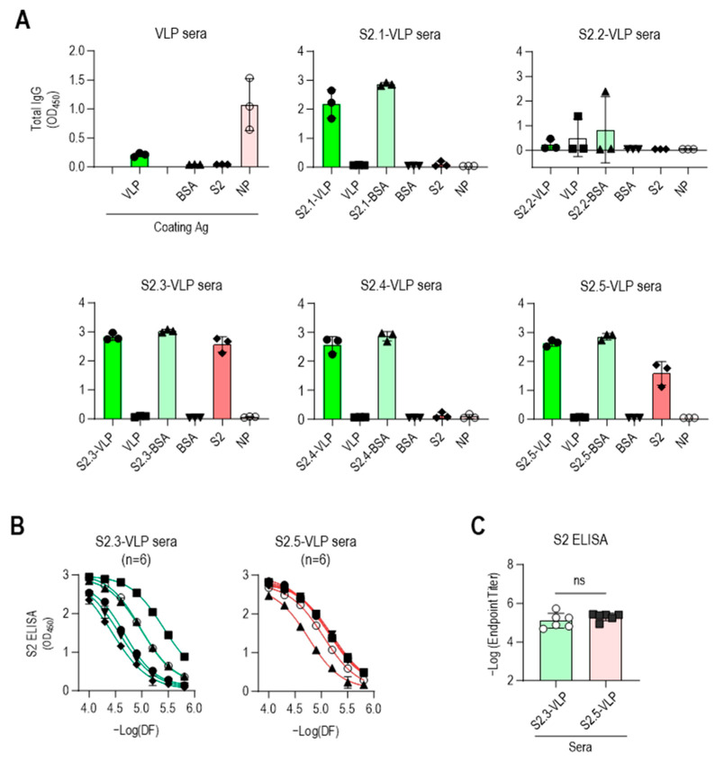
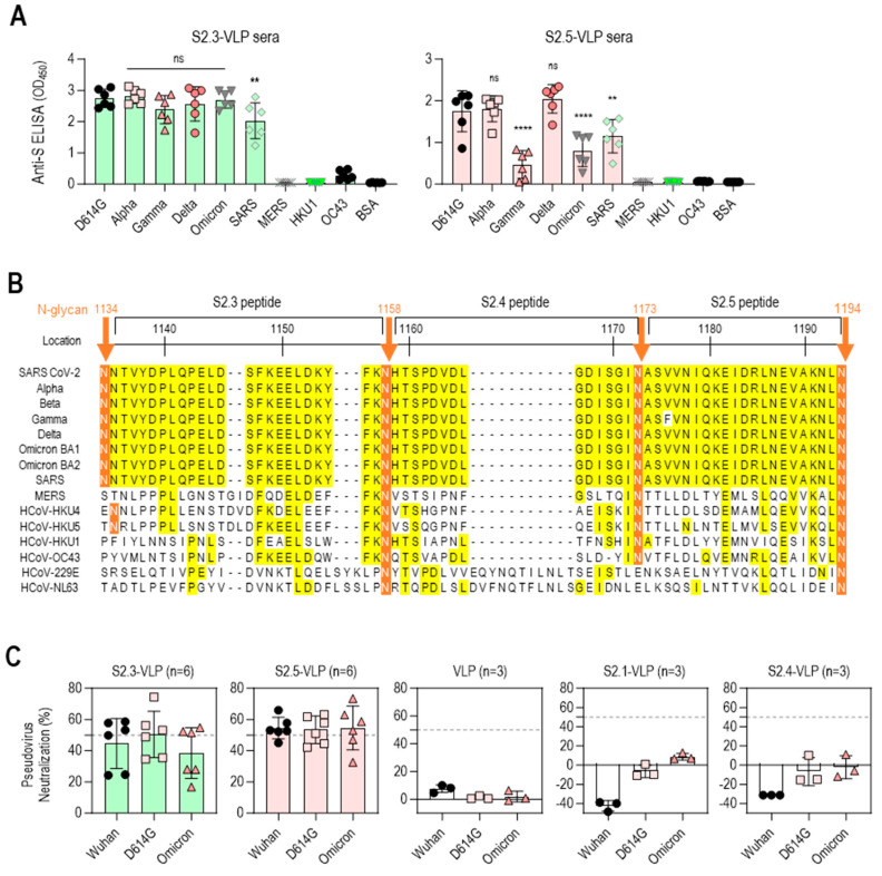
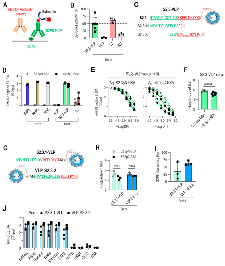
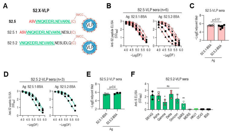
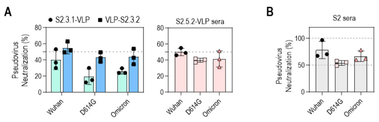
Approved COVID-19 vaccines primarily induce neutralizing antibodies targeting the receptor-binding domain (RBD) of the SARS-CoV-2 spike (S) protein. However, the emergence of variants of concern with RBD mutations poses challenges to vaccine efficacy. This study aimed to design a next-generation vaccine that provides broader protection against diverse coronaviruses, focusing on glycan-free S2 peptides as vaccine candidates to overcome the low immunogenicity of the S2 domain due to the N-linked glycans on the S antigen stalk, which can mask S2 antibody responses. Glycan-free S2 peptides were synthesized and attached to SARS-CoV-2 virus-like particles (VLPs) lacking the S antigen. Humoral and cellular immune responses were analyzed after the second booster immunization in BALB/c mice. Enzyme-linked immunosorbent assay revealed the reactivity of sera against SARS-CoV-2 variants, and pseudovirus neutralization assay confirmed neutralizing activities. Among the S2 peptide-conjugated VLPs, the S2.3 (N1135-K1157) and S2.5 (A1174-L1193) peptide-VLP conjugates effectively induced S2-specific serum immunoglobulins. These antisera showed high reactivity against SARS-CoV-2 variant S proteins and effectively inhibited pseudoviral infections. S2 peptide-conjugated VLPs activated SARS-CoV-2 VLP-specific T-cells. The SARS-CoV-2 vaccine incorporating conserved S2 peptides and CoV-2 VLPs shows promise as a universal vaccine capable of generating neutralizing antibodies and T-cell responses against SARS-CoV-2 variants.

Keywords: SARS-CoV-2; broadly neutralizing antibody; conserved S2; peptide conjugation; universal vaccine; virus-like particles.


Conflict of interest statement

The authors declare no conflicts of interest.

Figures

Figure 1
Figure 1
Designing antigenic peptide sequences for a broad-spectrum SARS-CoV-2 vaccine. (A) Diagram of the SARS-CoV-2 spike protein showing its structural components. The spike is organized into two primary domains, S1 and S2; these includes the N-terminal domain (NTD), receptor binding domain (RBD), fusion peptide (FP), heptad repeat 1 (HR1), heptad repeat 2 (HR2), transmembrane (TM), and cytoplasmic tail (CT). The specific segment boundaries of these regions are indicated, and locations of glycosylation identified through experimental methods are noted below each segment. The positions of the glycan-free S2 peptide sequences are shown. (B) SDS-PAGE and Western blot analysis of proteins following conjugation of S2 peptides to BSA. To confirm the formation of S2 peptide–BSA conjugates, they were probed with an anti-S2 antibody, NBP2. Anti-NP antibody probing was used as a control to assess nonspecific binding. (C) ELISA analysis of S2 peptide-conjugated BSA. Recombinant S2 antigen was used as a positive control. Antibodies were added at 100 ng/well. (D) Reactivity of antisera against inactivated SARS-CoV-2 (IAV), S2, or Strimer to S2 peptide-conjugated BSA. The antisera were diluted to 1:10,000. Each dot represents the antibody response of individual mice and displays the mean ± standard deviation (SD) of replicate wells.
Figure 2
Figure 2
Vaccination with S2 peptide-conjugated VLPs elicits robust humoral immune responses in BALB/c mice. (A) S2 peptide-conjugated VLP design and S2 peptide sequences. (B) SDS-PAGE and Western blot analysis of S2 peptide-conjugated VLPs. The VLPs were probed with an anti-NP monoclonal antibody (1G6) to detect the presence of VLPs. The conjugated S2 peptides on the VLPs were probed with an anti-S2 monoclonal antibody (NBP2) to confirm successful conjugation. (C) Transmission electron micrographs (TEM) of unmodified VLPs and S2 peptide-conjugated VLPs. Representative images are shown, with the scale bar representing 100 nm. (D) ELISA analysis of S2 peptide-conjugated VLPs. VLPs, VLP-SMCC, S2 peptide-conjugated VLPs, and BSA (negative control) were plated and probed with SARS-CoV-2 NP-specific antibody (1G6) or S2-specific antibody (NBP2). Antibodies were added at 100 ng/well. (E) BALB/c immunization regimen. Groups of 3 BALB/c mice were given three immunizations with S2 peptide–VLPs or control VLPs with an adjuvant. The VLP immunogens were administered via subcutaneous injection into the footpad at a dose of 10 μg per injection. Mice were immunized at weeks 0 (prime), 2 (first boost), and 4 (second boost). Serum collection was performed before prime injection and two weeks after the second boost (day 42). (F) Specific total IgG responses to VLPs or S2 peptide-conjugated VLPs (S2.X-VLP). ELISA against immunogens was performed using sera (1:10,000 dilution) collected before priming and two weeks after the second boost. Each dot represents the antibody response of individual mice to VLP or S2-VLP immunization and displays the mean ± SD of replicate wells. (G) IFNγ ELISpot assay for splenocytes from VLP-S2.3 immunized mice. BALB/c mice (n = 3 per group) were immunized with S2.3-VLP or naïve. Two weeks after the second boost (day 42), pooled splenocytes were harvested and added to anti-IFNγ antibody-precoated ELISpot plates with SARS-CoV-2-NEM VLPs in duplicate. After splenocyte stimulation for 48 h, anti-IFNγ responses were developed, and spots were counted using an ELISpot plate reader. (H) IL4 assay for splenocytes from VLP-S2.3 immunized and naïve mice. After stimulating the splenocytes with SARS-CoV-2-NEM VLPs, the cell culture supernatant was collected and added to plates precoated with anti-IL4 antibody. The anti-IL4 response was measured, and the IL4 concentration in the splenocyte culture medium was determined using a standard curve. Each dot represents the measurement of an individual mouse, and the graph displays the mean ± SD of replicate wells. ns: p > 0.05, **: p ≤ 0.01.
Figure 3
Figure 3
Vaccination with S2.3 or S2.5 peptide-conjugated VLPs induces SARS-CoV-2 S2 antigen-specific antibodies. (A) The specific immune responses of antisera against S2 peptides or SARS-CoV-2 S2 antigen were analyzed. Antisera collected after the second boost were diluted to 1:10,000 and used for analysis. Each dot represents the antibody response of an individual mouse. (B) Antisera obtained from immunization with S2.3-VLP or S2.5-VLP were analyzed for their specific immune responses to the S2 antigen. Antisera collected after the second boost were serially diluted and used for analysis. Serial dilutions were performed in two-fold dilutions starting at 1:10,000. Each curve represents the antibody response of an individual mouse (n = 6) to the S2 antigen, and each dot on the curve displays the mean ± SD of replicate wells. (C) SARS-CoV-2 S2 total IgG endpoint titers in serum obtained from mice vaccinated with S2.3 or S2.5 peptide-conjugated VLPs. ns: p > 0.05.
Figure 4
Figure 4
Vaccination with S2.3 or S2.5 peptide-conjugated VLPs induces broadly neutralizing antibodies against SARS-CoV-2 variants. (A) The total IgG response of sera from mice immunized with S2.3-VLP (left plot) or S2.5-VLP (right plot) against the spike proteins of SARS-CoV-2 variants (D614G, Alpha, Gamma, Delta, Omicron) as well as other betacoronaviruses (SARS, MERS, HKU1, and OC43) was evaluated. The immunized sera were diluted 1:10,000. ns: p > 0.05, **: p ≤ 0.01, ****: p ≤ 0.0001. (B) Amino acid alignment of the S2 peptide sequences of SARS-CoV-2 variants and related β-coronaviruses. The positions of the N-glycans are shown, and consensus sequences are highlighted in yellow. (C) The neutralization potential of sera obtained from mice immunized with S2.3-VLP (n = 6) and S2.5-VLP (n = 6) was assessed against the pseudotyped SARS-CoV-2 prototype (Wuhan) or variants (D614G and Omicron BA4.2). The neutralization assay was performed using sera collected on day 42 after immunization, with triplicate measurements for each sample. The pseudovirus infection without sera was used as the positive control. The unmodified VLP-immunized sera and sera from mice immunized with S2.1-VLP or S2.4-VLP were analyzed.
Figure 5
Figure 5
C-terminal exposure of S2.3 peptide confers an epitope that induces S2P6-like broadly neutralizing antibodies against SARS-CoV-2. (A) Competitive ELISA for quantifying S2P6-like activity of test sera. The recombinant S2 antigen was coated on a MaxiSorp plate. The test solution containing the sera or antibody of interest was added to the well along with HRP-conjugated S2P6. After incubation, S2P6-HRP activity was measured to determine the competitive binding between the test antibodies and HRP-conjugated S2P6 for the S2 antigen. (B) S2P6-like activity of VLP-S2.3, S2, or inactivated SARS-CoV-2 (IAV) immunized sera. Each dot represents the measurement of an individual mouse (n = 6 or 3) and displays the mean ± standard deviation (SD) of replicate wells. (CF) Fine mapping of antigenic epitopes of S2.3-reactive antibodies or VLP-S2.3 antisera using short peptides derived from the S2.3 peptide. Two short peptides (S2.3pN and S2.3pC) were synthesized (C), and their conjugates to BSA served as coating antigens for detecting epitope-specific antibodies. The reactivity of S2.3 reactive antibodies, including S2P6, NBP2, and MA5 monoclonal antibodies and antisera against VLP-S2.3 or S2 antigen, was measured against these short peptides. Antibodies were added at 100 ng/well. Antisera were treated at a 1:10,000 dilution (D) or two-fold serial dilutions starting at 1:10,000 (E). Each dot represents the measurement of an individual mouse (n = 6 or 3) and displays the mean ± standard deviation (SD) of replicate wells. S2.3 peptide total IgG endpoint titers were also analyzed (F). (GJ) C-terminal exposure of S2.3 peptide on VLP confers an epitope that induces S2P6-like broadly neutralizing antibodies against SARS-CoV-2. For the exposure of the C-terminus of the S2.3 peptide on peptide–VLP conjugates, two amino acid residues were added after the S2.3 peptide as a spacer (S2.3.1-VLP) or cysteine was added at the N-terminus of the S2.3 peptide and conjugated with SMCC-linked VLPs (VLP-S2.3.2) (G). These peptide-conjugated VLPs were immunized following the immunization regimen (n = 3/each immunogen), as shown in Figure 2E, and the reactivity of antisera after the second boost against S2.3pN or S2.3pC epitopes was analyzed (H). Their S2P6-like activities were analyzed using sandwich ELISA, as shown in Figure 5A (I). Additionally, their reactivity against the S antigen of SARS-CoV-2 S variants or other β-corona viruses were examined (J). The antisera were diluted at 1:10,000.
Figure 6
Figure 6
Immune sera elicited with peptides lacking an N-terminal sequence containing V1176 of the S2.5 peptide showed enhanced reactivity toward S antigen variants. (AC) The S2.5.1 peptide, an extended S2.5 peptide from S (1174–1193) to S (1174–1198), and the S2.5.2 peptide, an S2.5 peptide with a deletion of V1176 at the N-terminus, were synthesized and conjugated to VLP or BSA (A), and their reactivity to S2.5-VLP-immunized sera was analyzed by endpoint titer determination. (B,C) Antisera were treated with two-fold serial dilutions starting at 1:10,000. (DE) The reactivity of S2.5.2-VLP-immunized sera against S2.5.1 or S2.5.2 peptide was analyzed by endpoint titer determination. (F) Reactivity of S2.5.2-VLP-immunized sera against the SARS-CoV-2 S variants. Serum was diluted to 1:10,000. Statistical analysis was performed using one-way ANOVA, followed by Dunnett’s multiple comparison test. ns: no significance; * p = 0.042; ** p = 0.0097.
Figure 7
Figure 7
Neutralization potential of VLPs conjugated with peptides containing S2.3 or S2.5 epitopes against pseudotyped SARS-CoV-2. (A) Neutralization potential of S2.3 epitope (S2.3.1, S2.3.2, or S2.5.2)-conjugated VLPs. (B) Neutralization potential of S2 subunit-immunized sera. Pseudoviruses with SARS-CoV-2 spike proteins (Wuhan-Hu-1, D614G, or Omicron BA.2) and sera collected on day 14 after the second boost were used for neutralization assays.

Similar articles

Cited by

References

    1. Chow E.J., Uyeki T.M., Chu H.Y. The effects of the COVID-19 pandemic on community respiratory virus activity. Nat. Rev. Microbiol. 2023;21:195–210. doi: 10.1038/s41579-022-00807-9. - DOI - PMC - PubMed
    1. Kyriakidis N.C., López-Cortés A., González E.V., Grimaldos A.B., Prado E.O. SARS-CoV-2 vaccines strategies: A comprehensive review of phase 3 candidates. NPJ Vaccines. 2021;6:28. doi: 10.1038/s41541-021-00292-w. - DOI - PMC - PubMed
    1. Voysey M., Clemens S.A.C., Madhi S.A., Weckx L.Y., Folegatti P.M., Aley P.K., Angus B., Baillie V.L., Barnabas S.L., Bhorat Q.E., et al. Safety and efficacy of the ChAdOx1 nCoV-19 vaccine (AZD1222) against SARS-CoV-2: An interim analysis of four randomised controlled trials in Brazil, South Africa, and the UK. Lancet. 2021;397:99–111. doi: 10.1016/S0140-6736(20)32661-1. - DOI - PMC - PubMed
    1. Polack F.P., Thomas S.J., Kitchin N., Absalon J., Gurtman A., Lockhart S., Perez J.L., Pérez Marc G., Moreira E.D., Zerbini C., et al. Safety and Efficacy of the BNT162b2 mRNA Covid-19 Vaccine. N. Engl. J. Med. 2020;383:2603–2615. doi: 10.1056/NEJMoa2034577. - DOI - PMC - PubMed
    1. Sadoff J., Gray G., Vandebosch A., Cárdenas V., Shukarev G., Grinsztejn B., Goepfert P.A., Truyers C., Fennema H., Spiessens B., et al. Safety and Efficacy of Single-Dose Ad26.COV2.S Vaccine against COVID-19. N. Engl. J. Med. 2021;384:2187–2201. doi: 10.1056/NEJMoa2101544. - DOI - PMC - PubMed

LinkOut - more resources