Review
. 2022 May 13;3(6):1025-1038.
doi: 10.1016/j.fmre.2022.04.020. eCollection 2023 Nov.

Inflammation-associated ectopic mineralization

Affiliations
Review

Inflammation-associated ectopic mineralization

Jing-Han Song et al. Fundam Res. .

Abstract

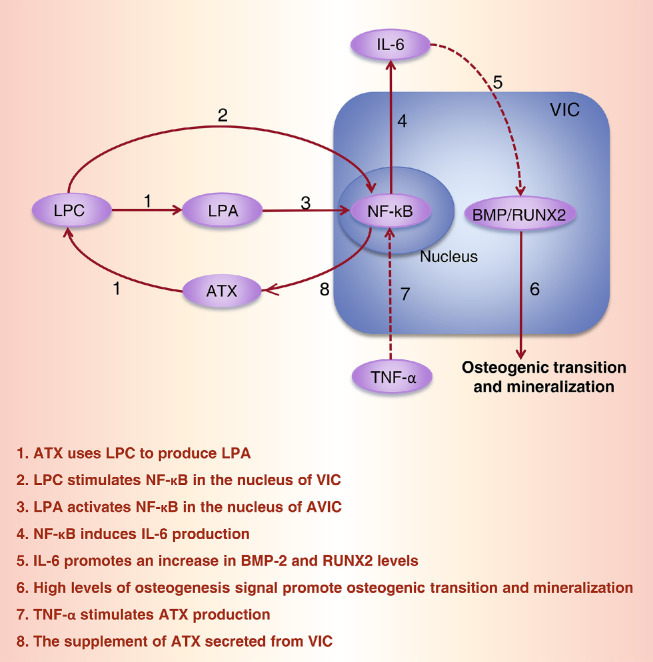
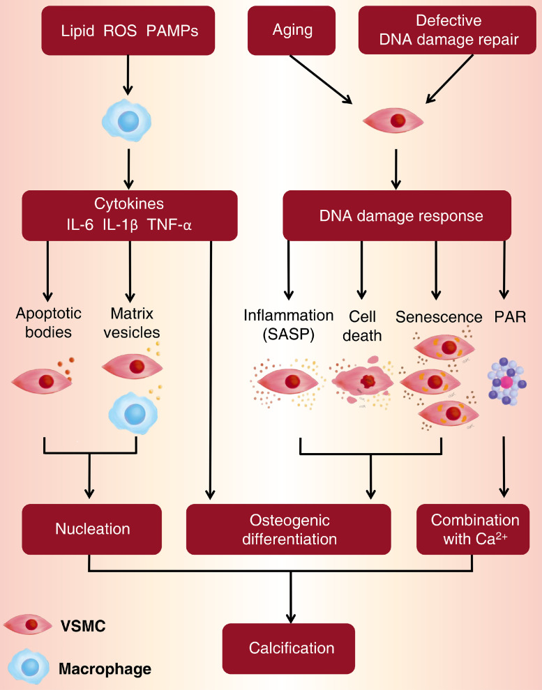
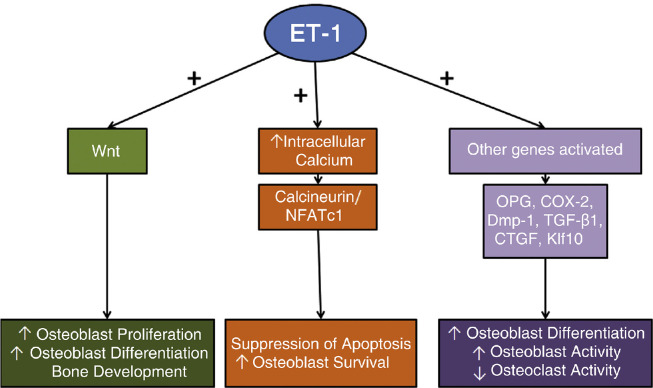
Ectopic mineralization refers to the deposition of mineralized complexes in the extracellular matrix of soft tissues. Calcific aortic valve disease, vascular calcification, gallstones, kidney stones, and abnormal mineralization in arthritis are common examples of ectopic mineralization. They are debilitating diseases and exhibit excess mortality, disability, and morbidity, which impose on patients with limited social or financial resources. Recent recognition that inflammation plays an important role in ectopic mineralization has attracted the attention of scientists from different research fields. In the present review, we summarize the origin of inflammation in ectopic mineralization and different channels whereby inflammation drives the initiation and progression of ectopic mineralization. The current knowledge of inflammatory milieu in pathological mineralization is reviewed, including how immune cells, pro-inflammatory mediators, and osteogenic signaling pathways induce the osteogenic transition of connective tissue cells, providing nucleating sites and assembly of aberrant minerals. Advances in the understanding of the underlying mechanisms involved in inflammatory-mediated ectopic mineralization enable novel strategies to be developed that may lead to the resolution of these enervating conditions.

Keywords: Anti-inflammatory treatments; Ectopic mineralization; Immune cells; Inflammatory conditions; Inflammatory mediators; Osteogenic signaling pathways.


Conflict of interest statement

The authors declare that they have no conflicts of interest in this work.

Figures

Image, graphical abstract
Graphical abstract
Fig 1
Fig. 1
Mechanisms of innate and adaptive immune cells involved in CAVD. Innate and adaptive immune cells promote CAVD through osteogenic differentiation of valve-resident cells, extracellular matrix remodeling, and apoptosis. In the innate immune system, M1 and M2 macrophages can up-regulate the expression of pro-inflammatory cytokines which link with receptors on AVICs to stimulate osteogenic differentiation. Macrophages and mast cells secret matrix metalloproteinases contributing to extracellular matrix remodeling. In the adaptive immune system, mechanisms of adaptive immune cells and CAVD are not clear enough. T regulatory cells can release transforming growth factor-beta (TGF-β) to induce VICs mineralization. Apart from T lymphocytes, antibodies secreted by B lymphocytes show a correlation with calcification in aortic valves. M1, M1 macrophage 1; M2, M2 macrophage; MC, mast cell; N, neutrophil; ECM, extracellular matrix; VSMC, vascular smooth muscle cell; VIC, valvular interstitial cell; ABs, apoptotic bodies; EVs, extracellular vesicles; IL, interleukin; TNF-α, tumor necrosis factor alpha; MMPs, matrix metalloproteinases; TGF-β, transforming growth factor beta; NETs, neutrophil extracellular traps; IFN-γ, interferon gamma. NKT, natural killer T cell; CTL, cytotoxic T lymphocyte; Th, T helper; Treg, T regulatory cell; P, plasmocyte; GzmB, Granzyme B; Prf, Perforin. (modified from [20] with permission from the publisher).
Fig 2
Fig. 2
The working model for PAMPs and DAMPs in CAVD. Caused by endothelial damage, PAMPs enter the subendothelial space. At the meantime, DAMPs assemble at the stricken areas as a response. They recognize and combine with Toll-like receptors on AVICs and stimulate NF-κB signaling to activate downstream signaling pathways. This sequence of events results in promoting osteogenic differentiation of AVICs. PAMP, pathogen-associated molecular pattern; DAMP, damage-associated molecular pattern; dsRNA, double-stranded RNA; LPS, lipopolysaccharide; HMGB1, high-mobility group box-1; TLR, toll-like receptor; NF-κB, nuclear factor κB; BMP, bone morphogenetic protein; RUNX2, Runt-related transcription factor 2; AVIC, aortic valve interstitial cell.
Fig 3
Fig. 3
The working model for the involvement of ATX in CAVD. Schematic of the pathway involved in ATX-mediated inflammation, osteogenic transition, and mineralization. ATX, autotaxin; BMP-2, bone morphogenetic protein-2; RUNX2, runt-related transcription factor 2; CAVD, calcific aortic valve disease; IL-6, interleukin 6; LPA, lysophosphatidic acid; LPC, lysophosphatidylcholine; NF-κB, nuclear factor κB; TNF-α, tumor necrosis factor α; VIC, valvular interstitial cell.
Fig 4
Fig. 4
Mechanisms in the initiation phase and the propagation phase of calcific aortic valve disease. BMP, bone morphogenetic protein; ENPP1, ectonucleotide pyrophosphate 1; LDL, low-density lipoprotein; RANK, receptor activator of nuclear factor κB; RANKL, receptor activator of nuclear factor κB ligand; RAS, renin-angiotensin system; VIC, valvular interstitial cell (modified from [46] with permission from the publisher).
Fig 5
Fig. 5
Osteogenic transition of vascular smooth muscle cells (VSMC) in intimal and medial calcification. A. Within the tunica media, in response to osteogenic stimuli, the VSMC cells differentiate into osteoblast-like cells that produce macroscopic calcification deposits. B. Lipid deposits between the tunica intimal and tunica media of the blood vessel stimulate macrophages infiltration and the differentiation of VSMCs into foam cells. Inflammation, apoptosis, and oxidative stress subsequently cause the VSMCs to differentiate into osteoblast-like cells which, in turn, lead to microcalcification deposits within the intimal wall. SMC, smooth muscle cells; MC, microcalcification. (reprinted from [12] with permission from the publisher).
Fig 6
Fig. 6
Macrophages and VSMCs are the main cells promoting the formation of vascular calcification.Lipid deposition, ROS, and PAMPs stimulate the release of IL-6, IL-1β, and TNF-α from macrophages. These pro-inflammatory cytokines directly promote osteogenic differentiation of VSMCs or promote the secretion of matrix vesicles and apoptotic bodies as nucleation sites for the deposition of Ca/P nanocrystals. Aging and defective DNA damage repair activate DNA damage signals in VSMCs. These damage signals stimulate a number of downstream events including inflammation in association with activation of the senescence-associated secretory phenotype (SASP), cell death, senescence, and formation of PAR (poly[ADP-ribose]). Together, these events provide an environment that is conducive to the calcification of the extracellular matrix. ROS, reactive oxygen species; PAMPs, pathogen-associated molecular patterns; IL-6, interleukin 6; IL-1β, interleukin 1β; TNF-α, tumor necrosis factor α; SASP, senescence-associated secretory phenotype; PAR, poly(ADP-ribose); VSMC, vascular smooth muscle cell.
Fig 7
Fig. 7
Mechanisms of M1 and M2 macrophages in formation and development of kidney stones. M1 and M2 macrophages are differentiated from monocytes and M0 by multiple cytokines, chemokines, and stimulation of calcium oxalate crystals. M1 macrophages act as nephrolithiasis promotors by increasing oxidative stress and pro-inflammatory cytokines such as IL-6, IL-10, and TGF-β; while M2 macrophages act as nephrolithiasis inhibitors by attenuating the development of CaOx crystals, and eliminating them by phagocytosis via lysosomes, clathrin, and anti-inflammatory mediators such as Arginase-1, Ym-1, and PPARγ. AR, androgen receptor; CSF-1, colony-stimulating factor-1; LPS, lipopolysaccharide GM-CSF, granulocyte-macrophage colony-stimulating factor; iNOS, inducible nitric oxide synthase; IRF1, interferon regulatory factor 1; Mφ, macrophage; M-CSF, macrophage colony-stimulating factor; NLRP3, nucleotide-binding oligomerization domain, leucine-rich repeat, and pyrin domain containing 3; FOXO1, forkhead box O1; HIF-1a, hypoxia-inducible factor-1 alpha; PPARγ, peroxisome proliferator-activated receptor-gamma; SIRT3, Sirtuin 3; TLR4, toll-like receptor 4. (reprinted from [87] with permission from the publisher).
Fig 8
Fig. 8
A positive feedback loop of IL-6 and chondrocyte calcification in osteoarthritis. Basic calcium phosphate (BCP) promotes the release of IL-6 from chondrocytes. Exogenous IL-6 increases the expression of genes involved in calcium and phosphorus deposition, including the pyrophosphate channel Ank, the calcium channel Annexin5 and the sodium/ phosphate cotransporter Pit-1. (modified from [97] with permission from the publisher).
Fig 9
Fig. 9
Endothelin-1 (ET-1) signaling pathways specific to osteoblasts. ET-1 activates the Wnt pathway, leading to osteoblast proliferation and differentiation. ET-1 stimulates the calcineurin/NFATc1 pathway which results in the suppression of apoptosis and encourages cell survival. Other genes are also activated to increase osteoblast proliferation and differentiation while inhibiting osteoclast activity. NFATc1, nuclear factor of activated T-cells 1; OPG, osteoprotegerin; COX-2, cyclooxygenase-2; Dmp-1, dentin matrix acidic phosphoprotein 1; TGF-β1, transforming growth factor-β1; CTGF, connective tissue growth factor; Klf10, kruppel-like factor 10. (reprinted from [108] with permission from the publisher).

References

    1. Li Q., Jiang Q., Uitto J. Ectopic mineralization disorders of the extracellular matrix of connective tissue: molecular genetics and pathomechanisms of aberrant calcification. Matrix Biol. 2014;33:23–28. - PMC - PubMed
    1. Maurer K.J., Carey M.C., Fox J.G. Roles of infection, inflammation, and the immune system in cholesterol gallstone formation. Gastroenterology. 2009;136(2):425–440. - PMC - PubMed
    1. Cherng J.H., Hsu Y.J., Liu C.C., et al. Activities of Ca2+-related ion channels during the formation of kidney stones in an infection-induced urolithiasis rat model. Am. J. Physiol. Ren. Physiol. 2019;317(5):F1342–F1349. - PubMed
    1. Lanzer P., Boehm M., Sorribas V., et al. Medial vascular calcification revisited: review and perspectives. Eur. Heart J. 2014;35(23):1515–1525. - PMC - PubMed
    1. Parente F., Pastore L., Bargiggia S., et al. Incidence and risk factors for gallstones in patients with inflammatory bowel disease: a large case-control study. Hepatology. 2007;45(5):1267–1274. - PubMed

LinkOut - more resources