A navigated, robot-driven laser craniotomy tool for frameless depth electrode implantation. An in-vivo recovery animal study
- PMID: 38933084
- PMCID: PMC11199345
- DOI: 10.3389/frobt.2024.1355409
A navigated, robot-driven laser craniotomy tool for frameless depth electrode implantation. An in-vivo recovery animal study
Abstract
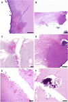
Objectives: We recently introduced a frameless, navigated, robot-driven laser tool for depth electrode implantation as an alternative to frame-based procedures. This method has only been used in cadaver and non-recovery studies. This is the first study to test the robot-driven laser tool in an in vivo recovery animal study. Methods: A preoperative computed tomography (CT) scan was conducted to plan trajectories in sheep specimens. Burr hole craniotomies were performed using a frameless, navigated, robot-driven laser tool. Depth electrodes were implanted after cut-through detection was confirmed. The electrodes were cut at the skin level postoperatively. Postoperative imaging was performed to verify accuracy. Histopathological analysis was performed on the bone, dura, and cortex samples. Results: Fourteen depth electrodes were implanted in two sheep specimens. Anesthetic protocols did not show any intraoperative irregularities. One sheep was euthanized on the same day of the procedure while the other sheep remained alive for 1 week without neurological deficits. Postoperative MRI and CT showed no intracerebral bleeding, infarction, or unintended damage. The average bone thickness was 6.2 mm (range 4.1-8.0 mm). The angulation of the planned trajectories varied from 65.5° to 87.4°. The deviation of the entry point performed by the frameless laser beam ranged from 0.27 mm to 2.24 mm. The histopathological analysis did not reveal any damage associated with the laser beam. Conclusion: The novel robot-driven laser craniotomy tool showed promising results in this first in vivo recovery study. These findings indicate that laser craniotomies can be performed safely and that cut-through detection is reliable.
Keywords: craniotomy; depth electrodes; epilepsy surgery; laser; navigated; robotic.
Copyright © 2024 Winter, Pilz, Kramer, Beer, Gono, Morawska, Hainfellner, Klotz, Tomschik, Pataraia, Hangel, Dorfer and Roessler.
Conflict of interest statement
DB, PG, and MM were employed by AOT, which is the company that provided the laser. The remaining authors declare that the research was conducted in the absence of any commercial or financial relationships that could be construed as a potential conflict of interest. The author(s) declared that they were an editorial board member of Frontiers, at the time of submission. This had no impact on the peer review process and the final decision.
Figures


References
-
- Augello M., Baetscher C., Segesser M., Zeilhofer H. F., Cattin P., Juergens P. (2018). Performing partial mandibular resection, fibula free flap reconstruction and midfacial osteotomies with a cold ablation and robot-guided Er:YAG laser osteotome (CARLO®) – a study on applicability and effectiveness in human cadavers. J. Craniomaxillofac. Surg. 46, 1850–1855. 10.1016/j.jcms.2018.08.001 - DOI - PubMed
-
- Baek K. W., Deibel W., Marinov D., Griessen M., Bruno A., Zeilhofer H. F., et al. (2015). Clinical applicability of robot-guided contact-free laser osteotomy in craniomaxillo-facial surgery: in-vitro simulation and In- vivo surgery in minipig mandibles. Br. J. Oral Maxillofac. Surg. 53, 976–981. 10.1016/j.bjoms.2015.07.019 - DOI - PubMed
-
- Dorfer C., Stefanits H., Pataraia E., Wolfsberger S., Feucht M., Baumgartner C., et al. (2014). Frameless stereotactic drilling for placement of depth electrodes in refractory epilepsy: operative technique and initial experience. Neurosurgery 10 (4), 582–591. 10.1227/NEU.0000000000000509 - DOI - PubMed
LinkOut - more resources
Full Text Sources

