. 2024 May 21;6(13):3410-3425.
doi: 10.1039/d4na00080c. eCollection 2024 Jun 25.

Multiscale modeling of surface enhanced fluorescence

Affiliations

Multiscale modeling of surface enhanced fluorescence

Pablo Grobas Illobre et al. Nanoscale Adv. .

Abstract

The fluorescence response of a chromophore in the proximity of a plasmonic nanostructure can be enhanced by several orders of magnitude, yielding the so-called surface-enhanced fluorescence (SEF). An in-depth understanding of SEF mechanisms benefits from fully atomistic theoretical models because SEF signals can be non-trivially affected by the atomistic profile of the nanostructure's surface. This work presents the first fully atomistic multiscale approach to SEF, capable of describing realistic structures. The method is based on coupling density functional theory (DFT) with state-of-the-art atomistic electromagnetic approaches, allowing for reliable physically-based modeling of molecule-nanostructure interactions. Computed results remarkably demonstrate the key role of the NP morphology and atomistic features in quenching/enhancing the fluorescence signal.


Conflict of interest statement

There are no conflicts to declare.

Figures

Fig. 1
Fig. 1. (a) Graphical depiction of PDI interacting with a spherical NP of radius R. PDI is placed at a distance d from the NP in longitudinal (top) and transversal (bottom) configurations. (b) PDI molecular orbitals involved in the electronic transition under study.
Fig. 2
Fig. 2. Longitudinal (a) and transversal (b) orientations of PDI interacting with a spherical Au NP studied at the QM/ωFQFμ (left) and QM/BEM (right) levels (see top panel). Radiative (middle) and nonradiative (bottom) decay rates (logarithmic scale) as a function of the PDI-NP distance as obtained by varying the NP radius (from 10 Å to 70 Å).
Fig. 3
Fig. 3. QM/ωFQFμ (left) and QM/BEM (right) ΦRB as a function of PDI-NP distance and NP radius for PDI in L orientation interacting with a spherical Au (a) and Ag (b) NP.
Fig. 4
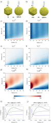
Fig. 4. Graphical representation of PDI interacting with Au Ih (a) and cTO (b) NPs, compared with spherical NPs. QM/ωFQFμ %Γnr (c and d), %Γr (e and f), and %ΦRB (g and h) 2D color plots as a function of the PDI-NP distance and NP radius for Ih vs. sphere and cTO vs. sphere systems, respectively. QM/ωFQFμ %Γnr, %Γr, and %ΦRB values for systems of 30 Å radius are displayed in panels (i) (Ih vs. sphere) and (j) (cTO vs. sphere).
Fig. 5
Fig. 5. QM/ωFQFμ ΦRB (top) and %ΦRB (bottom) as a function of the PDI-NP distance for tip, side, and face PDI adsorption geometries. Ih (a) and cTO (b) NP morphologies are studied and compared to spherical NPs.
Fig. 6
Fig. 6. (a) Geometrical representation of the studied SNRs interacting with PDI. (b) PRF of a SNRs with 18.5 Å radius (r) as function of the length (L). PDI emission (ωem) and absorption (ωabs) frequencies are marked with vertical red and green lines, respectively. (c) PDI ΦRB as a function of PDI-SNR distance for L = 149.5 Å and L = 154 Å.
Fig. 7
Fig. 7. (a) Graphical depiction of SNR pristine (a), r = 18.5 Å; L = 149.5 Å) and defected structures (c, e, g and i), as obtained by atomistic deletions at the SNR end (see red regions). The number of removed atoms is also reported for each structure. The associated ωFQFμ electric field enhancement 2D color maps, obtained at the PRF for a field polarization along the principal axis, are given in panels (b, d, f, h and j). (k) QM/ωFQFμ ΦRB and A (l) values as a function of the PDI-SNR distance.
Fig. 8
Fig. 8. (a) Graphical depiction of SNRs featuring an atomistic tip (a) and a bump (b). The number of added atoms compared to the pristine SNR (r = 18.5 Å; L = 149.5 Å) is also reported for each structure. The associated ωFQFμ electric field enhancement 2D color maps, obtained at the PRF for a field polarization along the principal axis, are given in panels (c and d). (e) QM/ωFQFμ ΦRB and A (f) values as a function of the PDI-SNR distance.
Fig. 9
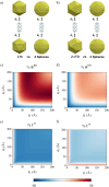
Fig. 9. Graphical representation of PDI interacting with Au Ih (a) and cTO (b) dimers, as compared with spherical NP dimers (radius 30 Å). QM/ωFQFμ %ΦRB (c and d), %Γnr (e and f) 2D color plots as a function of the distances between PDI and the two NPs, for Ih vs. sphere and cTO vs. sphere systems, respectively.
Fig. 10
Fig. 10. Graphical depiction of PDI interacting with two pristine Ag SNRs (a), two Ag SNRs featuring bumps (b) and tips (c). QM/ωFQFμ ΦRB (d–f) and ΦRB (g–i) 2D and 3D color maps as a function of the distances between PDI and the two SNRs.

References

    1. Li M. Cushing S. K. Wu N. Plasmon-enhanced optical sensors: a review. Analyst. 2015;140:386–406. doi: 10.1039/C4AN01079E. - DOI - PMC - PubMed
    1. Sharifi M. Attar F. Saboury A. A. Akhtari K. Hooshmand N. Hasan A. El-Sayed M. A. Falahati M. Plasmonic gold nanoparticles: Optical manipulation, imaging, drug delivery and therapy. J. Controlled Release. 2019;311–312:170–189. doi: 10.1016/j.jconrel.2019.08.032. - DOI - PubMed
    1. Yaraki M. T. Tan Y. N. Metal nanoparticles-enhanced biosensors: synthesis, design and applications in fluorescence enhancement and surface-enhanced Raman scattering. Chem.–Asian J. 2020;15:3180–3208. doi: 10.1002/asia.202000847. - DOI - PMC - PubMed
    1. Albrecht M. G. Creighton J. A. Anomalously intense Raman spectra of pyridine at a silver electrode. J. Am. Chem. Soc. 1977;99:5215–5217. doi: 10.1021/ja00457a071. - DOI
    1. Jeanmaire D. L. Van Duyne R. P. Surface Raman spectroelectrochemistry: Part I. Heterocyclic, aromatic, and aliphatic amines adsorbed on the anodized silver electrode. J. Electroanal. Chem. Interfacial Electrochem. 1977;84:1–20. doi: 10.1016/S0022-0728(77)80224-6. - DOI