. 2024 Jul 23;43(7):114375.
doi: 10.1016/j.celrep.2024.114375. Epub 2024 Jun 26.

The exocyst subunit EXOC2 regulates the toxicity of expanded GGGGCC repeats in C9ORF72-ALS/FTD

Affiliations

The exocyst subunit EXOC2 regulates the toxicity of expanded GGGGCC repeats in C9ORF72-ALS/FTD

Dilara O Halim et al. Cell Rep. .

Abstract

GGGGCC (G4C2) repeat expansion in C9ORF72 is the most common genetic cause of amyotrophic lateral sclerosis (ALS) and frontotemporal dementia (FTD). How this genetic mutation leads to neurodegeneration remains largely unknown. Using CRISPR-Cas9 technology, we deleted EXOC2, which encodes an essential exocyst subunit, in induced pluripotent stem cells (iPSCs) derived from C9ORF72-ALS/FTD patients. These cells are viable owing to the presence of truncated EXOC2, suggesting that exocyst function is partially maintained. Several disease-relevant cellular phenotypes in C9ORF72 iPSC-derived motor neurons are rescued due to, surprisingly, the decreased levels of dipeptide repeat (DPR) proteins and expanded G4C2 repeats-containing RNA. The treatment of fully differentiated C9ORF72 neurons with EXOC2 antisense oligonucleotides also decreases expanded G4C2 repeats-containing RNA and partially rescued disease phenotypes. These results indicate that EXOC2 directly or indirectly regulates the level of G4C2 repeats-containing RNA, making it a potential therapeutic target in C9ORF72-ALS/FTD.

Keywords: ALS; ASO; C9ORF72; CP: Cell biology; CP: Neuroscience; DPR; FTD; exocyst; iPSC; neurodegeneration; neuron; poly(GR).


Conflict of interest statement

Declaration of interests The authors declare no competing interests.

Figures

Figure 1.
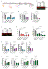
Figure 1.. Generation of a deletion in EXOC2 by the CRISPR-Cas9 technology in iPSC lines derived from two C9ORF72 patients
(A and B) Representative western blot images of EXOC2 in 3-week-old (A) and 3-month-old (B) isogenic control and C9ORF72 iPSC-MNs and densitometric quantification of EXOC2 protein level. Each data point represents one differentiation (n = 3 independent differentiations). Values are mean ± SEM. ns, not significant (one-way ANOVA with Tukey’s multiple comparisons test). (C) Schematic representation of the deletion in EXOC2 near exon 13 in C9ORF72 iPSC lines. (D) qRT-PCR analysis of EXOC2 mRNA levels in 3-week-old MNs differentiated from parental and EXOC2 deletion iPSC lines (EXOC2-del iPSC-MNs). (E) Representative western blot analysis of 3-week-old parental and EXOC2-del iPSC-MNs. Both samples in parental C9-26 or Del-1 came from the same iPSC line. Arrowhead indicates a truncated form of EXOC2, which is present but weaker in Del-1 lanes. (F) Representative immunostaining images for the dendrite-specific marker MAP2 in 3-week-old parental and EXOC2-del iPSC-MNs. (G) Quantification of the numbers of primary dendrites from C9ORF72 and EXOC2-del iPSC-MNs as illustrated in (F). Twenty randomly chosen fields were quantified for analysis. (H) Representative immunofluorescence image of surface NMDAR 2B staining in 3-week-old parental and EXOC2-del C9ORF72 iPSC-MNs. (I) Mean fluorescence values of NMDAR 2B from C9ORF72 and EXOC2-del iPSC-MNs as illustrated in (H). Ten randomly chosen fields were quantified for analysis. Neurons were transduced with lentivirus expressing GFP. Each data point represents one differentiation in (D) and one neuron in (G) (n = 3–4 independent differentiations). Values are mean ± SEM. **p < 0.01; ****p < 0.0001 by one-way ANOVA with Dunnett’s multiple comparisons (patient 1) and two-tailed Student’s t test (patient 2). Scale bar, 20 μm. See also Figures S1 and S2.
Figure 2.
Figure 2.. EXOC2 deletion rescues C9ORF72-ALS/FTD-related disease phenotypes
(A and B) Representative images (A) and densitometric quantification (B) for western blot analysis of CC3 in 3-month-old isogenic control, C9ORF72, and EXOC2-del iPSC-MNs. The upper half of the blot was probed for EXOC2 and actin, and the lower half of the blot was probed for CC3. (C) Representative images for immunostaining for TUJ1 showing neurite degeneration in 3-week-old isogenic control, C9ORF72, and EXOC2-del iPSC-MNs after neurotrophic factor withdrawal. Arrowheads indicate beading or fragmentation. Scale bar, 20 μm. (D) Quantification of images as shown in (C) for MN cultures obtained from the two C9ORF72 patients. Degeneration index was measured by the ratio of fragmented neurites over total area. Twenty randomly chosen fields were quantified for analysis. (E) Representative images for western blot analysis of p62 level in 2-month-old isogenic control, C9ORF72, and EXOC2-del iPSC-MNs. (F) Densitometric quantification of p62 protein level in 2-month-old cultures from isogenic control, C9ORF72, and EXOC2-del iPSC-MNs as shown in (E). Each data point in (B) and (F) represents one differentiation (n = 3–6 independent differentiations); each data point in (D) represents one field. Values are mean ± SEM. *p < 0.05; **p < 0.01; ***p < 0.001; ****p < 0.0001 by one-way ANOVA with Dunnett’s multiple comparisons test. See also Figure S2.
Figure 3.
Figure 3.. EXOC2 deletion reduces poly(GR) and C9ORF72 mRNA levels
(A) Poly(GR) level in 2-month-old isogenic control, parental, and EXOC2-del C9ORF72 iPSC-MNs. (B) Schematic representation of the different C9ORF72 transcript variants and location of the primer sets used to quantify each transcript. (C and D) qRT-PCR analysis of relative C9ORF72-V1 (C) and C9ORF72-V3 (D) mRNA levels in parental and EXOC2-del C9ORF72 iPSC-MNs. (E) Schematic representation of the location of the (A/G) SNP in C9ORF72. (F) Pyrosequencing quantification of the G allele in C9ORF72 and EXOC2-del iPSC-MNs from one patient. Each data point represents one differentiation (n = 3–6 independent differentiations). Values are mean ± SEM. **p < 0.01; ***p < 0.001; ****p < 0.0001 by one-way ANOVA with Dunnett’s multiple comparisons test (A and F and patient 1 in C and D) and two-tailed Student’s t test (patient 2 in B and C). See also Figure S3.
Figure 4.
Figure 4.. Treatment of mature iPSC-MNs with EXOC2 ASOs rescues disease phenotypes
(A) Schematic of ASO treatment and stress induction in mature iPSC-MNs. (B) Representative western blot image showing EXOC2 knockdown efficiency in mature isogenic control or C9ORF72 motor neurons. (C) Densitometric quantification of EXOC2 protein levels. (D) G4C2 repeats-containing pre-mRNA levels in C9ORF72 patient neurons treated with EXOC2 ASOs. (E and F) Representative western blot image (E) and densitometric quantification (F) of CC3 in mature isogenic control and C9ORF72 iPSC-MNs treated with control or EXOC2 ASOs. (G) ELISA showing pNfH in culture media from mature isogenic control and C9ORF72 iPSC-MNs treated with control or EXOC2 ASOs to assess neurodegeneration. pNfH levels were normalized to total protein concentration. (H and I) qRT-PCR analysis of relative EXOC2 mRNA and C9ORF72 V1/V3 pre-mRNA levels in 3-week-old C9ORF72 patient 4 (H) or patient 3 (I) iPSC-MNs left untreated or treated with 5 μM nontargeting control ASO or EXOC2 ASOs. (J) pNfH levels (normalized to total protein concentration) in culture media from mature iPSC-MNs of C9ORF72 patients and healthy subjects treated with control or EXOC2 ASOs to assess neurodegeneration. Each data point represents one differentiation (n = 3–4 independent differentiations). Values are mean ± SEM. **p < 0.01; ***p < 0.001; ****p < 0.0001 by one-way ANOVA with Dunnett’s multiple comparisons test. See also Figure S4.

References

    1. Ling SC, Polymenidou M, and Cleveland DW (2013). Converging mechanisms in ALS and FTD: disrupted RNA and protein homeostasis. Neuron 79, 416–438. 10.1016/j.neuron.2013.07.033. - DOI - PMC - PubMed
    1. Gitler AD, and Tsuiji H (2016). There has been an awakening: Emerging mechanisms of C9orf72 mutations in FTD/ALS. Brain Res. 1647, 19–29. 10.1016/j.brainres.2016.04.004. - DOI - PMC - PubMed
    1. Gao FB, Almeida S, and Lopez-Gonzalez R (2017). Dysregulated molecular pathways in amyotrophic lateral sclerosis-frontotemporal dementia spectrum disorder. EMBO J. 36, 2931–2950. 10.15252/embj.201797568. - DOI - PMC - PubMed
    1. DeJesus-Hernandez M, Mackenzie IR, Boeve BF, Boxer AL, Baker M, Rutherford NJ, Nicholson AM, Finch NA, Flynn H, Adamson J, et al. (2011). Expanded GGGGCC hexanucleotide repeat in noncoding region of C9ORF72 causes chromosome 9p-linked FTD and ALS. Neuron 72, 245–256. 10.1016/j.neuron.2011.09.011. - DOI - PMC - PubMed
    1. Renton AE, Majounie E, Waite A, Simón-Sánchez J, Rollinson S, Gibbs JR, Schymick JC, Laaksovirta H, van Swieten JC, Myllykangas L, et al. (2011).A hexanucleotide repeat expansion in C9ORF72 is the cause of chromosome 9p21-linked ALS-FTD. Neuron 72, 257–268. 10.1016/j.neuron.2011.09.010. - DOI - PMC - PubMed

Publication types

MeSH terms

Supplementary concepts

LinkOut - more resources