. 2024 Jun 28;384(6703):ado7082.
doi: 10.1126/science.ado7082. Epub 2024 Jun 28.

Brainwide silencing of prion protein by AAV-mediated delivery of an engineered compact epigenetic editor

Affiliations

Brainwide silencing of prion protein by AAV-mediated delivery of an engineered compact epigenetic editor

Edwin N Neumann et al. Science. .

Abstract

Prion disease is caused by misfolding of the prion protein (PrP) into pathogenic self-propagating conformations, leading to rapid-onset dementia and death. However, elimination of endogenous PrP halts prion disease progression. In this study, we describe Coupled Histone tail for Autoinhibition Release of Methyltransferase (CHARM), a compact, enzyme-free epigenetic editor capable of silencing transcription through programmable DNA methylation. Using a histone H3 tail-Dnmt3l fusion, CHARM recruits and activates endogenous DNA methyltransferases, thereby reducing transgene size and cytotoxicity. When delivered to the mouse brain by systemic injection of adeno-associated virus (AAV), Prnp-targeted CHARM ablates PrP expression across the brain. Furthermore, we have temporally limited editor expression by implementing a kinetically tuned self-silencing approach. CHARM potentially represents a broadly applicable strategy to suppress pathogenic proteins, including those implicated in other neurodegenerative diseases.


Conflict of interest statement

Competing interests:

J.S.W. declares outside interest in 5 AM Venture, Amgen, Chroma Medicine, KSQ Therapeutics, Maze Therapeutics, Tenaya Therapeutics, Tessera Therapeutics, Ziada Therapeutics and Third Rock Ventures. S.M.V. acknowledges speaking fees from Ultragenyx, Illumina, Biogen, Eli Lilly; consulting fees from Invitae and Alnylam; research support from Ionis, Gate, Sangamo. E.V.M. acknowledges speaking fees from Eli Lilly; consulting fees from Deerfield and Alnylam; research support from Ionis, Gate, Sangamo, and Eli Lilly. A.S.K. is a scientific advisor for and holds equity in Senti Biosciences and Chroma Medicine, and is a co-founder of Fynch Biosciences and K2 Biotechnologies. E.N.N., T.M.B., and J.S.W. are co-inventors on U.S. patent application no. 18/444,590 filed by the Whitehead Institute, relating to CHARM and self-silencing AAVs. E.N.N, T.M.B., J.S.W., E.V.M., and S.M.V. are co-inventors on provisional U.S. patent application no. 63/623,787 filed jointly by the Whitehead Institute and Broad Institute relating to ZFcharms for prion disease. B.E.D. declares outside interest in Apertura Gene Therapy and Tevard Biosciences and is an inventor on U.S. patent application no. US11499165B2 relating to the PHP.eB AAV capsid. All other authors declare that they have no competing interests.

Figures

Fig. 1.
Fig. 1.. PRNP is a viable target for epigenetic silencing, but existing technologies are not suitable for therapeutic use.
(A) A HEK293T cell line was made by integrating a lentiviral vector containing mU6-sgRNA targeting the PRNP TSS. Transfected cells were sorted by FACS (TagBFP) two days post-transfection and monitored for PRNP silencing by Alexa Fluor 647 anti-PrP staining and flow cytometry. (B) PrP and effector expression time course of HEK293T cells transiently transfected with plasmids encoding CRISPRi and CRISPRoff effectors. Data are mean ± SEM of n=2 replicates. (C) DNA methylation assessment by targeted nanopore long read sequencing of native genomic DNA extracted from HEK293T cells 50 days post-transfection. (D) Mouse N2a cells co-transfected with plasmids encoding CRISPRi/CRISPRoff and three sgRNAs targeting the TSS of Prnp were assessed for PrP expression and DNA methylation. (E) Schematic depicting AAV genome packaging constraints with CRISPRoff and ZFPoff to scale. (F) HEK293T cells were transiently transfected with ZFPoff and D3L-ZFP-KRAB and imaged after 6 days.
Fig. 2.
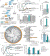
Fig. 2.. A histone H3 tail fused to the Dnmt3l C-terminal domain acts as a potent mediator of DNA methylation and transcriptional silencing.
(A) Cartoon depiction of endogenous DNMT3A recruitment and activation by the CHARM system. (B) Time course of effector (TagBFP) and mScarlet-CLTA reporter expression after transient transfection with effector-containing plasmids. Data are mean ± SEM of n=2 replicates. (C) First pass histone H3 tail fusion test on the mScarlet-CLTA reporter using different linkers to D3L. (D) Refinement of linker sequence between H3 tail and D3L. (E) Phylogenetic tree of DNMT3L orthologs and ancestral reconstruction nodes. Orthologs with measured silencing activity >5% by 14 days are labeled. (F) Repression of mScarlet-CLTA reporter 2 weeks post-transfection with different DNMT3L ortholog C-terminal domains fused to dCas9. (G) Transient transfection and repression of mScarlet-CLTA reporter with different length histone isoform H3.1 domains (FL; full length). A mismatched sgRNA against CLTA TSS is used to improve dynamic range. (H) Transient transfection of sgRNA and effector fused to dCas9 targeting the CLTA reporter in a methyltransferase knockout background. Data are mean ± SEM of n=3 replicates. (I) Time course of effector expression and mScarlet-CLTA silencing comparing CRISPRoff and CRISPRi against the series of optimized CHARM constructs. Data are mean ± SEM of n=2 replicates. (J) Comparison of CRISPRi, CRISPRoff, and the optimized CRISPRcharm effectors in silencing cell surface markers. Vectors encoding mU6-sgRNAs were transduced via lentivirus and effector plasmids were transiently transfected.
Fig. 3.
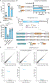
Fig. 3.. CHARM is flexible and specific.
(A) Mouse N2a cells were transiently transfected with plasmids encoding ZFcharm Kv1 constructed with the mouse Prnp-targeting ZFP 81187, ZFP 81201, or a non-targeting ZFP and stained with Alexa Fluor 647 anti-PrP. (B) Mouse N2a cells were transiently transfected with TALEcharm and TALEcharm Kv2 composed of engineered TALE proteins targeting the mouse Prnp TSS or a non-targeting TALE, then measured using Alexa Fluor 647 anti-PrP. (C) Schematic of AAV packaging using space-saving techniques like split-inteins (65) or a self-silencing approach including the tamoxifen-inducible engineered estrogen receptor ERT2 (66). WPRE3 is a structured 3’ element for mRNA stability (67). (D) HEK293T cells were transiently transfected with plasmids encoding ZFP81187 alone or the fusions ZFPoff, D3L-ZFP-KRAB, and ZFcharm Kv1 and then counted by flow cytometry after cell viability staining with LIVE/DEAD near-IR dye. Data are mean ± SEM of n=3 replicates. Statistical analyses are one-way ANOVAs followed by Tukey’s multiple comparisons test (**** p<0.0001; ns, not significant). (E and F) ZFcharm Kv1 (E) and CRISPRcharm Kv1 (F) were introduced into N2a cells by lentiviral transduction and assessed for Prnp knockdown and specificity by RNA sequencing 4 weeks later. CRISPRcharm Kv1 was evaluated in N2a cells expressing sgRNA targeting the mouse Prnp TSS or a non-targeting sgRNA. Log2 fold changes (upper) and volcano plots (lower) were generated from DESeq2.
Fig. 4.
Fig. 4.. AAV-delivered ZFcharms repress and methylate Prnp in vivo.
(A) Schematic of experimental design. ZFcharm Kv1 and ZFcharm were delivered to mice via AAV and whole brains were harvested 6 weeks later. Unless otherwise noted, the AAV dose was 1.5e13 vg/kg. (B) PrP ELISA and Prnp RT-qPCR data generated from brain hemisphere homogenate 6 weeks post injection. Data are mean ± SD of n=5–6 replicates. (C) ZFcharm Kv1 AAV dose-response analysis. Data are mean ± SD of n=6–8 replicates. (D) Quantification of DNA methylation via nanopore sequencing of the Prnp promoter. (E and F) Visualization of Prnp (yellow) and pan-neuronal marker Uchl1 (magenta) expression in coronal brain sections via HCR RNA-FISH (DAPI staining in blue). (E) Representative maximum-intensity projections of coronal brain hemisphere tile scans. White boxes indicate brain regions shown in panel F. Scale bar, 1mm. (F) Zoomed-in views of the cortex (CTX), hippocampus (HP), and thalamus (TH). Scale bar, 100 µm. (G) Machine learning classification of Prnp+ (yellow) and Prnp- (magenta) neurons using QuPath software (71). Uchl1- cells are shown in gray. Cell boundaries represent 4 µm expansions from DAPI-detected nuclei. Scale bar, 1mm. (H) Representative histograms of mean Prnp intensity in neurons. (I) Bar chart showing % Prnp+ neurons in treated and untreated brains based on QuPath classification. Data are mean ± SD of n=3 replicates. Statistical analyses are one-way ANOVAs followed by Tukey’s multiple comparisons test for panels B and C, and unpaired t test for panel I (* p < 0.05; ** p < 0.005; *** p < 0.0005; **** p<0.0001; ns, not significant).
Fig. 5.
Fig. 5.. Transient CHARM expression through self-silencing is sufficient for persistent PrP repression.
Self-silencing kinetics were quantified by measuring CHARM (orange; TagBFP or mCherry) and PrP (navy; Alexa Fluor 647 anti-PrP) expression over time via flow cytometry after lentiviral transduction of N2a cells. (A) Schematic of self-silencing CHARM constructs. (B) Schematic of experimental design. (C) Representative flow cytometry histograms of ZFcharm Kv1 and PrP expression at days 6, 14, and 60 post transduction. Dashed line indicates separation between expressing (‘ON’) and silenced (‘OFF’) cells. (D) Clonal bisulfite sequencing of EFS promoters driving ZFcharm Kv1-SCR and ZFcharm Kv1-SPM expression 5 and 25 days post transduction of N2a cells. % 5mCpG (black) across PCR clones is depicted as a pie chart. Sequence elements within the EFS promoter are shown in the schematic under the data. CpGs between the TATA box and TSS are highlighted in gray. (E) 60-day flow cytometry time course monitoring ZF editor and PrP expression across ZF-SPM constructs. Data are mean ± SD of n=2 replicates. (F) ZFcharm Kv1-SPM and PrP expression 6 months post transduction (n=1). (G) Schematic of experimental strategy to engineer a modular self-silencing ZFcharm Kv2 using two distinct ZF domains. (H) Tuning of self-silencing kinetics using an allelic series of ZF3 backbone RtoA mutations. ZFcharm Kv2 and PrP expression were quantified 9 and 22 days post transduction. Data are mean ± SD of n=2 replicates.
Fig. 6.
Fig. 6.. Self-silencing ZFcharm is functional in vivo.
(A) Schematic of experimental design. (B and C) PrP ELISA and Prnp RT-qPCR data generated from brain hemisphere homogenate 6 weeks post injection of 1.5e13 vg/kg AAV. AAV capsids were packaged with self-silencing ZFcharm constructs containing (B) or lacking (C) the KRAB domain. Data are mean ± SD of n=6–8 replicates. Statistical analyses are one-way ANOVAs followed by Tukey’s multiple comparisons test (* p < 0.05; ** p < 0.005; *** p < 0.0005; **** p<0.0001; ns, not significant). (D and E) Clonal bisulfite sequencing of the EFS promoter driving expression of self-silencing ZFcharm constructs containing (D) or lacking (E) the KRAB domain. % 5mCpG (black) is calculated for each CpG site across PCR clones and depicted as a pie chart. Sequence elements within the EFS promoter are shown in the schematic under the data. CpGs between the TATA box and TSS are highlighted in gray and the ZF binding site is highlighted in orange.

Comment in

References

    1. Prusiner SB, Prions. Proc. Natl. Acad. Sci. U. S. A 95, 13363–13383 (1998). - PMC - PubMed
    1. Vallabh SM, Minikel EV, Schreiber SL, Lander ES, Towards a treatment for genetic prion disease: trials and biomarkers. Lancet Neurol 19, 361–368 (2020). - PubMed
    1. Maddox RA, Person MK, Blevins JE, Abrams JY, Appleby BS, Schonberger LB, Belay ED, Prion disease incidence in the United States: 2003–2015. Neurology 94, e153–e157 (2020). - PubMed
    1. Büeler A. Aguzzi, Sailer A, Greiner RA, Autenried P, Aguet M, Weissmann C, Mice devoid of PrP are resistant to scrapie. Cell 73, 1339–1347 (1993). - PubMed
    1. Mallucci G, Dickinson A, Linehan J, Klöhn PC, Brandner S, Collinge J, Depleting Neuronal PrP in Prion Infection Prevents Disease and Reverses Spongiosis. Science 302, 871–874 (2003). - PubMed

Publication types