. 2024 Jun 28;384(6703):eadm8693.
doi: 10.1126/science.adm8693. Epub 2024 Jun 28.

A neutralizing antibody prevents postfusion transition of measles virus fusion protein

Affiliations

A neutralizing antibody prevents postfusion transition of measles virus fusion protein

Dawid S Zyla et al. Science. .

Abstract

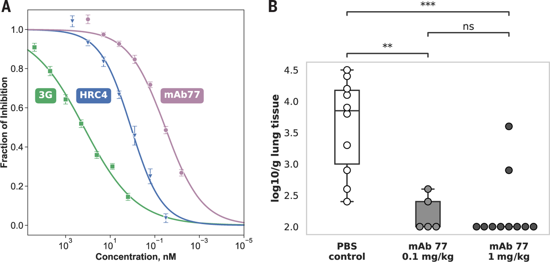
Measles virus (MeV) presents a public health threat that is escalating as vaccine coverage in the general population declines and as populations of immunocompromised individuals, who cannot be vaccinated, increase. There are no approved therapeutics for MeV. Neutralizing antibodies targeting viral fusion are one potential therapeutic approach but have not yet been structurally characterized or advanced to clinical use. We present cryo-electron microscopy (cryo-EM) structures of prefusion F alone [2.1-angstrom (Å) resolution], F complexed with a fusion-inhibitory peptide (2.3-Å resolution), F complexed with the neutralizing and protective monoclonal antibody (mAb) 77 (2.6-Å resolution), and an additional structure of postfusion F (2.7-Å resolution). In vitro assays and examination of additional EM classes show that mAb 77 binds prefusion F, arrests F in an intermediate state, and prevents transition to the postfusion conformation. These structures shed light on antibody-mediated neutralization that involves arrest of fusion proteins in an intermediate state.


Figures

Fig. 1.
Fig. 1.. mAb 77 displays robust antiviral activity in vitro and in vivo.
(A) Antiviral potency of mAb 77 (violet) in a viral entry assay to measure inhibition of infection in Vero cells expressing SLAM/CD150. The activity was compared with that of established MeV inhibitors, 3G [green; (51)] and HRC4 [blue; (53)]. Values for IC50 and Hill slope were estimated using logistic regression. 3G: IC50 = 150.1 ± 16.2 nM, Hill slope = 0.37 ± 0.01; HRC4: IC50 = 1.17 ± 0.27 nM, Hill slope = 0.62 ± 0.07; mAb 77: IC50 = 0.034 ± 0.005 nM, Hill slope = 0.52 ± 0.04. Each point with an error bar shows the average of three separate biological tests ± SE. (B) Antibody transfer experiment to assess in vivo protective efficacy of mAb 77. Cotton rats received intraperitoneal (IP) injections of 0.1 mg/kg or 1 mg/kg mAb 77 12 hours before challenge with MeV. Animals in the control group received PBS before the challenge. Values correspond to MeV titers in lung tissue collected 4 days after infection. The line inside the box represents the median value. Significance is shown using the Mann-Whitney-Wilcoxon test. P value annotations: ns, nonsignificant; P ≤ 1.00; *: 0.01 < P ≤ 0.05; **: 0.001 < P ≤ 0.01; ***: 0.0001 < P ≤ 0.001.
Fig. 2.
Fig. 2.. mAb 77 mirrors the action of the HRC4 inhibitor and stabilizes the extended state of F protein.
(A) Schematic representation of the RBC fusion assay conducted in HEK293T cells expressing a chimeric HN-H receptor binding protein with MeV F protein. (1) Without fusion inhibitors, the HN-H binds sialic acid on the RBCs. The MeV H stalk region subsequently activates the F protein, facilitating its refolding to a fusion intermediate that anchors the fusion peptide in the RBC membrane, culminating in the fusion of RBC and HEK293T membranes. (2) In the presence of zanamivir, a small-molecule inhibitor that competitively binds the HN sialic acid receptor binding site, reversible binding of RBCs to HN is observed (2.1). Adding 3G locks the F protein in its prefusion conformation, resulting in only reversible binding of most RBCs to the HN-H (2.2). (3) The subsequent checkpoint is evaluated through combined administration of zanamivir and ACK buffer. Zanamivir acts as described in (2), whereas ACK buffer induces lysis of RBCs (dashed line). At this checkpoint, fusion intermediates with anchored fusion peptides within RBC membranes, such as those stabilized by HRC4, resist zanamivir-induced dissociation (left, 3.1) but are lysed in the presence of ACK buffer (right, 3.2). (4) At the last checkpoint, complete fusion of RBC and HEK293T membranes is achieved. Here, ACK buffer addition has no impact on the fused RBCs (4.1). (B) Bar chart summarizing the results of a mechanistic assay comparing various known inhibitors and mAb 77. For each condition, cells were categorized on the basis of their interaction with RBCs: reversibly bound (dark blue), in which RBCs were adhered only by chimeric H and could be removed by treatment with zanamivir; irreversibly bound (pink), in which RBCs were initially attached by chimeric H, but subsequent F protein refolding anchored the fusion peptide in the RBC membrane, making RBC attachment insensitive to zanamivir, with RBC retained on the HEK293T cell surface; and fused (light blue), in which RBCs underwent membrane fusion with HEK293T cells expressing the fusion complex.
Fig. 3.
Fig. 3.. Cryo-EM structure of mAb 77 Fab fragment engaging FECTO at the base of the head domain.
(A) Primary structure annotation of FECTO. Functional domains are delineated as color-coded rectangular regions: SP, signal peptide; FP, fusion peptide; HRN, N-terminal heptad repeat; DIII, domain III; DI, domain I; DII, domain II; HRC, C-terminal heptad repeat; 2xST, Twin Strep-Tag II. White hexagons denote glycosylation sites within the F2 region. A black loop represents natural disulfide bonds. Black scissors indicate the furin cleavage site that divides the precursor F0 into F2 and F1 subunits. Arrows indicate the locations of two stabilizing mutations. (B) A 2.1-Å-resolution map of FECTO, showing F1 (green) and F2 (gray) with low-resolution map overlay showing details included in the map (silhouette). (C) Ribbon representation of the 2.3-Å-resolution structure of FECTO in the presence of FIP with a low resolution map as a silhouette. (D) Domain description of FECTO, colored from N terminus to C terminus (red to purple), as in (A). (E) A ribbon representation of FECTO in complex with three copies of Fab 77. The Fabs interact symmetrically with the lower section of the head domain in a cleft between domains I and II. The FECTO F1 chain is represented in green, the F2 chain in gray, the 77 Fab heavy variable domain (VH) in violet, and the light variable domain (VL) in pale pink. The cryo-EM map, delineated as a silhouette, also encompasses the masked-out region, including the constant light and heavy segments (CL and CH) of the Fab 77. (F) CDR loops H1 and H2 of Fab 77 establish hydrogen bonds with three distinct FECTO polypeptide chains: F2A (light gray), F1A (light green), and F1B (dark green). The interacting residues on FECTO and Fab 77 are shown in the respective colors of their chains. Interface residues F1B 383 and 384 and CRD-H1 32 and 33 are omitted for better visibility. Dashed lines depict the selected hydrogen bonds. (G) The VH region of Fab 77 contributes the highest number of interactions with F, and all three heavy chain complementarity-determining regions (CDRs H1, H2, and H3) engage with the head domain. Only one CDR L2 from the VL domain contributes to the interaction. Black rectangles show regions enlarged in (F) and (H). (H) The CDR H3 and L2 interact with only a single FECTO chain, F1A. The interacting residues are highlighted in the color of the respective chains, as in (E). Dashed lines represent chosen hydrogen bonds; a solid line indicates a salt bridge. Single-letter abbreviations for the amino acid residues are as follows: A, Ala; C, Cys; D, Asp; E, Glu; F, Phe; G, Gly; H, His; I, Ile; K, Lys; L, Leu; M, Met; N, Asn; P, Pro; Q, Gln; R, Arg; S, Ser; T, Thr; V, Val; W, Trp; and Y, Tyr.
Fig. 4.
Fig. 4.. The postfusion conformation of FECTO shows significant structural domain reconfiguration that facilitates membrane fusion.
(A) Cryo-EM structure of FECTO in its postfusion conformation revealing a structural rearrangement of the HRN and HRC, forming a canonical postfusion six-helix bundle. F1 and F2 are in shades of blue and pink, respectively. The cryo-EM map is displayed as a silhouette, with two notable glycosylation sites (N61 and N67) indicated and visualized only at the map level. (B) Domain-based coloring of FECTO in the postfusion conformation, as in Fig. 3D. (C) Global alignment comparison between FECTO in prefusion (depicted in white with silhouette) and postfusion (in domain color scheme) conformations. Optimal global alignment per chain was attained by aligning FECTO on domain I (highlighted in light green). Regions that undergo conformational changes are surrounded in boxes and shown in (D) and (E). (D) Domain III undergoes the most drastic shift between pre- and postfusion, moving its center of mass over by 11 Å and rotating by 52°. Most of the fold remains unchanged when local alignment is performed on the sequence region corresponding to the domain region, with the most notable changes seen in the HRN, which releases the fusion peptide and refolds from multiple disjoined α helices and β strands into an ~100 Å extended helix. The furin-containing loop is depicted as a dashed line. Scissors symbolize the furin cleavage site. (E) Domain II undergoes a twisting motion during the refolding step. Only slight differences are observed in the fold after local alignment for the residue range corresponding to the domain. The C-terminal region of the domain undergoes the most change. The HRC changes direction by nearly 180° around residue G433. HRC in black and color corresponds to the prefusion and postfusion structure, respectively. (F) Mapping of residues comprising the mAb 77 epitope on both the pre- and postfusion states of FECTO reveals displacement of the epitope in the postfusion state that explains the lack of mAb 77 binding to FECTO in the postfusion conformation.
Fig. 5.
Fig. 5.. Structural analysis of MeV F sequence conservation and viral evolution assay reveals consistent conservation across the protein.
(A) Conservation map of the MeV F full-length primary sequence derived from >830 F sequences in the NCBI protein database. The sequences were aligned using Clustal Omega (88), and the AL2CO algorithm (89) was used to calculate the conservation score. MeV F demonstrates uniform sequence conservation, with only two regions having lower conservation: the signal peptide and the cytoplasmic tail, which are highlighted by black bars at the top of the plot. (B) Mapping the AL2CO score onto the tertiary pre- and postfusion structure of FECTO shows no conservation hotspots. In general, less exposed residues show higher sequence conservation compared with solvent-exposed residues. (C) Most residues forming the mAb 77 epitope demonstrate high sequence conservation (light gray), but G37 and R437 (shown in shades of yellow) have been found in natural variants. (D) Cell-based fusion inhibition assay to examine the impact of G37R and R436K mutations on mAb 77 binding affinity. The R436K mutation is common but has minimal influence on inhibition of antibody-mediated fusion. Conversely, the rarer G37R mutation could evade fusion inhibition by mAb 77.
Fig. 6.
Fig. 6.. Cryo-EM to elucidate mAb 77 mechanism of action.
(A) 2D classes show conformational heterogeneity in the FECTO–Fab 77 dataset. Using an extensive 2D classification procedure, we could isolate 2D classes that constituted <1% of the dataset. The leftmost panel, panel 1, shows a 2D projection of the final volume of FECTO–Fab 77, followed by classes that depict intermediate refolding steps during Fab 77–induced FECTO refolding. Panel 2 shows a refolding intermediate, clearly showing a missing density corresponding to the HRN domain and a lack of density between Fab 77–bound domain II (DII, cyan) and domain I (DI, light green) of neighboring protomers. Panel 3 shows a conformational state with protomer separation, which finally leads to a stabilized F fragment (domains I and II) by Fab 77 in panel 4. The upper and lower circles correspond to the projection and annotation based on the projection, respectively. Colors correspond to the domains: purple, Fab 77; light green, domain I; dark green, domain II; and orange, HRN. Scale bars correspond to 100 Å (first three panels from left) and 50 Å (rightmost panel). F2 was omitted from the annotated image for clarity. (B) Cryo-EM 2D class averages of Fab 77 stabilizing a fragment of FECTO. (C) Approximately 3.6-Å-resolution density map of the Fab 77–FECTO fragment superimposed on the trimeric FECTO–Fab 77 complex. A zoomed-in view of the corresponding density associated with only a stabilized portion of FECTO is shown on the right. The interaction between Fab 77 and FECTO maintains stabilization of the previously characterized epitope to inhibit further refolding. Domain coloring follows the scheme presented in Fig. 3. The left map was Gaussian-blurred to 1.5 SD to simplify visualization. Residues corresponding to the FECTO fragment are indicated. (D) The proposed mechanism of action for mAb 77 depicts the observed populations of FECTO–Fab 77 complexes across the reported experiments. Numbers for each stage of refolding correspond to those in (A).

Comment in

References

    1. Katz SL, Hinman AR, Summary and conclusions: Measles elimination meeting, 16–17 March 2000. J. Infect. Dis 189 (suppl. 1), S43–S47 (2004). doi: 10.1086/377696 - DOI - PubMed
    1. Fiebelkorn AP et al. A comparison of postelimination measles epidemiology in the United States, 2009–2014 versus 2001–2008. J. Pediatric Infect. Dis. Soc 6, 40–48 (2017). - PMC - PubMed
    1. Gastañaduy PA et al. Measles—United States, January 1–May 23, 2014. MMWR Morb. Mortal. Wkly. Rep 63, 496–499 (2014). doi: 10.1111/ajt.12902 - DOI - PMC - PubMed
    1. Fill M-MA et al. Notes from the field: Measles outbreak of unknown source — Shelby County, Tennessee, April–May 2016. MMWR Morb. Mortal. Wkly. Rep 65, 1039–1040 (2016).doi: 10.15585/mmwr.mm6538a3 - PubMed
    1. Orenstein W, Seib K, Mounting a good offense against measles. N. Engl. J. Med 371, 1661–1663 (2014). doi: 10.1056/NEJMp1408696 - DOI - PubMed

Publication types

MeSH terms

LinkOut - more resources