. 2024 Jun 27;20(6):e1011337.
doi: 10.1371/journal.pgen.1011337. eCollection 2024 Jun.

Golgi associated RAB2 interactor protein family contributes to murine male fertility to various extents by assuring correct morphogenesis of sperm heads

Affiliations

Golgi associated RAB2 interactor protein family contributes to murine male fertility to various extents by assuring correct morphogenesis of sperm heads

Haoting Wang et al. PLoS Genet. .

Abstract

Sperm heads contain not only the nucleus but also the acrosome which is a distinctive cap-like structure located anterior to the nucleus and is derived from the Golgi apparatus. The Golgi Associated RAB2 Interactors (GARINs; also known as FAM71) protein family shows predominant expression in the testis and all possess a RAB2-binding domain which confers binding affinity to RAB2, a small GTPase that is responsible for membrane transport and vesicle trafficking. Our previous study showed that GARIN1A and GARIN1B are important for acrosome biogenesis and that GARIN1B is indispensable for male fertility in mice. Here, we generated KO mice of other Garins, namely Garin2, Garin3, Garin4, Garin5a, and Garin5b (Garin2-5b). Using computer-assisted morphological analysis, we found that the loss of each Garin2-5b resulted in aberrant sperm head morphogenesis. While the fertilities of Garin2-/- and Garin4-/- males are normal, Garin5a-/- and Garin5b-/- males are subfertile, and Garin3-/- males are infertile. Further analysis revealed that Garin3-/- males exhibited abnormal acrosomal morphology, but not as severely as Garin1b-/- males; instead, the amounts of membrane proteins, particularly ADAM family proteins, decreased in Garin3 KO spermatozoa. Moreover, only Garin4 KO mice exhibit vacuoles in the sperm head. These results indicate that GARINs assure correct head morphogenesis and some members of the GARIN family function distinctively in male fertility.


Conflict of interest statement

The authors have declared that no competing interests exist.

Figures

Fig 1
Fig 1. GARINs show predominant expression in mouse testes and interact with RAB2A/B.
(A) Schematic representation of GARIN family members. The RAB2-binding domain is shown in blue. (B) RT-PCR for GARIN family members was performed with cDNA obtained from mouse tissues. Actb was used as a loading control. BR, brain; TH, thymus; LU, lung; HE, heart; LI, liver; SP, spleen; KI, kidney; TE, testis; OV, ovary. (C) RT-PCR was performed with cDNA obtained from postnatal testes with primers for GARIN family members. Actb was used as a loading control. (D) Schematic representation of the domains of RAB2A/B used in Fig 1E-I. The PA tag is shown in green and the Small GTP-binding protein domain is shown in magenta. (E—I) Co-IP of GARINs and RAB2A/B. Anti-FLAG and anti-PA antibodies were used to detect GARINs-FLAG and PA-RAB2A/B, respectively. In Fig 1E, the input PA bands were saturated because the signals were strong. Anti-ACTB antibody was used to detect ACTB as an endogenous control.
Fig 2
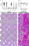
Fig 2. In vivo fertility test and histological analyses of Garin2-5b-/- males.
(A) Pups per plug for WT males and Garin2-5b (Garin2, 3, 4, 5a, and 5b) KO males. A vaginal plug was considered as a sign of coitus. For each genotype, 5 males were tested. Pups/plug of Garin2-5b-/- males were compared with WT males (One-way ANOVA, c, P < 0.001). (B) Testis weight of WT and Garin2-5b-/- males. The difference between testis weight of WT and Garin2-5b-/- males was not significant (One-way ANOVA, P > 0.05). (C) PAS-staining of testis and epididymis sections, from WT and Garin2-5b-/- males.
Fig 3
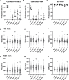
Fig 3. In vitro fertilization and motility of Garin2-5b KO spermatozoa.
(A—C) In vitro fertilization analyses of WT and Garin2-5b (Garin2, 3, 4, 5a, and 5b) KO males in cumulus-intact condition (A), cumulus-free condition (B), and zona pellucida-free (ZP-free) condition (C). (D—I) straight line velocity (VSL), curvilinear velocity (VCL), and average path velocity (VAP) of WT spermatozoa and Garin2-5b KO spermatozoa after incubating for 10 minutes (D–F) or 120 minutes (G–I). Velocity parameters of Garin2-5b KO spermatozoa were compared with WT (One-way ANOVA, a, P < 0.05; b, P < 0.01; c, P < 0.001). For each KO mouse line and WT mouse, more than 3 males were analyzed.
Fig 4
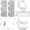
Fig 4. Sperm head morphology examination by elliptic Fourier descriptors (EFDs) and principal component (PC) analysis.
(A) Representative images of sperm head morphology of WT and Garin2-5b (Garin2, 3, 4, 5a, and 5b) KO spermatozoa. (B) The percentage of abnormal sperm heads examined under a microscope. (C) Two PCs were generated against different aspects of the sperm heads. (D) PC1 and PC2 scores of WT and Garin2-5b KO sperm heads. PC2 distinguished sperm morphology but not PC1 between WT and Garin2-5b KO. (E) PC2 scores from (D) were plotted solely. Garin2-5b-/- groups were compared with the WT group (One-way ANOVA, a, P < 0.05; c, P < 0.001). For each KO mouse line and WT mouse, more than 3 males were analyzed.
Fig 5
Fig 5. Acrosome morphology of Garin2-5b KO spermatozoa and acrosome reaction of Garin3 KO spermatozoa.
(A) Acrosomal morphology of WT and Garin2-5b (Garin2, 3, 4, 5a, and 5b) KO mice. The acrosome was stained with PNA (red) while nuclei were stained with Hoechst33342 (Blue). (B) PNA positive area of WT and Garin2-5b KO spermatozoa was quantified. Garin2-5b KO were compared with WT (One-way ANOVA, a, P < 0.05). For each KO mouse line and WT mouse, 3 males were analyzed. (C) Acrosome reaction rates of WT and Garin3 KO spermatozoa after incubating for 10 minutes, 4 hours, and after adding Ca2+ ionophore A23187. The difference between acrosome reaction rates of WT and Garin3 KO mice in each condition was not significant (Student’s t-test, P > 0.05).
Fig 6
Fig 6. KO of Garin3 led to the loss of ADAMs and aberrant ZP-binding ability.
(A) GO analysis of proteins significantly downregulated in Garin3 KO spermatozoa (S2 and S6 Tables). MF, CC, and BP are abbreviations for molecular function, cellular component, and biological process, respectively. (B) Western blotting analyses revealed the amounts of ADAM2, ADAM3, and ADAM32 decreased in Garin3 KO spermatozoa. IZUMO1 and ACTB were detected as endogenous controls. (C) Representative image of ZP-binding analyses of WT and Garin3 KO spermatozoa. (D) Numbers of ZP-bound spermatozoa per egg. Garin3 KO spermatozoa exhibited lower ZP-binding ability compared to WT spermatozoa. For Garin3-/- and WT mice, 3 males were used for the ZP-binding assay (Student’s t-test, c, P < 0.001).
Fig 7
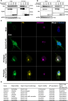
Fig 7. Differences in the interaction between GARIN1B/3 and RAB2A/B.
(A and B) Co-IP of GARIN1B/3 and constitutively active (CA)/constitutively negative (CN) forms of RAB2A/B. GARIN3 interacts with both CA and CN forms of RAB2A/B, while GARIN1B only interacts with the CA form of RAB2A/B. (C) Localization of GARIN3-FLAG (green) with or without PA-RAB2A/B (yellow) in COS-7 cells. The Golgi apparatus and nuclei were stained with anti-GOLGA2 (magenta) and Hoechst33342 (Blue), respectively. (D) Summary of phenotypes observed on male reproduction for all Garins KO male mice.

References

    1. Baibakov B, Gauthier L, Talbot P, Rankin TL, Dean J. Sperm binding to the zona pellucida is not sufficient to induce acrosome exocytosis. Development. 2007;134: 933–943. doi: 10.1242/dev.02752 - DOI - PubMed
    1. Hirohashi N, Yanagimachi R. Sperm acrosome reaction: its site and role in fertilization. Biol Reprod. 2018;99: 127–133. doi: 10.1093/biolre/ioy045 - DOI - PubMed
    1. Morohoshi A, Miyata H, Tokuhiro K, Iida-Norita R, Noda T, Fujihara Y, et al.. Testis-enriched ferlin, FER1L5, is required for Ca2+activated acrosome reaction and male fertility. Sci Adv. 2023;9. doi: 10.1126/sciadv.ade7607 - DOI - PMC - PubMed
    1. Brucker C, Lipford GB. The human sperm acrosome reaction: physiology and regulatory mechanisms. An update. Hum Reprod Update. 1995;1: 51–62. doi: 10.1093/humupd/1.1.51 - DOI - PubMed
    1. Okabe M. Sperm–egg interaction and fertilization: past, present, and future. Biol Reprod. 2018;99: 134–146. doi: 10.1093/biolre/ioy028 - DOI - PubMed