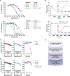
Preclinical proof of principle for orally delivered Th17 antagonist miniproteins
- PMID: 38936360
- PMCID: PMC11316638
- DOI: 10.1016/j.cell.2024.05.052
Preclinical proof of principle for orally delivered Th17 antagonist miniproteins
Abstract
Interleukin (IL)-23 and IL-17 are well-validated therapeutic targets in autoinflammatory diseases. Antibodies targeting IL-23 and IL-17 have shown clinical efficacy but are limited by high costs, safety risks, lack of sustained efficacy, and poor patient convenience as they require parenteral administration. Here, we present designed miniproteins inhibiting IL-23R and IL-17 with antibody-like, low picomolar affinities at a fraction of the molecular size. The minibinders potently block cell signaling in vitro and are extremely stable, enabling oral administration and low-cost manufacturing. The orally administered IL-23R minibinder shows efficacy better than a clinical anti-IL-23 antibody in mouse colitis and has a favorable pharmacokinetics (PK) and biodistribution profile in rats. This work demonstrates that orally administered de novo-designed minibinders can reach a therapeutic target past the gut epithelial barrier. With high potency, gut stability, and straightforward manufacturability, de novo-designed minibinders are a promising modality for oral biologics.
Keywords: IL-17; IL-23R; Th17; autoinflammation; computational protein design; inflammatory bowel disease; oral biologics; protein engineering.
Copyright © 2024 The Author(s). Published by Elsevier Inc. All rights reserved.
Conflict of interest statement
Declaration of interests S.B., T.-Y.Y., I.S.P., L.S., and D.B. are co-founders and shareholders of Mopac Biologics, Inc. S.B., F.S., T.-Y.Y., and D.B. are co-inventors on a patent describing the IL-23R minibinders (PCT/US2021/039122), licensed to Mopac Biologics. S.B. is a board member and paid consultant of Mopac Biologics.
Figures


Comment in
-
Oral miniproteins treat IBD.Nat Rev Drug Discov. 2024 Sep;23(9):660. doi: 10.1038/d41573-024-00126-z. Nat Rev Drug Discov. 2024. PMID: 39054399 No abstract available.
Similar articles
-
atRA Attenuates High Salt-Driven EAE Mainly Through Suppressing Th17-Like Regulatory T Cell Response Mediated by the Inhibition of IL-23R Signaling Pathway.Inflammation. 2025 Jun;48(3):1403-1419. doi: 10.1007/s10753-024-02130-2. Epub 2024 Aug 21. Inflammation. 2025. PMID: 39167321
-
BATF-dependent Th17 cells act through the IL-23R pathway to promote prostate adenocarcinoma initiation and progression.J Natl Cancer Inst. 2024 Oct 1;116(10):1598-1611. doi: 10.1093/jnci/djae120. J Natl Cancer Inst. 2024. PMID: 38833676 Free PMC article.
-
The roles of interleukin (IL)-17A and IL-17F in hidradenitis suppurativa pathogenesis: evidence from human in vitro preclinical experiments and clinical samples.Br J Dermatol. 2025 Mar 18;192(4):660-671. doi: 10.1093/bjd/ljae442. Br J Dermatol. 2025. PMID: 39531733 Clinical Trial.
-
The Black Book of Psychotropic Dosing and Monitoring.Psychopharmacol Bull. 2024 Jul 8;54(3):8-59. Psychopharmacol Bull. 2024. PMID: 38993656 Free PMC article. Review.
-
Systemic pharmacological treatments for chronic plaque psoriasis: a network meta-analysis.Cochrane Database Syst Rev. 2017 Dec 22;12(12):CD011535. doi: 10.1002/14651858.CD011535.pub2. Cochrane Database Syst Rev. 2017. Update in: Cochrane Database Syst Rev. 2020 Jan 9;1:CD011535. doi: 10.1002/14651858.CD011535.pub3. PMID: 29271481 Free PMC article. Updated.
Cited by
-
Re-engineering of a carotenoid-binding protein based on NMR structure.Protein Sci. 2024 Dec;33(12):e5216. doi: 10.1002/pro.5216. Protein Sci. 2024. PMID: 39548819
-
The Effects of Fisetin on Gene Expression Profile and Cellular Metabolism in IFN-γ-Stimulated Macrophage Inflammation.Antioxidants (Basel). 2025 Feb 4;14(2):182. doi: 10.3390/antiox14020182. Antioxidants (Basel). 2025. PMID: 40002369 Free PMC article.
-
Emergence of specific binding and catalysis from a designed generalist binding protein.bioRxiv [Preprint]. 2025 Jul 22:2025.01.30.635804. doi: 10.1101/2025.01.30.635804. bioRxiv. 2025. PMID: 39975260 Free PMC article. Preprint.
-
Advances in the research of intestinal fungi in Crohn's disease.World J Gastroenterol. 2024 Oct 21;30(39):4318-4323. doi: 10.3748/wjg.v30.i39.4318. World J Gastroenterol. 2024. PMID: 39492826 Free PMC article.
-
One-shot design of functional protein binders with BindCraft.Nature. 2025 Aug 27. doi: 10.1038/s41586-025-09429-6. Online ahead of print. Nature. 2025. PMID: 40866699
References
-
- Feagan BG, Sandborn WJ, Gasink C, Jacobstein D, Lang Y, Friedman JR, Blank MA, Johanns J, Gao L-L, Miao Y, et al. (2016). Ustekinumab as Induction and Maintenance Therapy for Crohn’s Disease. N. Engl. J. Med. 375, 1946–1960. - PubMed
-
- Sands BE, Sandborn WJ, Panaccione R, O’Brien CD, Zhang H, Johanns J, Adedokun OJ, Li K, Peyrin-Biroulet L, Van Assche G, et al. (2019). Ustekinumab as Induction and Maintenance Therapy for Ulcerative Colitis. N. Engl. J. Med. 381, 1201–1214. - PubMed
-
- Yang H, Li B, Guo Q, Tang J, Peng B, Ding N, Li M, Yang Q, Huang Z, Diao N, et al. (2022). Systematic review with meta-analysis: loss of response and requirement of ustekinumab dose escalation in inflammatory bowel diseases. Aliment. Pharmacol. Ther. 55, 764–777. - PubMed
-
- Roblin X, Duru G, Papamichael K, Cheifetz AS, Kwiatek S, Berger A-E, Barrau M, Waeckel L, Nancey S, and Paul S (2023). Development of Antibodies to Ustekinumab Is Associated with Loss of Response in Patients with Inflammatory Bowel Disease. J. Clin. Med. Res 12. 10.3390/jcm12103395. - DOI - PMC - PubMed
-
- Janssen Biotech, Inc. (2016). Stelara (ustekinumab) [package insert]. www.accessdata.fda.gov/drugsatfda_docs/label/2016/761044lbl.pdf. Accessed May 26, 2024.
MeSH terms
Substances
Grants and funding
LinkOut - more resources
Full Text Sources
Molecular Biology Databases
Miscellaneous

