. 2024 Jul 16;11(7):ENEURO.0093-24.2024.
doi: 10.1523/ENEURO.0093-24.2024. Print 2024 Jul.

Chronic Social Defeat Stress Increases Brain Permeability to Ghrelin in Male Mice

Affiliations

Chronic Social Defeat Stress Increases Brain Permeability to Ghrelin in Male Mice

Andrea Smith et al. eNeuro. .

Abstract

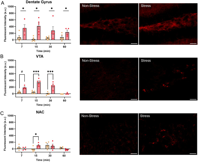
Ghrelin is a stomach-derived hormone that increases feeding and is elevated in response to chronic psychosocial stressors. The effects of ghrelin on feeding are mediated by the binding of ghrelin to the growth hormone secretagogue receptor (GHSR), a receptor located in hypothalamic and extrahypothalamic regions important for regulating food intake and metabolic rate. The ability of ghrelin to enter the brain, however, seems to be restricted to circumventricular organs like the median eminence and the brainstem area postrema, whereas ghrelin does not readily enter other GHSR-expressing regions like the ventral tegmental area (VTA). Interestingly, social stressors result in increased blood-brain barrier permeability, and this could therefore facilitate the entry of ghrelin into the brain. To investigate this, we exposed mice to social defeat stress for 21 d and then peripherally injected a Cy5-labelled biologically active ghrelin analog. The results demonstrate that chronically stressed mice exhibit higher Cy5-ghrelin fluorescence in several hypothalamic regions in addition to the ARC, including the hippocampus and midbrain. Furthermore, Cy5-ghrelin injections resulted in increased FOS expression in regions associated with the reward system in chronically stressed mice. Further histologic analyses identified a reduction in the branching of hypothalamic astrocytes in the ARC-median eminence junction, suggesting increased blood-brain barrier permeability. These data support the hypothesis that during metabolically challenging conditions like chronic stress, ghrelin may be more able to cross the blood-brain barrier and diffuse throughout the brain to target GHSR-expressing brain regions away from circumventricular organs.

Keywords: blood–brain barrier; chronic social stress; dopamine; energy balance; food intake; ghrelin; hypothalamus; nucleus accumbens; ventral tegmental area.


Conflict of interest statement

The authors declare no competing financial interests.

Figures

Figure 1.
Figure 1.
Peripherally administered Cy5-ghrelin accesses the brain via median eminence. A, Experimental timeline of 21 d chronic social defeat stress paradigm, followed by timed Cy5-ghrelin administration and perfusion. B, Chronic social defeat stress increased total food consumed during the stress paradigm but did not alter weight gain. C, Validation of Cy5-ghrelin labeling revealed similar patterns of entry via the median eminence, which was unique from vehicle-injected controls. Fluorescent intensity from vehicle-injected controls was used to normalize the Cy5-ghrelin fluorescence results. Panels D–F show Cy5-ghrelin signal in the OVLT (panels a and b), SFO (panels c and d), and choroid plexus (CP; panels e and f) of control and socially defeated mice. As shown in this image, mice exposed to chronic social defeat appeared to have increased Cy5-ghrelin signal compared with signal in the same regions compared with nonstressed mice. Scale bars: C, 100 μm; DF, 50 μm. ***p < 0.001.
Figure 2.
Figure 2.
Chronic social defeat stress increases detectable ghrelin signals in hypothalamic nuclei. A, Cy5-ghrelin fluorescent signal 7 min following peripheral injection shows increased ghrelin infiltration into the hypothalamus in stressed mice. The arrowheads highlight the areas of Cy5-ghrelin labeling in nonstressed and stressed mice. Scale bar, 100 µm. Cy5-ghrelin fluorescent signal was elevated in stressed mice in the (B) ARC at 7 and 15 min following injection and in the (C) VMH at 7 min following injection. D, Cy5-ghrelin fluorescent intensity was increased in the LH of stressed mice, regardless of time since injection. E, There were no significant differences in the DMH. *p < 0.05, **p < 0.01, ***p < 0.001.
Figure 3.
Figure 3.
Chronic social defeat enhances detectable ghrelin signals in nonhypothalamic, ghrelin-responsive brain regions. A, Stress increased Cy5-ghrelin fluorescent signal in the dentate gyrus of the hippocampus, which was not altered by time following injection. B, Cy5-ghrelin signal was nearing a significant increase into the VTA at 7 min and was significantly elevated at 15 and 30 min, as displayed in microscopy images. C, Cy5-ghrelin fluorescent intensity in the NAc increased in stressed mice at 15 min following injection. Images show Cy5-ghrelin signal 15 min following peripheral administration. Scale bar, 50 µm. #p < 0.07, *p < 0.05, ***p < 0.001.
Figure 4.
Figure 4.
Ghrelin increases c-fos expression throughout the mediobasal hypothalamus and midbrain of socially defeated mice. A, Ghrelin increased the number of c-fos+ cells in the ARC of both nonstressed and stressed mice. Socially defeated mice had an increase in c-fos+ cells in the (B) VMH and (C) LH in response to ghrelin administration. D, There was no difference in c-fos–expressing cells in the DMH. E, Images of the mediobasal hypothalamus highlight stress-related changes in ghrelin-induced c-fos expression. Scale bar, 100 µm. F, Stress enhanced ghrelin's activation of c-fos in the VTA, while in the (G) NAc, ghrelin increased c-fos+ cells compared with vehicle-injected controls. H, Midbrain images highlight differences in c-fos expression in the VTA of nonstressed and stressed mice. Scale bar, 50 µm. *p < 0.05, **p < 0.01, ***p < 0.001.
Figure 5.
Figure 5.
Chronic stress induces morphological changes to astrocytes in the ARC surrounding areas of ghrelin entry. A, B, Images highlight the changes in astrocyte morphology including a reduction in the number of branches and branch length (empty arrowhead) in stressed mice, compared with nonstressed controls that displayed extensive branching and radial morphology (filled arrowhead). Reduction in astrocyte branching was identified near the Cy5-ghrelin signal. Scale bar, 100 µm. C, Cumulative branch length of astrocytes in stressed mice was significantly decreased, compared with nonstressed controls. D, There was no difference in the total number of astrocyte branches between the stressed and nonstressed groups. E, Sholl analysis revealed a significant difference in the number of astrocyte branch intersections measured at a constant radial distance from the cell body, with stressed mice exhibiting a significant reduction in branches at 44 and 46 µm from the soma. F, Dextran fluorescent signal in neurovasculature (tomato lectin) of the ARC was visible in stressed mice. G, Chronic stress was associated with an increase in the mean fluorescent intensity in the median eminence and ARC junction, compared with nonstressed controls, suggesting leakiness of the blood–brain barrier. *p < 0.05, **p < 0.01.

Similar articles

Cited by

References

    1. Abbott NJ, Rönnbäck L, Hansson E (2006) Astrocyte–endothelial interactions at the blood–brain barrier. Nat Rev Neurosci 7:41–53. 10.1038/nrn1824 - DOI - PubMed
    1. Abizaid A, et al. (2006) Ghrelin modulates the activity and synaptic input organization of midbrain dopamine neurons while promoting appetite. J Clin Invest 116:3229–3239. 10.1172/JCI29867 - DOI - PMC - PubMed
    1. Abizaid A (2009) Ghrelin and dopamine: new insights on the peripheral regulation of appetite. J Neuroendocrinol 21:787–793. 10.1111/j.1365-2826.2009.01896.x - DOI - PubMed
    1. Arshadi C, Günther U, Eddison M, Harrington KIS, Ferreira TA (2021) SNT: a unifying toolbox for quantification of neuronal anatomy. Nat Methods 18:374–377. 10.1038/s41592-021-01105-7 - DOI - PubMed
    1. Balland E, et al. (2014) Hypothalamic tanycytes are an ERK-gated conduit for leptin into the brain. Cell Metab 19:293–301. 10.1016/j.cmet.2013.12.015 - DOI - PMC - PubMed

LinkOut - more resources